STIMULATION AND AROUSAL
Arousal Responses to Olfactory or Trigeminal Stimulation During Sleep Boris A. Stuck, MD1; Kathrin Stieber1; Sabine Frey1; Christopher Freiburg1; Karl Hörmann, MD1; Joachim T. Maurer, MD1; Thomas Hummel, MD2 Department of Otorhinolaryngology, Head and Neck Surgery, University Hospital Mannheim, Germany; 2 Smell & Taste Clinic, Department of Otorhinolaryngology, University of Dresden Medical School, Germany 1
Study Objectives: The interaction of sensory physiology and sleep has been studied for various sensory systems. Nevertheless, the question whether chemosensory (especially olfactory) stimuli may lead to arousals during sleep remains under discussion. Specifically, the central processing of olfactory information shows fundamental differences compared to other sensory systems. Design: Prospective controlled trial. Setting: Sleep research facility, University Hospital. Participants: Five young healthy, normosmic volunteers. Intervention: Intranasal chemosensory stimulation during sleep was based on air-dilution olfactometry. For olfactory stimulation H2S (smell of rotten eggs) was used in 4 concentrations (1, 2, 4, and 8 ppm). For trigeminal stimulation CO2 (stinging sensation) was also administered in 4 concentrations (10%, 20%, 40%, and 60% v/v) while odorless stimuli were used for control. Measurements: Arousal reactions due to chemosensory stimulation were
assessed during overnight polysomnography 30 seconds after the presentation of every stimulus during 23 nights of testing. Results: For olfactory testing, an average number of 703 olfactory stimuli and 157 odorless controls were used for analysis per subject. Even the highest stimulus concentration did not produce an increase in arousal frequency. For trigeminal testing, an average number of 405 stimuli and 79 controls were used for analysis per subject, and an increase in arousal frequency was observed following the increase of stimulus concentration. Conclusions: With the present results we were able to demonstrate that, in contrast to trigeminal stimulation, the presentation of a strong but selective olfactory stimulus does not lead to arousals during nocturnal sleep in humans. Keywords: chemosensation, arousals, olfaction, trigeminal Citation: Stuck BA; Stieber K; Frey S et al. Arousal responses to olfactory or trigeminal stimulation during sleep. SLEEP 2007;30(4):506-510.
INTRODUCTION
question remains whether chemosensory stimuli, and especially olfactory stimuli, may lead to arousals during sleep. Considering the limited data available, awakening reactions seem to occur following olfactory stimulation.6,7 Yet the odorants used for stimulation in these trials were either peppermint6,7 or pyridine,7 both of which activate not only the olfactory but also the trigeminal system which is a characteristic of most odorants.8 Only a limited number of odorants such as hydrogen sulfide (H2S), vanillin or phenylethyl alcohol have little or no trigeminal impact.8,9 Although both systems interact at several levels of processing,10 we investigated these sensations separately as they represent 2 different sensory systems. Arousals during sleep may be triggered by a number of internal and external stimuli such as acoustic,11-14 vibrotactile,14 nociceptive,15,16 and thermal stimuli.17 This process may be different for olfaction. The central processing of olfactory stimuli shows fundamental differences compared with other sensory systems. These differences include: (1) the predominantly ipsilateral processing of the olfactory stimuli, (2) the almost direct anatomical connectivity to the “limbic system” and thus to centers for the processing of memories and emotions, and (3) the fact that olfactory information processing largely bypasses the thalamus1,18,19 which typically gates information from other sensory systems during sleep.20 Hence, the question arises whether olfactory information is gated at all. Recent observations by Marukami et al suggest that the processing in the olfactory cortex is gated through activation in the ascending reticular activating system (ARAS).21 Thus, for the olfactory system the ARAS seems to take the place of the thalamus. The aim of the present study was to investigate whether trigeminal or olfactory stimuli induce arousals during sleep and to investigate the relation between stimulus concentration and arousal frequency.
THE INTERACTION OF SENSORY PHYSIOLOGY AND SLEEP HAS BEEN STUDIED FOR VARIOUS SENSORY SYSTEMS INCLUDING VISION, AUDITION, AND somatosensation, both in humans and in animals. The vast majority of studies, however, have been performed on the basis of auditory stimuli. This may be explained at least in part by the fact that acoustic stimuli can be controlled more easily than, for example, somatosensory stimuli and chemosensory stimuli. The orbitofrontal cortex is closely involved in the processing of olfactory information1 that has also been implied in sleep physiology.2 In addition, recent work suggests that sleep deprivation produces a specific decrease of odor identification, indicating a functional connectivity between olfaction and sleep via the orbitofrontal cortex.3,4 Whether olfactory stimuli are processed on a cortical level during sleep is still debated. Recent investigations suggest that chemosensory event-related potentials can indeed be recorded during sleep and that those chemosensory event-related potentials are significantly altered compared to wakefulness.5 Nevertheless, the Disclosure Statement Drs. Stuck, Maurer, Hummel, Hormann, Ms. Steiber, Ms. Frey, and Mr. Freiburg have indicated no financial conflicts of interest. Submitted for publication August 2006 Accepted for publication December 2006 Address correspondence to: Boris A. Stuck, MD; Department of Otorhinolaryngology, Head and Neck Surgery, University Hospital Mannheim, 68135 Mannheim, Germany; phone +49 621 383 1600; fax: +49 621 383 3827;
[email protected] SLEEP, Vol. 30, No. 4, 2007
506
Chemosensory Induced Arousals—Stuck et al
METHODS
Moreover, this constant airstream ensures that the influence of breathing patterns on stimulus presentation to the oflactory epithelium is minimized. For specific olfactory stimulation, the unpleasant H2S, typically described as smelling like rotten eggs, was presented at 1, 2, 4, and 8 ppm, all concentrations being clearly above threshold. To evaluate trigeminal function, CO2 was administered at 10%, 20%, 40%, and 60% v/v, with the last 2 concentrations producing strong stinging painful sensations at the nasal mucosa.32 Stimulus duration was one second. Application of a limited number of these short CO2 stimuli at this long interstimulus interval to the surface of the respiratory epithelium is not likely to produce changes in blood pH. Longer stimuli were thought to lead to desensitization which appears to be especially pronounced in the olfactory system.33,34 Odorless stimuli were presented as a control. Stimuli were presented in a randomized fashion during every night according to a computer based randomization protocol. The interstimulus interval was set to 30 seconds. To allow mobility during sleep, a tube approximately 60 cm long was used to connect the subject’s nostril with the olfactometer outlet. This ensured that changes in body position had little influence on stimulus presentation. The tube was secured with tape to the nostril. A curtain separated the subject’s bed from the olfactometer and the investigator. Ear plugs were given to subjects to dampen external sounds. The hypnogram was constantly observed by the same trained investigator during all nights (KS) to detect when the subjects were falling asleep. As soon as subjects reached a stable sleep stage, chemosensory stimulation started. Stimuli were presented in both light (sleep stages 1 and 2) and slow wave sleep (sleep stages 3 and 4). With regard to the experimental setting, the sleep time available for testing was limited. As REM sleep predominantly occurs during the second half of the night, sleep time available in REM sleep was minimal. For this reason, stimuli were presented in NREM sleep only. Arousals were registered online by the investigator (KS) and confirmed within a few hours by 2 experienced observers (BAS and JTM), independently from each other. An arousal was defined as being associated with the stimulus if it appeared within 30 s after chemosensory stimulation. The number of arousal reactions in relation to the number of stimuli was then calculated for the different stimulus concentrations and sleep stages for every subject (arousal frequency in %), and a cumulative frequency was calculated by summarizing all stimuli and all arousals observed at a specific stimulus concentration for the entire group. If a change in sleep stage during the measurement was detected, the measurement was continued. In those cases in which an awakening occurred, measurements were stopped and restarted as soon as the subject again reached a stable sleep stage. When measurements were started or interrupted, marks were set in the hypnogram to be able to re-evaluate and potentially reclassify sleep stages by 2 independent sleep specialists (BAS, JTM). Statistical analyses were based on SPSS (Statistical Product and Service Solution) v. 12.0 (SPSS Inc, Chicago, IL, USA). Data were submitted to analyses of variance for repeated measures (rm-ANOVA), with “stimulus concentration” as within-subject factor, separately for light and slow wave sleep, and for olfactory and trigeminal stimuli. Degrees of freedom are presented following the F-value. The alpha level was set at 0.05.
The study was conducted at the Sleep Disorders Center at the Department of Otorhinolaryngology, Head and Neck Surgery Mannheim. The study protocol was approved by the local ethics board of the Medical Faculty Mannheim of the University of Heidelberg; written informed consent was obtained from all participants. Participants Since young females have demonstrated the best olfactory performance among human subjects,22-24 5 young healthy female volunteers were included in this prospective study (mean age 24.6±1.5 years, range: 23–26 years), and 23 nights of testing were performed. Exclusion criteria were actual/previous history of smell or taste disorders, use of medications known to affect chemosensory function,25 and a history of sleep disorders. At the screening visit, relevant nasal pathology such as mucosal inflammation, significant septal deviation, and nasal polyposis were ruled out with a detailed clinical examination including nasal endoscopy. Patency of the nasal airways was additionally ascertained using active anterior rhinomanometry (Rhinomanometer 300, ATMOS Medizintechnik GmbH & Co KG, Lenzkirch, Germany). Psychophysical Testing of Olfactory Function All participants underwent olfactory testing using the “Sniffin’ Sticks” test kit to establish normal olfactory function.26,27 Odorants were presented in odor dispensers similar to felt-tip pens. Testing involved assessment of n-butanol odor thresholds, odor discrimination, and odor identification. In order to categorize olfactory function in terms of functional anosmia, hyposmia, and normosmia, the sum of the 3 scores for odor thresholds, odor discrimination, and odor identification was used (“TDI score”).28 Sleep Recordings To assess arousal reactions to chemosensory stimulation during sleep, subjects were admitted to the sleep disorders center. An overnight polysomnogram was performed to assess nocturnal sleep, including sleep stages and arousal reactions. Monitoring included 2 electroencephalographic channels (C3-A2, C4-A1), 2 electrooculograms (left, right), 2 submental and 2 leg electromyograms (left, right). Sleep stages were scored according to Rechtschaffen and Kales.29 Arousals were defined as an abrupt shift in EEG frequency, which may include theta, alpha and/or frequencies greater than 16 Hz lasting 3 seconds or longer after at least 10 seconds of any sleep stage, as defined by the Atlas task force of the American Sleep Disorders Association.30 Chemosensory stimulation For stimulation, a dynamic olfactometer based on air-dilution olfactometry was used (OM6b; Burghart instruments, Wedel, Germany). This allows the presentation of odorous stimuli within a continuous airstream of 8 L/min; this does not alter the mechanical or thermal conditions at the nasal mucosa.31 SLEEP, Vol. 30, No. 4, 2007
507
Chemosensory Induced Arousals—Stuck et al
90 80
10
arous al- freque ncy in %
arous al- freque ncy in %
12
8 6
light sleep
4
slow wave sleep
2 0 -2
0
1
2
4
70 60 50
light sleep
40
slow wave sleep
30 20 10 0
8
-10
0
10
20
ppm H2S
40
60
v/v % CO2
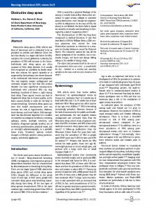
Figure 1—Arousal frequency (in %) for the different concentrations of H2S in both light and slow wave sleep (mean±SD)
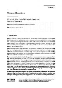
Figure 2—Arousal frequency (in %) for the different concentrations of CO2 in both light and slow wave sleep (mean±SD)
RESULTS
to arousal reactions during sleep. In contrast, for trigeminal testing an increase in arousal frequency was observed in relation to an increase in stimulus concentration (Table 2; Figure 2). This was true for both sleep stages (light sleep: F4,16 = 18.4, P