bs_bs_banner
Entomological Research 45 (2015) 345–353
SH ORT COM M U NICAT IO N
Molecular identification of four Panonychus species (Acari: Tetranychidae) in Korea, including new records of P. caglei and P. mori Tin Moe KHAING1*, Jae-Kyoung SHIM1,2 and Kyeong-Yeoll LEE1,2,3 1
School of Applied Biosciences, Kyungpook National University, Daegu, Republic of Korea Institute of Plant Medicine, Kyungpook National University, Daegu, Republic of Korea 3 Sustainable Agriculture Research Center, Kyungpook National University, Gunwi, Republic of Korea 2
Correspondence Kyeong-Yeoll Lee, School of Applied Biosciences, Kyungpook National University, Sankyukdong 1370, Daegu 702-701, Republic of Korea. Email:
[email protected] Received 31 August 2015; accepted 24 September 2015. *Present address: Food Security Working Group, Yangon, Myanmar. doi: 10.1111/1748-5967.12135
Abstract Four species of the genus Panonychus are identified in Korea including two previously known species P. citri (McGregor) and P. ulmi (Koch) and two newly identified species P. mori Yokoyama and P. caglei Mellot. Morphological diagnostics were observed among the four species in the color of dorsal tubercles and in the shape of male aedeagus. The dorsal tubercles of P. citri, P. ulmi, P. mori and P. caglei are red, white, whitish red and light red, respectively. The aedeagi of male adults are diagnostic in sigmoid-shaped at the distal end and its shape and length was different among four species. Molecular comparison of the internal transcribed spacer 2 (ITS2) sequence of nuclear DNA and cytochrome-c oxidase subunit I (COI) nucleotide sequence of mitochondrial DNA among the four species showed divergences 8–12% and 9–12%, respectively. Molecular analysis of the ITS2 and COI sequences revealed their divergences were slightly different among four species. In addition, species-specific primer sets were designed at the base on ITS2 sequences to precisely diagnose these four species at the molecular level. Key words: COI, ITS2, molecular diagnosis, spider mites.
Introduction The genus Panonychus Yokoyama is a small group in the family Tetranychidae that includes at least 16 species globally (Bolland et al. 1998; De Vis & De Moraes 2002). Previously, seven species have been identified in Japan, four in China and four in the USA; however, only two species including the citrus red mite, P. citri (McGregor) and the European red mite, P. ulmi (Koch), are known in Korea (Han 1969, 1970; Bolland et al. 1998; Ohno et al. 2011). These two species are cosmopolitan and cause serious damage to citrus and apple trees, and also to a variety of economically important agricultural crops (Jeppson et al. 1975; Gotoh & Noguchi 1990; Kitashima & Gotoh 1995; Bolland et al. 1998; Gotoh et al. 2003; Lei et al. 2004; Osakabe et al. 2005). © 2015 The Entomological Society of Korea and Wiley Publishing Asia Pty Ltd
The genus Panonychus can be distinguished from other genera in the family Tetranychidae by a dorsal idiosomal setae set on strong tubercles (Bolland et al. 1998). In addition, the male aedeagus is the most important key character for speciation in the Tetranychidae (Jeppson et al. 1975). However, the limitation of any other morphological characters in nymphs and female adults make difficulties in species diagnosis in this genus. Therefore, studies using various molecular markers are highly valuable to determine inter- or intra-specific relationships in mites. The internal transcribed space 2 (ITS2) sequences of nuclear DNA have been used to infer species diagnostics among a wide range of insects and mites (Hillis & Dixon 1991; Hinomoto & Takafuji 2001; Navajas et al. 2001; Rojas et al. 2001; Noge et al. 2005; Ben-David et al. 2007). Noge et al. (2005) analyzed ITS2 sequences of 73 specimens of astigmatid mites and
T. M. Khaing et al.
suggested their sequences are species-specific and useful for studying species diagnostics. Navajas et al. (1994) suggest ITS2 sequences of nuclear DNA show better resolution in species identification of tetranychid groups. In addition, the cytochrome-c oxidase subunit I (COI) sequence of mitochondrial DNA (mtDNA) is commonly used to identify species in the family Tetranychidae (Lunt et al. 1996; Navajas et al. 1996). Both P. citri and P. ulmi are recognized as economically important pests of various crops, including citrus and apple; however, molecular identification has not been determined on other species of the genus Panonychus. Navajas et al. (1996) reported that the molecular identification based on the COI sequences of 20 species of phytophagous mites including P. citri and P. ulmi belonging to nine genera and two families is concordant with the morphological characters. Toda et al. (2000) determined molecular identification among four Japanese Panonychus species, by analyzing the COI gene. Prior to this report, P. citri and P. ulmi were identified in Korea only based on morphological characters (Lee et al. 1986). In the present study, we identified four Panonychus species including two new records of P. mori and P. caglei in Korea by using morphological and molecular analysis. In addition, we designed species-specific primer sets based on ITS2 sequences.
Materials and methods Sample collection and morphological analysis We collected P. citri from citrus trees and P. ulmi from apple trees. Panonychus mori was collected from jujube plants, Zizyphus jujube, at Gyeongsan, Gyeongbuk Province, in June 2011 and P. caglei was collected from kudzu vines, Pueraria thunbergiana, at Byunsan peninsula, located in the western part of Korea, in September 2011. Samples were preserved in a 70% ethanol solution for further investigation. For morphological identification, we mounted slides in Hoyer’s medium (Gutierrez 1985). Mites mounted on the slides were observed under a high-power phase contrast microscope (Olympus, Tokyo, Japan). Species identification was performed based on the key characters listed in Ehara and Gotoh (1992). DNA extraction and PCR amplification Genomic DNA was extracted from non-mashed samples of a single adult female mite, according to Khaing et al. (2013). First, a single mite was in a 1.5 mL sample tube containing 200 μL of digestion buffer and 20 μL of proteinase K (50 μg/mL), for 12 h at 55°C. The mite exoskeleton was then transferred into another tube, which was filled with 200 μL of 70% ethanol for further morphological identification. 346
Genomic DNA was extracted from the remaining solution using a PureLink Genomic DNA Mini Kit (Invitrogen, Waltham, MA, USA). Concentrations of purified DNA samples were determined by using a Nanophotometer (Implen, Munich, Germany). The ITS2 sequence was amplified by using the following pair of primers designed by Ben-David et al. (2007): 5′-GTCACATCTGTCTGAGAGTTGAGA-3′ and 5′-GTAR CCTCACCTRMTCTGAGATC-3′. Polymerase chain reaction (PCR) was performed in 20 μL of Smart Taq Pre-Mix (Solgent, Daejeon, Korea) containing 30 ng of DNA as a template and 10 pmol of each primer using a PTC-200 thermal cycler (MJ Research, Hercules, CA, USA). The cycle conditions were denaturation at 95°C for 5 min, 40 cycles (95°C for 60 s, 51°C for 40 s and 72°C for 60 s) and a final extension at 72°C for 10 min. The COI sequence was amplified by using a pair of primers designed by Navajas et al. (1996): an unvT primer set 5′-TGATTTTTTGGTCA CCCAGAAG-3′ and 5′-TACAGCTCCTATAGATAAAAC3′. The PCR cycle conditions were initial denaturation at 95°C for 5 min, 35 cycles (95°C for 60 s, 51°C for 60 s, 72°C for 60 s) and a final extension at 72°C of 10 min. The PCR products were separated by using 1% agarose gel electrophoresis, visualized under UV light after staining with ethidium bromide solution and purified by using spin columns (Wizard PCR Preps DNA Purification System; Promega, WI, USA). The purified products were directly sequenced by using an Applied Biosystems 3100 Capillary DNA Sequence and a BigDye® Terminator Cycle Sequencing Kit (Applied Biosystems, Foster City, CA, USA) at the Solgent Sequencing Facility (Solgent, Daejeon, Korea). Identification of species-specific primer sequences We designed species-specific primer sets based on ITS2 sequences. The ITS2 sequences determined in the present study and those obtained from NCBI GenBank were aligned and differentiated regions were selected for each species (Table 1). The species-specific primers from each sample were analyzed using PCR. The cycle conditions were denaturation at 95°C for 5 min, 30 cycles (95°C for 60 s, 46°C for 40 s and 72°C for 60 s) and a final extension at 72°C for 10 min. The PCR products were separated by 1% agarose gel electrophoresis as described above. DNA sequencing and species identification analysis The partial ITS2 and COI sequences were compared with those of spider mites in NCBI database (Altschul et al. 1997; Schaffer et al. 2001). The sequences were aligned and edited using the Clustal W multiple alignments in BioEdit v7.0 Entomological Research 45 (2015) 345–353 © 2015 The Entomological Society of Korea and Wiley Publishing Asia Pty Ltd
Panonychus species in Korea
Table 1 Species-specific primers of four Panonychus species
Species P. citri P. ulmi P. mori P. caglei
†
Table 2
Primer set
Sequence (5′–3′)
Length (bp)
Tm†
Pc-ITS2 (F) Pc-ITS2 (R) Pu-ITS2 (F) Pu-ITS2 (R) Pm-ITS2 (F) Pm-ITS2 (R) Pca-ITS2 (F) Pca-ITS2 (R)
5′-GCAGTAGTTTAACACCTACTCTGC-3′ 5′-AATGGAAGACGGGATACATGCG-3′ 5′-GCTTAGCAGAGTTTAAATACTCTGC-3′ 5′-TGGAGATGTGTGATACACGCTAC-3′ 5′-GTTCGGTCTTAATTGATTCGAACC-3′ 5′-TGCTAAGGATACAACCAACAGTC-3′ 5′-ACAGCAATATCAGCAGCAACAG-3′ 5′-AGGGAGATGCGTGATACATGC-3′
414
55
405
55
291
55
260
55
Tm, melting temperature (°C).
Panonychus species from Korea GenBank accession no. Species
Population
Host-plant
Collection site
ITS2
COI
Suwon Busan CBNU, Cheongju KNU, Daegu Suwon KNU, Daegu CBNU, Cheongju Gyeongsan, Hayang Byunsan peninsula CBNU, Cheongju
JF774169
KC502919
JF776969
KC502918
KC502915 KC502916 JF774165
KC502917 KC502920 AF131105
Panonychus citri
Pc-BS1
Citrus unshiu Poncirus trifoliate Morus alba
P. ulmi
Pu-GH1
P. mori P. caglei T. urticae†
Pm-GH1 Pcag-DMC1 Tu-CBNU1
Malus pumila Pyrus ussuriensis Glycine max Ziziphus zizyphus Pueraria lobata Malus pumila
†
Tetranychus urticae was used as an outgroup. CBNU, Chungbuk National University; KNU, Kyungpook National University.
(Thompson et al. 1994; Hall 1999). Sequence divergences were calculated using Molecular Evolutionary Genetic Analysis (MEGA) software v4.0.0 (Tamura et al. 2007) among taxa, based on Kimura 2-parameter (K2P) distances (Kimura 1980). Bootstrap values were obtained from 1000 replicates (Felsenstein 1985). Sequences were deposited in the GenBank database. Accession numbers of ITS2 and COI sequences are shown in Table 2.
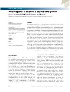
Results and discussion In the present study, we identified morphological and molecular differences among four species of the genus Panonychus in Korea, including two previously known species, P. citri and P. ulmi, and two newly identified species, P. mori and P. caglei. In addition, we designed species-specific primers to precisely diagnose identity these four species. Species in the genus Panonychus are called red mites because the body color is generally red; however, different species show slight variations in the overall body and also in the color of specific body parts, such as dorsal tubercles, which are projected humps at the dorsal surface of the body; Entomological Research 45 (2015) 345–353 © 2015 The Entomological Society of Korea and Wiley Publishing Asia Pty Ltd
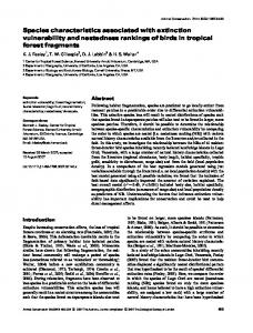
the center of each tubercle contains a single hair (Ehara 1999; Zhang 2003). This character is found in the genus Panonychus but not in other genera of the family Tetranychidae (Bolland et al. 1998). We compared colors of body, dorsal tubercles and their hairs among female adults of four species: P. citri was bright red in both body and tubercles (Fig. 1A), P. ulmi was dark red in body but white in dorsal tubercles and their setae (Fig. 1B), P. mori was red in body but whitish red in tubercles and their setae (Fig. 1C) and P. caglei was light red to pink in body but white in tubercles and their setae (Fig. 1D) (Ehara & Gotoh 1992; Ehara 1999). Thus, each species is unique in the color patterns of the body and their dorsal tubercles. The number of setae in genu IV can be used to identify species within this genus. For example, P. citri and P. ulmi have three setae, whereas P. caglei has only two setae in the same segment (Jeppson et al. 1975). The aedeagus of Panonychus species is typically sigmoidshaped at the distal end. The aedeagus of P. citri is longer and more slender at the proximal part and gently sigmoidshaped at the distal end than the aedeagus of P. ulmi (Fig. 2A, B) (Baker & Tuttle 1994; Ehara 1999). The aedeagus of P. mori is more strongly sigmoid-shaped at the 347
T. M. Khaing et al.
Figure 1 Dorsal setae set on different colors of tubercles in four Panonychus species: P. citri (A), P. ulmi (B), P. mori (C) and P. caglei (D).
Figure 2
Different shapes of male aedeagi in four Panonychus species: P. citri (A), P. ulmi (B), P. mori (C), and P. caglei (D).
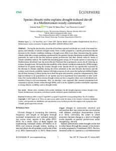
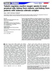
distal end than the aedeagi of P. citri and P. ulmi (Fig. 2A, C). The aedeagus of P. caglei is slender and gently sigmoidshaped at the distal end; further, the proximal end is shorter than P. citri and longer than that of P. ulmi (Fig. 2D). Nucleotide sequences of both ITS2 and COI regions were determined from four Panonychus species and those fragment lengths were 478 and 38 bp, respectively (Table 2). In this study, we first report ITS2 sequences of both P. mori and P. caglei and the COI of P. caglei. The alignment of ITS2 sequences showed high divergence among the four species, with insertions and deletions of some nucleotides (Fig. 3). Otherwise, the alignment of the COI sequences showed lower divergence, without insertions or deletions of nucleotides (Fig. 4). Sequence divergence of ITS2 and COI among the four species was 8–12% and 9–12%, respectively. The nucleotide differences and pairwise distances based on ITS2 and COI nucleotide sequences revealed that P. mori
and P. caglei were located more closely to P. ulmi than to P. citri (Table 3). We determined the genetic distance of ITS2 and COI sequences obtained for the four Panonychus species in Korea and other countries (Figs 5, 6). The sequences of Tetranychus urticae were used as outgroups in this study. Genetic distances of two sequences were slightly different among the four species. Analysis of the ITS2 sequences using the neighbor-joining method revealed the relationship P. ulmi–P. caglei–P. citri–P. mori; on the other hand, analysis of the COI sequences revealed the relationship P. citri–P. caglei–P. ulmi–P. mori. Our results are in accordance with those of two previous studies (Osakabe & Sakagami 1994; Toda et al. 2000). Panonychus mori was previously known to be a diapause strain of P. citri but it was subsequently re-described as P. mori based on specimens collected from mulberry and pear (Ehara & Gotoh 1992). Restriction fragment length polymorphism (RFLP) analysis
348
Entomological Research 45 (2015) 345–353 © 2015 The Entomological Society of Korea and Wiley Publishing Asia Pty Ltd
Panonychus species in Korea
Figure 3 Alignment of partial ITS2 sequences in four species of the genus Panonychus (JF774169, JF776969, KC502915 and KC502916) and in Tetranychus urticae (JF774165). Identity with the first sequence is indicated by dots. Gaps are indicated by dashes.
of the rDNA sequence, which has the ITS2 region, showed that P. mori was closely related to P. citri than P. ulmi (Osakabe & Sakagami 1994). In contrast, COI sequence analysis showed that P. mori was more closely related to P. ulmi than P. citri (Toda et al. 2000). Thus, further investigation with more molecular data is required to determine the precise species identification among Panonychus species. We designed species-specific primer sets of four species based on 29 sequences determined in the present study and obtained from the NCBI database. Each primer set identified its own species but not any of the other species (Fig. 7). Thus, our primer sets can be used to precisely diagnose species within the genus Panonychus. In many countries, both P. citri and P. ulmi are serious pests in citrus and apple orchards, respectively (Shi & Feng 2006). These two species are also known to infest
various other crops. In Korea, P. citri infests bitter orange, spindle tree and Japanese yew. Panonychus ulmi infests apple, pear, plum, grape, persimmon, white mulberry, strawberry, Chinese wolfberry and soybean (Lee et al. 1986). Few studies regarding the host range of P. mori have been conducted. Gotoh and Higo (1997) collected this species from muka, peach, Japanese pear, moonseed and mulberry in Japan. In the present study, we identified a serious infestation of P. mori in jujube orchards in Hayang, Gyeongbuk province, Korea. Panonychus caglei was first identified from raspberry plants (Rosaceae) including Potentilla norvegica, Rosa sp. and Rubus idaeus (Mellot 1968). The species is distributed in USA, Brazil, Taiwan, China and Japan (Bolland et al. 1998; Ohno et al. 2011). In the present study, we collected P. caglei from kudzu vines on the Byunsan peninsula, located in the western part of Korea.
Entomological Research 45 (2015) 345–353 © 2015 The Entomological Society of Korea and Wiley Publishing Asia Pty Ltd
349
T. M. Khaing et al.
Figure 4 Alignment of partial COI sequences in four species of the genus Panonychus (KC502919, KC502918, KC502917 and KC502920) and in Tetranychus urticae (AF131105). Identity with the first sequence is indicated by dots.
Ribosomal ITS2 sequences P. citri P. ulmi P. mori P. caglei Mitochondrial COI sequences P. citri P. ulmi P. mori P. caglei
P. citri
P. ulmi
P. mori
P. caglei
– 0.133 0.133 0.136
63 – 0.107 0.060
66 55 – 0.093
66 35 51 –
– 0.094 0.126 0.094
32 – 0.091 0.088
42 31 – 0.117
32 30 39 –
In conclusion, we compared four species in the genus Panonychus in Korea. This is the first record of P. mori and P. caglei in Korea. We identified the four species based on morphological differences in the color of the dorsal tubercles and in the shape of the male aedeagus. Molecular analysis of the ITS2 and COI sequences revealed their diversities were slightly different among four species. In addition, we designed species-specific primer sets based on the ITS2 sequences to precisely diagnose these four Panonychus species at the molecular level. 350
Table 3 Number of nucleotide differences (above the diagonal) and pairwise distances among four Panonychus species
Acknowledgements We thank insightful comments of Dr Won-Goo Lee, an emeritus professor at Chunbuk National University, and Dr Jong-Ho Lee at National Plant Quarantine Service, Anyang, Republic of Korea. This research was supported by grants from the Animal and Plant Quarantine Agency, Anyang, Republic of Korea and Agricultural Biotechnology Program at Ministry of Agriculture, Food and Rural Affairs, the Republic of Korea. Entomological Research 45 (2015) 345–353 © 2015 The Entomological Society of Korea and Wiley Publishing Asia Pty Ltd
Panonychus species in Korea
Figure 5 Neighbor-joining tree for Panonychus species based on ITS2 sequences. Tetranychus urticae was used as an outgroup. Numbers adjacent to branches denote bootstrap values (>50%) of 1000 replicates.
Figure 6 Neighbor-joining tree for Panonychus species based on COI sequences. Tetranychus urticae was used as an outgroup. Numbers adjacent to branches denote bootstrap values (>30%) of 1000 replicates.
Figure 7 Agarose gel electrophoresis profile of PCR products, obtained by using species specific primer sets: DNA marker (M), Panonychus citri (1), P. ulmi (2), P. mori (3), P. caglei (4).
Entomological Research 45 (2015) 345–353 © 2015 The Entomological Society of Korea and Wiley Publishing Asia Pty Ltd
351
T. M. Khaing et al.
References Altschul SF, Madden TL, Schaffer AA et al. (1997) Gapped BLAST and PSI-BLAST: a new generation of protein database search programs. Nucleic Acids Research 17: 3389–3402. Baker EW, Tuttle DM (1994) A Guide to the Spider Mites (Tetranychidae) of the United States. Indira Publish House, West Bloomfield, MI. Ben-David T, Melamed S, Gerson U, Morin S (2007) ITS2 sequences as barcodes for identifying and analyzing spider mites (Acari: Tetranychidae). Experimental & Applied Acarology 41: 169–181. Bolland HR, Gutierrez J, Flechtmam CHW (1998) World Catalogue of the Spider Mite Family (Acari: Tetranychidae). Koninklijke Brill Press, Leiden, Boston, Koln. De Vis RM, De Moraes GJ (2002) A new species of Panonychus (Acari: Tetranychidae) from Peru. Zootaxa 48: 1–6. Ehara S (1999) Revision of the spider mite family Tetranychidae of Japan (Acari, Prostigmata). Species Diversity 4: 63–141. Ehara S, Gotoh T (1992) Descriptions of two Panonychus spider mites from Japan, with a key to species of the genus in the world (Acari: Tetranychidae). Applied Entomology and Zoology 27: 107–115. Felsenstein J (1985) Confidence limits on phylogenies: an approach using the bootstrap. Evolution 39: 783–791. Gotoh T, Higo Y (1997) Differences in host range and reproductive compatibility among populations of Panonychus mori Yokoyama (Acari: Tetranychidae). International Journal of Acarology 23: 119–125. Gotoh T, Noguchi O (1990) Developmental success and reproductive incompatibility among populations of the European red mite, Panonychus ulmi (Acari: Tetranychidae). Experimental & Applied Acarology 10: 157–165. Gotoh T, Ishikawa Y, Kitashima Y (2003) Life-history traits of the six Panonychus species from Japan (Acari: Tetranychidae). Experimental & Applied Acarology 29: 241–252. Gutierrez J (1985) Mounting techniques. In: Helle W, Sabelis MW (eds) Spider Mites. Their Biology, Natural Enemies and Control, pp 351–353. Science Publishers, Amsterdam. Hall TA (1999) Bioedit, a user-friendly biological sequences alignment editor and analysis program for windows 95/98/NT. Nucleic Acids Symposium Series 41: 95–98. Han KP (1969) Studies on mites (II). On some mites of the apple trees. Korean Journal of Plant Protection 8: 29–35. Han KP (1970) Studies on mites (III). On some mites of persimmon and citrus. Korean Journal of Plant Protection 9: 33–35. Hillis DM, Dixon MT (1991) Ribosomal DNA: molecular evolution and phylogenetic inference. Quarterly Review of Biology 66: 411–453. Hinomoto N, Takafuji A (2001) Genetic diversity and phylogeny in the kanzawai spider mite, Tetranychus kanzawai, in Japan. Experimental & Applied Acarology 25: 355–370. Jeppson LR, Keifer HH, Baker EW (1975) Mites Injurious to Economic Plants, pp 155–163. University of California Press, Berkeley, CA.
352
Khaing TM, Lee JH, Lee WG, Lee KY (2013) A new record of Amphitetranychus quercivorus (Acari: Tetranychidae) in Korea and molecular comparison with A. viennensis. Journal of AsiaPacific Entomology 16: 155–160. Kimura M (1980) A simple method for estimating evolutionary rate of base substitutions through comparative studies of nucleotide sequences. Journal of Molecular Evolution 16: 111– 120. Kitashima Y, Gotoh T (1995) Host range difference and reproductive incompatibility among five populations of the citrus red mite, Panonychus citri (McGregor) (Acari: Tetranychidae). Journal of Acarology Society (Japan) 4: 91–101. Lee WK, Lee BH, Kim BJ (1986) Taxonomic studies on spider mites (Tetranychidae: Acarina) of Korea. Korean Journal of Systematic Zoology 2: 13–16. Lei HD, Hu JH, Li HJ et al. (2004) Performances of the citrus red mite, Panonychus citri. (McGregor) (Acarina: Tetranychidae) on various citrus varieties. Acta Entomologica Sinica 47: 607– 611. Lunt DH, Zhang DX, Szymura JM, Hewitt GM (1996) The insect cytochrome oxidase I gene: evolutionary patterns and conserved primers for phylogenetic studies. Insect Molecular Biology 5: 153–165. Mellot JL (1968) Panonychus caglei, new species, the Raspberry red mite (Acarina: Tetranychidae). Acarologia 10: 230–244. Navajas M, Gutierrez J, Bonato O, Bolland HR, Mapangoudivassa S (1994) Intraspecific diversity of the cassava green mite Mononycellus progresivus (Acari, Tetranychidae) using comparisons of mitochondrial and nuclear ribosomal DNA-sequence and cross-breeding. Experimental & Applied Acarology 18: 351–360. Navajas M, Gutierrez J, Lagnel J (1996) Mitochondrial cytochrome oxidase I in tetranychid mites: a comparison between molecular phylogeny and changes of morphological and life history traits. Bulletin of Entomological Research 86: 407–417. Navajas M, Gutierrez J, Williams M, Gotoh T (2001) Synonymy between two spider mite species, Tetranychus kanzawai and T. hydrangea (Acari: Tetranychidae), shown by ribosomal ITS2 sequences and cross-breeding experiments. Bulletin of Entomological Research 91: 117–123. Noge K, Mori N, Tanaka C, Nishida R, Tsuda M, Kuwahara Y (2005) Identification of astigmatid mites using the second internal transcribed spacer (ITS2) region and its application for phylogenetic study. Experimental & Applied Acarology 35: 29–46. Ohno S, Miyagi A, Gotoh T et al. (2011) Wild host plants of four spider mite species (Acari: Tetranychidae) infesting fruit crops in Okinawa. Journal of Asia-Pacific Entomology 14: 281–284. Osakabe M, Sakagami Y (1994) RFLP analysis of ribosomal DNA in sibling species of spider mite genus Panonycus (Acari: Tetranychidae). Insect Molecular Biology 3: 63–66. Osakabe M, Goka K, Toda S, Shintaku T, Amano H (2005) Significance of habitat type for the genetic population structure
Entomological Research 45 (2015) 345–353 © 2015 The Entomological Society of Korea and Wiley Publishing Asia Pty Ltd
Panonychus species in Korea
of Panonychus citri (McGregor) (Acarina: Tetranychidae). Experimental & Applied Acarology 36: 25–40. Rojas MDE, Mora MD, Ubeda JM, Cutillas C, Navajas M, Guevara DC (2001) Phylogenetic relationships in rhinonyssid mites (Acari: Rhinonyssidae) based on mitochondrial 16S rDNA sequences. Experimental & Applied Acarology 25: 957– 967. Schaffer S, Weil B, Nguyen VD et al. (2001) A high-resolution reference map for cytoplasmic and membrane associated proteins of Corynebacterium glutamicum. Electrophoresis 22: 4404–4422. Shi WB, Feng MG (2006) Field efficacy of application of Beauveria bassiana formulation and low rate pyridaben for sustainable control of citrus red mite Panonychus citri (Acari: Tetranychidae) in orchards. Biological Control 39: 210–217.
Tamura K, Dudley J, Nei M, Kumar S (2007) MEGA4: molecular evolutionary genetics analysis (MEGA) software version 4.0. Molecular Biology and Evolution 24: 1596–1599. Thompson JD, Higgins DG, Gibson TJ (1994) CLUSTAL W: improving the sensitivity of progressive multiple sequence alignments through sequence weighting, positions specific gap penalties and weight matrix choice. Nucleic Acids Research 22: 4673–4680. Toda S, Osakabe M, Komazaki S (2000) Interspecific diversity of mitochondrial COI sequences in Japanese Panonychus species (Acari: Tetranychidae). Experimental & Applied Acarology 24: 821–829. Zhang ZQ (2003) Mites of Greenhouses: Identification, Biology and Control, pp 71–73. CABI publishing, Wallingford.
Entomological Research 45 (2015) 345–353 © 2015 The Entomological Society of Korea and Wiley Publishing Asia Pty Ltd
353