WScJ 1: 25-30, 2016
Spinopelvic Reconstruction Following Lumbosacral Tumor Resection Wataru Ishida, Benjamin D. Elder, Sheng-Fu L. Lo, Timothy F. Witham Department of Neurosurgery, The Johns Hopkins University, School of Medicine
Abstract The surgical treatment of tumors near the lumbosacral junction or involving the sacrum is challenging due to the potential for both neurological injury and biomechanical instability postoperatively. The objective of this manuscript is to review the biomechanics and complications of different lumbosacropelvic reconstruction techniques. Standard iliac bolt techniques are reviewed and compared with newer techniques such as S2alar-iliac screws and minimally invasive techniques. Following resection of tumors in the lumbosacral region, spinopelvic reconstruction is technically feasible and can be accomplished in a biomechanically stable fashion. Key words: S2-alar-iliac screws, biomechanics, iliac bolt, lumbosacral junction, pelvic fixation, spinal tumor
Introduction
T
umors involving sacral nerve roots and/or bony structures at the lumbosacral junction are challenging to treat surgically due to potential neurological deficits and biomechanical instability following resection (16, 26). However, with the advent of new modern spinal instrumentation, more biomechanically stable lumbosacropelvic constructs can be used. Additionally, with the recent advances in adjuvant chemotherapy and radiation therapy, patients with both primary and some metastatic tumors may be candidates for aggressive surgical intervention. Although metastatic tumors are uncommon in the sacrum or at the lumbosacral junction, there are instances when these complex spinopelvic reconstruction techniques should be used, including treatment of local spread of rectal tumors. For instance, Colibaseanu et al. (5) performed a retrospective study which included 30 patients who had locally aggressive rectal cancer and underwent curative-intent sacropelvic resection. They demonstrated that with the median follow-up of 2.7 years, overall survival at 2 and 5 years was 86% and 46% and disease-free survival at 2 and 5 years was 79% and 43%, which was comparable to patients undergoing nonsacropelvic resections. Additionally, there are other reports World Spinal Column Journal, Volume 7 / No: 1 / January 2016
of treating metastatic lumbosacral tumors surgically, including myofibroblastic sarcoma (11), neurofibrosarcoma (19), malignant fibrous histiocytoma (8), low-grade malignant peripheral nerve sheath tumor (13), ependymoma and meningioma (6). The objective of this manuscript is to review the biomechanics and complications of different reconstruction techniques. Additionally, the functional and oncological outcomes of these surgical procedures will be discussed.
Metastatic Spinal Tumors in the Lumbosacral Region The aggressive en bloc resection of tumors in the lumbosacral region, with procedures such as total sacrectomy or L5 spondylectomy, are typically indicated for patients with locally invasive primary sacral tumors, such as chordomas, sarcomas, chondrosarcomas, or giant cell tumors, and can be a potentially curative procedure. However, this strategy has been applied in limited cases to metastatic diseases, mainly due to recent advances in adjuvant treatment in surgical oncology. The indication still remains to be controversial but locally invasive colorectal cancer without any signs of distant metastases may be one indication, given its relatively better overall survival and direct attachment to the sacrum. Other locally invasive tumors originating from pelvic organs 25
Spinopelvic Reconstruction for Spinal Tumors
may also be candidates for surgical treatment. Additionally, treatment of metastatic tumors in the lumbar spine near the lumbosacral junction often requires pelvic fixation even with separation-surgery procedures, to allow for adequate stabilization of the lumbosacral junction, as these patients often have poor bone quality.
General Surgical Techniques for Spinopelvic instrumentation There are several techniques for spinopelvic instrumentation, which were originally described for spinal deformity or trauma surgery. These include sacral sublaminar wires and hooks, S1 tricortical screws, the Galveston rod technique, intrasacral rods, transiliac bars, iliac screws, and S2-alar-iliac screws. Any of the aforementioned sacropelvic instrumentation can be coupled with lumbar pedicle screws and rod constructs in order to support the entire spine column after resection of spinal tumors in the lumbosacral region. Of these, iliac screws and S2-alar-iliac screws are the techniques most frequently utilized for spinopelvic reconstruction due to better biomechanical strength and low complication rates. Iliac Screws Iliac screws (Figs. 1a and 2a) are almost always applicable unless a hemi- or partial pelvectomy is required, which is really only required for primary tumors. They are essentially a modified version of the Galveston technique and have three-times more biomechanical strength than the Galveston technique (24). To place an iliac screw, the posterior superior iliac spine (PSIS) must be exposed, and a rongeur is used to create a recess in the ilium to lessen the prominence of the screw head. The screw trajectory is then delineated using a probe, at an angle between 20-45 degrees caudal and 30-45 degrees lateral. Careful probe advancement allows it to remain in the cancellous bone between the cortical tables of the ilium. Two iliac screws can be placed on each side when additional biomechanical strength is required. These screws can be placed freehand or under fluoroscopic guidance or navigation. If freehand placement is used, it is recommended that a preoperative CT scan be obtained to better delineate the bony anatomy. The optimal trajectory results in iliac screw placement ~1 cm above the greater sciatic notch to allow for optimal screw purchase in the ilium. Additionally, it is important to confirm radiographically that the screw does not violate the hip joint. 26
Due to the enhanced biomechanical strength, Kuklo et al. (14) demonstrated that in 81 patients with high-grade spondylolisthesis, bilateral iliac screws combined with bilateral S1 screws resulted in excellent distal fixation and a high fusion rate of 95.1%. Others reported improved fusion rates in comparison to conventional techniques such as the Galveston technique and intrasacral rods as well (7, 23). However, there are several drawbacks of this technique, which include the requirement of offset use to line up with lumbar and sacral pedicle screws, extensive dissection of paraspinal muscles and adjacent skin, potential wound dehiscence, implant prominence and pain due to its high profile nature, and potential limitations in iliac crest bone graft harvesting (22). S2-alar-iliac Screws To overcome some of these limitations, the S2-alar-iliac screw technique (Figs. 1b and 2b) has been described and utilized in both the adult and pediatric populations (25). This technique is advantageous as implant prominence is minimized. Additionally, it allows for easier rod placement as the insertion site sits in line with the tulips of the lumbar and S1 pedicle screws without requiring offset use. For instance, Chang et al. (2) identified that the starting point, which was “approximately 25 mm caudal to the superior endplate of S1 and 22 mm lateral to the midline,” was 15 mm deeper than the entry point of iliac screws. In this technique, the starting point is “approximately 1 mm inferior and 1 mm lateral to the S1 dorsal foramen” (22). A trajectory towards the greater trochanter was described, which is “directed lateral approximately 40 to 50° angulation relative to the horizontal line connecting the PSIS and 20 to 30° caudal from straight lateral.” A pilot hole can be created using either a pelvic drill or a probe. Using this technique, O’Brien et al. (22) placed screws with mean length 84 mm. Minimally invasive percutaneous approaches for S2alar-iliac screw placement have also been described (17, 21). A starting point is selected “1 mm inferior and 1 mm lateral to the S1 dorsal foramen using fluoroscopy,” with angulation slightly above the sciatic notch on anteroposterior fluoroscopy. This technique may also provide greater biomechanical strength than the iliac screw technique (27). A recently published retrospective article by Mazur et al. (18), which included 23 patients who received S2-alar-iliac screws and 37 patients who received iliac screws, revealed that the use World Spinal Column Journal, Volume 7 / No: 1 / January 2016
W Ishida et al.
of S2-alar-iliac screws was an independent predictor of fewer unplanned reoperations for instrumentation-related complications such as hardware prominence, loosening or breakage of instrumentation, or wound dehiscence compared to iliac screws (8.7% and 35.1 %, respectively). Similarly, Ilyas et al. (12) retrospectively compared both techniques and reported significant differences including 13% absolute risk reduction (ARR) in acute infections, 18.1% ARR in implant loosening, 14.5% ARR in revision surgery, 18.7% ARR in late pain, and a 10.8% ARR in delayed wound issues, by using S2-alar-iliac screws. As long as the S2-alar segment is intact, this technique is recommended
a
since it has a relatively low complication rate and provides more rigid reconstruction than other available techniques. Furthermore, in tumor cases where the S2-alar structures and the ilia are completely intact after the tumor resection, for instance, a case with an aneurysmal bone cyst of the L5 vertebra, S2-alar-iliac screw techniques are feasible.
Spinopelvic Reconstruction Following Total Sacrectomy Although this type of reconstruction is not really needed for the treatment of metastatic tumors, a discussion of these techniques is included for completeness. Generally, there are
B
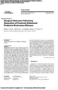
Figure 1: (a) Schematic demonstrating iliac screw placement with a recess created at the PSIS, with requirement for offset in order to attach to the rod. (b) Schematic demonstrating S2-alar-iliac screw placement allowing for attachment to the rod without offset use.
a
B
Figure 2: (a) Postoperative computed tomography scan demonstrating successful placement of iliac bolt. (b) Postoperative computed tomography scan demonstrating successful placement of S2-alar-iliac screws.
World Spinal Column Journal, Volume 7 / No: 1 / January 2016
27
Spinopelvic Reconstruction for Spinal Tumors
three components in spinopelvic reconstruction; spinopelvic fixation, posterior pelvic ring fixation, and anterior spinal column support. There are several biomechanical studies on spinopelvic fixation techniques. Yu et al. (28) evaluated the effects of the extent of sacrectomy on the stability of the lumbopelvic fixation construct using single and dual iliac screw techniques. They concluded that the single iliac screw technique for L3-iliac fixation could effectively restore the local stability for sacrectomy below the cut-line which passed through the superior border of the S1 foramen at the level of 1 cm below the sacral promontory. However, for instabilities caused by one-sided sacroiliac joint resection or total sacrectomy, the dual iliac screw technique should be considered. A human cadaveric study by Mindea et al. (20) examined the biomechanical strength of (1) doublerod, double iliac screw (DDS) constructs (Figure 3a), (2) single-rod, single iliac screw (SSS) constructs (Figure 3b), (3) double iliac screw (DIS) constructs (Figure 3c), and (4) modified Galveston technique (MGT) constructs in the setting of total sacrectomy (Figure 3d). Among them, DDS constructs demonstrated the highest strength in terms of flexion-extension, lateral bending, and axial rotation. In terms of selecting iliac screw length, according to a biomechanical study by Zheng et al (29), short iliac screws (7mm in diameter and 70mm in length) are susceptible to loosening after cyclic loading. Bone cement augmentation of short screws has demonstrated a significant increase in the fixation strength of short screws to an extent similar to that of long iliac screws (7mm in diameter and 120mm in length). Thus, given the potential complications of longscrew breach, short iliac screw fixation with augmentation with bone cement may be a viable clinical option for lumbopelvic reconstruction, although much larger screw diameters are now available and more commonly used. There are essentially three techniques for posterior pelvic ring reconstruction, which include allografts (femur or tibia) with screw fixation to bilateral ilia, transiliac bars, and cages. Gokaslan et al. (9) reported two cases of the resection of malignant tumors, using the Galveston L-rod reconstruction technique combined with a transiliac bar and tibial allograft between two ilia (Figure 3e). At the 1-year follow-up, one patient could walk independently, and the other ambulated independently using a cane. This combination technique is preferentially used given the potential benefit of fusion between the native ilia and the allograft in place of the sacrum. 28
Recently, the importance of anterior spinal column support in lumbopelvic reconstruction after total sacrectomy has been discussed extensively. A cadaveric biomechanical study by Cheng et al. (3) investigated the following 4 constructs: sacral rod reconstruction (Figure 3f); bilateral fibular flap reconstruction (Figure 3g); four-rod reconstruction (Figure 3h), and improved compound reconstruction. (Figure 3i) Among these, improved compound reconstruction, which utilized the sacral rod and the fibular triangular construct in the anterior approach, produced optimal structural stability after total sacrectomy. Similarly, Clark et al. (4) examined the biomechanical strength of three constructs, which included femoral strut allograft reconstruction, where a femoral allograft was placed between ilia and secured with bone screws (Figure 3j), L5–iliac cage strut reconstruction, where two titanium cages were placed obliquely, each wedged between the inferior L5 endplate and the iliac bone (Figure 3k), and S1 body replacement with expandable cage reconstruction, in which a rod was placed from the inferior L5 endplate and fixed to a transiliac bar and a 22 mm expandable cage was placed between the L5 endplate and the transiliac bar (Figure 3l). They concluded that S1 body replacement with expandable cage reconstruction provided the most biomechanically stable structure among them. Furthermore, minimally invasive techniques for anterior support have also been discussed by Le et al. (15), who tested the biomechanics of supplemental percutaneous lumbo-sacral-iliac screw fixation (Figure 3m) in human cadavers. They used an insertion point approximately 2 cm superior to the superior point of the greater sciatic notch and 2 cm anterior to the posterior superior iliac spine, ending at the inferior endplate of L5. Although they were not able to show statistically significant differences between constructs with or without lumbo-sacral-iliac screws, given their less invasiveness and theoretical advantages for biomechanical strength, further studies related to this novel technique are warranted.
Complications Due to the complex anatomy of the lumbosacropelvic region and local invasiveness and vascularity of metastatic tumors, there is a wide variety of complications using these reconstructive techniques. These include visceral injury, neurovascular injury, screw breach, excessive blood loss, wound infection, formation of hematoma and seroma in the surgical defect, and postoperative ileus. Additionally, while both iliac screws and S2-alar-iliac screws cross the World Spinal Column Journal, Volume 7 / No: 1 / January 2016
W Ishida et al.
B
C
F
G
H
I
J
K
L
M
a
D
E
Figure 3: Complex lumbo-sacro-pelvic reconstruction techniques. (a) Double-rod, double iliac screw (DDS) construct, (b) single-rod, single iliac screw (SSS) construct, (c) double iliac screw (DIS) construct, and (d) modified Galveston technique construct in the setting of total sacrectomy [20]. (e) Galveston L-rod reconstruction technique combined with a transiliac bar and tibial allograft between two ilia. (f) Sacral rod reconstruction, (g) bilateral fibular flap reconstruction (h) four-rod reconstruction, and (i) improved compound reconstruction, proposed by Cheng et al. [3]. (j) femoral strut allograft reconstruction, (k) L5–iliac cage strut reconstruction, and (l) S1 body replacement with expandable cage reconstruction proposed by Clark et al. [4]. (m) supplemental percutaneous lumbo-sacral-iliac screw fixation proposed by Le et al. [15].
SI articulation, S2-alar iliac screws often result in articular cartilage damage (22), but do not actually fuse the joint, so some patients may develop SI joint osteoarthritis and pain. If these patients fail conservative treatment, removal of the pelvic screws can be considered if the L5-S1 segment is adequately fused. Additionally, since these pelvic screws do not intend to fuse the SI joint, lucency around the pelvic screws or evidence of screw loosening if often noted on follow-up radiographs, but this is of unclear clinical significance and requires further study for better elucidation of the impact of this complication.
Radiographical Outcomes Gottfried et al. (10) measured the pre- and postoperative modified pelvic incidence (mPI) of 22 patients who underwent sacrectomy and revealed that the mean change in mPI was statistically different (p < 0.001) for patients undergoing subtotal versus those undergoing total sacrectomy (1.6° ± 0.9° vs. 13.6° ± 4.9° (± SD)). Changes in PI influence spinopelvic balance and may have postoperative clinical importance. Thus, they concluded that attention to spinopelvic alignment during lumbopelvic reconstruction and fixation after tumor resection is necessary and longterm studies are warranted to evaluate the impact of the change in PI on sagittal balance, pain, and ambulation after total sacrectomy. World Spinal Column Journal, Volume 7 / No: 1 / January 2016
None of the current literature has focused on pseudarthrosis in the setting of lumbopelvic reconstruction. Hence, it was impossible to assess the non-union rate related to this procedure. Future retrospective studies might be warranted with special attention to bone allografts as a part of the reconstruction. Likewise, there is limited data on the mechanical failure rate, but according to a systematic review by Bederman et al. (1), 5 out of 31 patients with total sacrectomy had mechanical failure, including rod breakage or screw pull-out. Although there was no statistically significant difference, patients without anterior column support tended to have high mechanical failure rates (17.4% vs. 12.5%). Again, this is suggestive of the potential benefit of adding anterior column support to spinopelvic reconstruction after total sacrectomy, but since extensive instrumentation both anteriorly and posteriorly requires more operative time and more sophisticated techniques, the potential complications must be thoroughly discussed with the patients.
Conclusions The resection of tumors in the lumbosacral region and subsequent spinopelvic reconstruction are both technically feasible and can be accomplished in a biomechanically stable fashion. Pelvic fixation is often required for reconstruction of the lumbo-sacro-pelvic region following sacrectomy, and 29
Spinopelvic Reconstruction for Spinal Tumors
should be considered in the treatment of metastatic disease following resection of tumors near the lumbosacral junction for added biomechanical support, particularly in patients with poor bone quality. The use of S2-alar-iliac screws offers several advantages over iliac screws, and can be placed as long as the S2-alar segment is intact following oncologic resection. Finally, minimally invasive techniques can be utilized for pelvic screw placement as well.
REFERENCES 1. Bederman SS, Shah KN, Hassan JM, Hoang BH, Kiester PD, Bhatia NN: Surgical techniques for spinopelvic reconstruction following total sacrectomy: A systematic review. Eur Spine J 23:305–319, 2014 2. Chang T-L, Sponseller PD, Kebaish KM, Fishman EK: Low profile pelvic fixation: Anatomic parameters for sacral alar-iliac fixation versus traditional iliac fixation. Spine (Phila Pa 1976) 34:436–40, 2009 3. Cheng L, Yu Y, Zhu R, Lv H, Jia Y, Zeng Z, et al: Structural stability of different reconstruction techniques following total sacrectomy: A biomechanical study. Clin Biomech (Bristol, Avon) 26:977–981, 2011 4. Clark AJ, Tang J a, Leasure JM, Ivan ME, Kondrashov D, Buckley JM, et al: Gait-simulating fatigue loading analysis and sagittal alignment failure of spinal pelvic reconstruction after total sacrectomy: Comparison of 3 techniques. J Neurosurg Spine 20:364–370, 2014 5. Colibaseanu DT, Dozois EJ, Mathis KL, Rose PS, Ugarte MLM, Abdelsattar ZM, et al: Extended Sacropelvic Resection For Locally Recurrent Rectal Cancer. Dis Colon Rectum 57:47–55, 2014 6. Dickey ID, Hugate RRJ, Fuchs B, Yaszemski MJ, Sim FH: Reconstruction after total sacrectomy: Early experience with a new surgical technique. Clin Orthop Relat Res 438:42–50, 2005 7. Emami A, Deviren V, Berven S, Smith J a, Hu SS, Bradford DS: Outcome and complications of long fusions to the sacrum in adult spine deformity: Luque-galveston, combined iliac and sacral screws, and sacral fixation. Spine (Phila Pa 1976) 27:776–786, 2002 8. Gallia GL, Suk I, Witham TF, Gearhart SL, Black JH 3rd, Redett RJ, et al: Lumbopelvic reconstruction after combined L5 spondylectomy and total sacrectomy for en bloc resection of a malignant fibrous histiocytoma. Neurosurgery 67:E498–502, 2010 9. Gokaslan ZL, Romsdahl MM, Kroll SS, Walsh GL, Gillis TA, Wildrick DM, et al: Total sacrectomy and Galveston L-rod reconstruction for malignant neoplasms. Technical note. J Neurosurg 87:781–787, 1997 10. Gottfried ON, Omeis I, Mehta VA, Solakoglu C, Gokaslan ZL, Wolinsky JP: Sacral tumor resection and the impact on pelvic incidence. J Neurosurg Spine 14:78–84, 2011 11. Humphries WE 3rd, Satyan KB, Relyea K, Kim ES, Adesina AM, Chintagumapala M, et al: Low-grade myofibroblastic sarcoma of the sacrum. J Neurosurg Pediatr 6:286–290, 2010 12. Ilyas H, Place H, Puryear A: A comparison of early clinical and radiographic complications of iliac screw fixation versus S2 alar iliac (S2AI) fixation in the adult and pediatric populations. J Spinal Disord Tech 28:199–205, 2015 13. Kiatisevi P, Piyaskulkaew C, Sukunthanak B, Thanakit V, Bumrungchart S: Total sacrectomy for low-grade malignant peripheral nerve sheath tumour: A case report. J Orthop Surg 22:409–414, 2014 14. Kuklo TR, Bridwell KH, Lewis SJ, Baldus C, Blanke K, Iffrig TM, et al: Minimum 2-year analysis of sacropelvic fixation and L5-S1 fusion using S1 and iliac screws. Spine (Phila Pa 1976) 26:1976–1983, 2001
15. Le VH, Heckmann N, Jain N, Wang L, Turner AWL, Lee TQ, et al: Biomechanical evaluation of supplemental percutaneous lumbosacro-iliac screws for spinopelvic fixation following total sacrectomy. J Spinal Disord Tech 28:E181–185, 2015 16. Maricevich M, Maricevich R, Chim H, Moran SL, Rose PS, Mardini S: Reconstruction following partial and total sacrectomy defects: An analysis of outcomes and complications. J Plast Reconstr Aesthet Surg 67:1257–1266, 2014 17. Martin CT, Witham TF, Kebaish KM: Sacropelvic fixation: Two case reports of a new percutaneous technique. Spine (Phila Pa 1976) 36:E618–621, 2011 18. Mazur MD, Ravindra VM, Schmidt MH, Brodke DS, Lawrence BD, Riva-cambrin J, et al: Unplanned reoperation after lumbopelvic fixation with S-2 alar-iliac screws or iliac bolts. J Neurosurg Spine 23:67–76, 2015 19. Min K, Espinosa N, Bode B, Exner GU: Total sacrectomy and reconstruction with structural allografts for neurofibrosarcoma of the sacrum. A case report. J Bone Joint Surg Am 87:864–869, 2005 20. Mindea SA, Chinthakunta S, Moldavsky M, Gudipally M, Khalil S: Biomechanical comparison of spinopelvic reconstruction techniques in the setting of total sacrectomy. Spine (Phila Pa 1976) 37:E1622– 1627, 2012 21. O’Brien JR, Matteini L, Yu WD, Kebaish KM: Feasibility of minimally invasive sacropelvic fixation: percutaneous S2 alar iliac fixation. Spine (Phila Pa 1976) 35:460–464, 2010 22. O’Brien JR, Yu WD, Bhatnagar R, Sponseller P, Kebaish KM: An anatomic study of the S2 iliac technique for lumbopelvic screw placement. Spine (Phila Pa 1976) 34:E439–442, 2009 23. Peelle MW, Lenke LG, Bridwell KH, Sides B: Comparison of pelvic fixation techniques in neuromuscular spinal deformity correction: Galveston rod versus iliac and lumbosacral screws. Spine (Phila Pa 1976) 31:2392–2398; discussion 2399, 2006 24. Schwend RM, Sluyters R, Najdzionek J: The pylon concept of pelvic anchor- age for spinal instrumentation in the human cadaver. Spine (Phila Pa 1976) 28:542–547, 2003 25. Sponseller PD: The S2 Portal to the ilium. Semin Spine Surgery 2:8387, 2007 26. Verlaan JJ, Kuperus JS, Slooff WB, Hennipman A, Oner FC: Complications, secondary interventions and long term morbidity after en bloc sacrectomy. Eur Spine J 24:2209-2219, 2015 27. Wong CCH, Corn C, Crosby C, Even J, Mencio GA, Devin CJ, et al: Biomechanical Analysis of Iliac Screws versus S2 Alar-Iliac Screws. Abstract at 27th Annual Meeting of the AANS/CNS Section on Disorders of the Spine and Peripheral Nerves, 2011 28. Yu B-S, Zhuang X-M, Li Z-M, Zheng Z-M, Zhou Z-Y, Zou X-N, et al: Biomechanical effects of the extent of sacrectomy on the stability of lumbo-iliac reconstruction using iliac screw techniques: What level of sacrectomy requires the bilateral dual iliac screw technique? Clin Biomech (Bristol, Avon) 25:867–72, 82010 29. Zheng ZM, Zhang KB, Zhang JF, Yu BS, Liu H, Zhuang XM: The effect of screw length and bone cement augmentation on the fixation strength of iliac screws: A biomechanical study. J Spinal Disord Tech 22:545–550, 2009 Manuscript submitted November 03, 2015. Accepted November 27, 2015. Address correspondence to: Benjamin D. Elder, Department of Neurosurgery, The Johns Hopkins University School of Medicine, Baltimore, USA Phone: (410) 955 50 00 email:
[email protected]
30
World Spinal Column Journal, Volume 7 / No: 1 / January 2016