2515
Development 126, 2515-2525 (1999) Printed in Great Britain © The Company of Biologists Limited 1999 DEV8592
Sprouty: a common antagonist of FGF and EGF signaling pathways in
Drosophila Susanne Kramer1,*, Masataka Okabe2, Nir Hacohen4,‡, Mark A. Krasnow4 and Yasushi Hiromi1,2,3,§ 1Department 2Department
of Molecular Biology, Princeton University, Princeton, NJ 08544, USA of Developmental Genetics, National Institute of Genetics, and 3Department of Genetics, Graduate University for Advanced Studies, Mishima, Shizuoka 411-8540, Japan 4Department of Biochemistry, and Howard Hughes Medical Institute, Stanford University School of Medicine, Stanford, CA 94305, USA *Present address: Institut für Genetik, Universität Würzburg, Biozentrum, Am Hubland, D-97074 Würzburg, Germany ‡Present address: Whitehead Institute, Cambridge, MA 02142, USA §Author for correspondence (e-mail:
[email protected])
Accepted 16 March; published on WWW 4 May 1999
SUMMARY Extracellular factors such as FGF and EGF control various aspects of morphogenesis, patterning and cellular proliferation in both invertebrates and vertebrates. In most systems, it is primarily the distribution of these factors that controls the differential behavior of the responding cells. Here we describe the role of Sprouty in eye development. Sprouty is an extracellular protein that has been shown to antagonize FGF signaling during tracheal branching in Drosophila. It is a novel type of protein with a highly conserved cysteine-rich region. In addition to the embryonic tracheal system, sprouty is also expressed in other tissues including the developing eye imaginal disc, embryonic chordotonal organ precursors and the midline glia. In each of these tissues, EGF receptor signaling is known to participate in the control of the correct number INTRODUCTION FGF and EGF are extracellular signaling factors that control various aspects of morphogenesis, patterning and cellular proliferation in both invertebrates and vertebrates. These ligands act through high-affinity transmembrane receptors with an intracellular tyrosine kinase moiety. In Drosophila, a single EGF receptor homolog, encoded by the Egfr gene, and two FGF receptor homologs, encoded by breathless and heartless, have been identified (Beiman et al., 1996; Gisselbrecht et al., 1996; Glazer and Shilo, 1991; Klämbt et al., 1992; Livneh et al., 1985). Upon reception of the extracellular signals, these receptor tyrosine kinases (RTKs) activate the ras/MAPK signaling pathway. Many of the molecular components of this signaling pathway, such as ras GTPase, raf kinase and MAPK are shared among different RTKs. Thus information from multiple extracellular signals are interpreted using the same molecular cassette (reviewed by Schlessinger, 1993). While the intracellular tyrosine kinase domain is conserved, the extracellular domains of the RTKs are specialized for their diverse inputs. Of these three receptors, the EGF receptor is the
of neurons or glia. We show that, in all three tissues, the loss of sprouty results in supernumerary neurons or glia, respectively. Furthermore, overexpression of sprouty in wing veins and ovarian follicle cells, two other tissues where EGF signaling is required for patterning, results in phenotypes that resemble the loss-of-function phenotypes of Egf receptor. These results suggest that Sprouty acts as an antagonist of EGF as well as FGF signaling pathways. These receptor tyrosine kinase-mediated pathways may share not only intracellular signaling components but also extracellular factors that modulate the strength of the signal. Key words: Eye development, Chordotonal organ, ras signaling, Sprouty, FGF, EGF, Drosophila
most complex, as it is responsive to three tissue-specific activating ligands, Spitz, Gurken and Vein (reviewed by Perrimon and Perkins, 1997; Schweitzer and Shilo, 1997). Spitz acts in the ventral ectoderm, the midline of the central nervous system (CNS), the chordotonal organs and imaginal discs. Gurken is the key determinant in patterning the ovarian follicle cells. The third ligand, Vein, functions during wing vein formation and in the attachment of the embryonic muscles. Unlike Spitz and Gurken, which are members of the TGFα family, Vein shows similarity to neuregulins (NeumanSilberberg and Schüpbach, 1993; Rutledge et al., 1992; Schnepp et al., 1996). The only known ligand for the FGF receptors is the Branchless FGF, which acts through Breathless during tracheal branching (Sutherland et al., 1996). In most of these systems, the distribution of the activating ligands, which is governed mainly by their synthesis, processing and diffusion, is the primary factor controlling the differential behavior of the responding cells. In addition, the EGF signaling pathway is subject to negative feedback regulation. The production of the EGF-like antagonist Argos is dependent on EGFR signaling (Schweitzer et al., 1995; Golembo et al., 1996)
2516 S. Kramer and others and a hyperactivation of EGFR results in a downregulation of receptor expression (Sturtevant et al., 1994). Elucidating the mechanisms that regulate RTK signaling is key to the understanding of how extracellular signals achieve precise cellular responses. Recently, we have reported that Sprouty (SPRY) acts as a novel extracellular antagonist of FGF signaling during tracheal development (Hacohen et al., 1998). Similar to Argos, which is induced by EGFR signaling (Golembo et al., 1996), spry expression is induced by the FGF pathway that it inhibits. Here we show that spry is also expressed in other developmental systems such as the eye imaginal disc, the embryonic chordotonal organ precursors and the midline glia. In all of these systems, EGF receptor signaling is known to participate in the control of the correct number of neurons or glia. We examine spry function in several of these tissues, focusing on its regulation of EGF-induced neuronal differentiation in the eye. The Drosophila compound eye is a stereotyped array of 800 unit eyes or ommatidia, each of which comprises an invariant number of cells; 8 photoreceptor neurons (named R1 through R8) and 12 non-neuronal accessory cells. The photoreceptor cells are recruited by a stereotyped sequence of inductive interactions, mediated by two RTKs, EGFR and Sevenless (SEV). EGFR signaling is required for the recruitment or maintenance of all ommatidial cells (Freeman, 1996; Kumar et al., 1998; Xu and Rubin, 1993), whereas SEV is required specifically for the neuronal specification of the R7 cell (Tomlinson and Ready, 1986). Activation of EGFR by its ligand Spitz (Freeman, 1994; Tio et al., 1994) or SEV by BOSS (Van Vactor et al., 1991) activates the ras/MAPK pathway, which ultimately impinges on a number of nuclear targets such as the Pointed Ets-domain transcription factor (Brunner et al., 1994; O’Neill et al., 1994) and results in the initiation of neuronal development (for review see Dickson, 1995; Zipursky and Rubin, 1994). In addition to the presumptive photoreceptor neurons, two cell types within an ommatidium can be induced to become neurons upon ectopic activation of the RTK signaling pathways: the lens-secreting non-neuronal cone cells and the mystery cells, two cells transiently associated with the ommatidial precluster (Tomlinson and Ready, 1987; Wolff and Ready, 1991a). Ectopic activation of the ras/MAPK pathway in these cells is sufficient to elicit their differentiation as neurons (Basler et al., 1991; Dickson et al., 1992; Fortini et al., 1992). In normal development, ectopic activation of the pathway is prevented by the action of several negative regulators of RTK signaling. Gap1 encodes a GTPase activating protein that is thought to act by decreasing the levels of activated Ras1 in the eye, whereas yan encodes for an Etsdomain protein that interferes with Pointed signaling (Gaul et al., 1992; Lai and Rubin, 1992). Mutations in either of these loci causes the cone cells and mystery cells to differentiate as photoreceptor neurons. Absence of a third factor, Argos, a secreted antagonist of EGF signaling, results in a transformation of mystery cells into outer photoreceptor cells and in the recruitment of extra cone and pigment cells, but does not affect the development of the presumptive cone cells (Freeman et al., 1992b; Kretzschmar et al., 1992; Okano et al., 1992). Here we show that mutation in spry causes a transformation
of non-neuronal cone cells into R7 cells and of mystery cells into outer photoreceptor cells, a phenotype identical to the one seen with hyperactivation of EGFR signaling. SPRY can also antagonize EGFR signaling in other tissues such as the midline glia, the chordotonal organs, the wing and the ovarian follicle cells. Since SPRY acts as an antagonist of FGFR-mediated signaling during tracheal development, the EGFR and FGFR signaling pathways appear to share an unexpected degree of conservation in the extracellular compartment.
MATERIALS AND METHODS Genetics We identified five EMS-induced alleles of spry in a genetic screen for dominant suppressors of the ro-svp eye phenotype (Kramer et al., 1995). Misexpression of svp in R2/R5 by the ro-svp transgene causes misspecification of these cells, resulting in compromised neuronal induction of R1/R6. spry dominantly suppresses this phenotype by rescuing the neuronal differentiation of R1/R6, without restoring the misspecification of R2/R5 (data not shown). The screen was carried out essentially as described previously for suppressors of the sev-svp phenotype (Kramer et al., 1995), except that mutagenized st e males were crossed to CyO, 2 × P[ro-svp]/Sp virgin females. 50,000 F1 progeny were screened and Su(ro-svp) on the third chromosome were balanced over TM3, ry, 2 × P[ro-svp]. Mutations on the third chromosome were mapped meiotically using the rucuca chromosome (ru, h, th, st, cu, sr, e, ca). Deficiency mapping of spry was carried out with Df(3L)HR119, Df(3L)HR232, Df(3L)HR370 (Wohlwill and Bonner, 1991), Df(3L)A466 (Kulkarni et al., 1994) and Df(3L)1226 (gift of S. Paine-Saunders and J. Fristrom). Df(3L)HR119 (63C1; 63D3), Df(3L)HR232 (63C6; 63E) and Df(3L)1226 (63C6; 63E1) failed to complement the lethality of spry EMS alleles, whereas Df(3L)HR370 (63A1; 63D1) and Df(3L)A466 (63D1-2; 64B1-2) complemented it. The eye phenotypes of various spry alleles in trans to each other were indistinguishable from those over a deficiency of the locus, suggesting that all spry alleles tested were amorphs or strong hypomorphs (Table 1). spry254 clones and clones doubly mutant for spry254 and a strong hypomorphic allele of sina (sina2; Carthew and Rubin, 1990) or a null allele of argos (argos257; Okabe et al., 1996) were generated using the FRT technique (Xu and Rubin, 1993). spry∆5 clones in a sev− background were generated in animals of the genotype w1118, sevd2, P[hs-FLP]; spry∆5, P[FRT]80B/P[w+]70C, P[FRT]80B. Control animals were raised without heat shock. Gap1B2 and yan1 are described in Gaul et al. (1992), and Lai and Rubin (1992), respectively. pointed8B is an amorph (Klämbt, 1993) and Ras1e2F/Ras1C33 is a combination of Ras1 hypomorphic alleles that survives to the third instar larval stage (our unpublished observation). breathless∆10, heartless80 and branchlessP1 are amorphs or strong hypomorphs (Klämbt et al., 1992; Shishido et al., 1997; Sutherland et al., 1996). For ectopic expression studies, UAS-spry was placed under the control of elav-GAL4 C155 (Lin and Goodman, 1994), sevE-GAL4 K25 (Brunner et al., 1994), en-GAL4 (gift of A. Brand and N. Perrimon), CY2 (Queenan et al., 1997) and BH1 (Schüpbach and Wieschaus, 1998). sevE-Ras1N17, UAS-spry and GMR-argos lines are described in Allard et al. (1996), Hacohen et al. (1998) and Sawamoto et al. (1998), respectively. Histology and histochemistry Fixation and sectioning of adult heads and antibody staining of imaginal discs were performed as described (Tomlinson and Ready, 1987), except that, in some instances, the peripodial membrane was not removed from imaginal discs. SPRY protein was detected using two polyclonal rabbit antisera; 26A was raised against full-length
Sprouty antagonizes FGF and EGF signaling 2517 Table 1. Comparison of recessive phenotypes of different spry allelic combinations Genotype spry∆5/spry∆5 spry∆5/Df(3L)1226 spry226/Df(3L)1226 spry∆5/spry226 spryF7/spry226 spry254 clone sevd2, spry254 clone
Average no. of outer PRCs
Average no. of R7 cells
Ommatidia scored
6.09 6.09 6.17 5.96 6.04 6.14 6.15
3.09 2.71 3.26 3.04 3.25 3.08 2.96
120 82 115 190 114 126 96
Retinular phenotypes of animals of the indicated genotypes were examined in apical tangential sections and average numbers of outer PRCs and R7 cells per ommatidium calculated for each genotype. For the wild type, the average number of outer PRCs would be 6.0 and the average number of R7 cells would be 1.0.
SPRY protein and 32C against a peptide extending from amino acid 63 through 462 (Hacohen et al., 1998). An affinity-purified antibody against BarH1/BarH2 proteins (Higashijima et al., 1992), a monoclonal antibody against ELAV protein and monoclonal antibody 22C10 (Fujita et al., 1982) were gifts of the Saigo, Rubin and Goodman laboratories, respectively. Monoclonal antibodies against βgalactosidase were purchased from Promega. The expression pattern of enhancer trap insertion 9143 was detected using anti-βgalactosidase mAb 40-1a (obtained from Developmental Studies Hybridoma Bank) and the Vectastain elite kit (Vector laboratories). Embryo stainings were carried out according to Patel (1994). The following lacZ enhancer trap lines were used as cell-typespecific markers; X81 and AE127 are enhancer trap insertion in the rhomboid gene (Freeman et al., 1992a) and the svp gene (Mlodzik et al., 1990), respectively. P82 expresses lacZ in R3, R4 and R7 (Kramer et al., 1995), H214 is an enhancer trap insertion that expresses βgalactosidase at high levels only in the R7 cell (Mlodzik et al., 1992) and AA142 is an enhancer trap insertion with expression in the midline glia (Klämbt et al., 1991). In situ hybridizations to eye imaginal discs and embryos were performed essentially as described by O’Neill and Bier (1994), and Lehmann and Tautz (1994). RNA probes were transcribed from a 1 kb and a 2.2 kb EcoRI fragment of the longest spry cDNA cloned into BSSK+ (Hacohen et al., 1998) using the Boehringer Mannheim kit according to the instructions of the manufacturer.
RESULTS
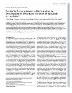
spry is required to prevent neuronal induction of non-neuronal cells in the retina Animals homozygous for any of the EMS-induced alleles of spry or spry∆5 died as pharate adults. The rare escapers had eyes that were similar in size to wild-type eyes but had a disorganized exterior. A majority of the ommatidia in these animals contained supernumerary photoreceptor neurons, which by their morphology were R7 cells. In addition, some of the extra photoreceptors resembled outer photoreceptor neurons (Fig. 1C; Table 1). Examination of the early stages of neuronal development in the eye imaginal disc with molecular markers revealed that the supernumerary photoreceptors originated from non-neuronal cone cells and mystery cells that had assumed R7 and R3/R4 fates, respectively (Fig. 2). Neuronal markers were inappropriately activated in cone and mystery cells at the same time in development as in the normal photoreceptors, implying that the defect in the mutant occurs
Fig. 1. The eye phenotype of spry loss-of-function alleles. (A) A tangential section of a wild-type eye at the apical level shows a regular array of ommatidia. Each ommatidium contains eight photoreceptor neurons (R1-8), which can be identified by the size and position of their darkly stained light-gathering structure (rhabdomeres). Based on their morphology, the photoreceptor cells (PRCs) can be subdivided into three classes. The outer PRCs (R1 through R6) have large rhabdomeres that extend through the entire depth of the retina and are arranged in a trapezoidal array. R7 and R8 have small rhabdomeres that project into the center of the ommatidium. In this apical section, only the rhabdomere of the R7 cell is visible. R8 is underlying the R7 cell in a more basal position. (B) spry loss-of-function alleles had a weak dominant phenotype, shown here is an apical tangential section of a spry226/+ eye. In eyes of animals heterozygous for a spry EMS allele, on average 3.4% of ommatidia had an extra R7 cell (arrowhead) with some variation between different alleles (spry226: 7.2%, spryG5: 5.5%, spryF7: 1.7%, spry254: 1.2%, spry211: 1.0%). spry∆5 showed the weakest dominant phenotype; 0.8% of ommatidia had an extra outer PRC. (C) An apical tangential section of an eye of an animal homozygous for spry∆5, a putative amorph of spry. Most ommatidia contained between one and four extra R7-like photoreceptor cells, and 27% of ommatidia had one or two extra outer photoreceptor cells. In addition, a small percentage of ommatidia had less than the normal number of photoreceptor cells.
at the normal time of photoreceptor induction. Thus, spry functions in the eye imaginal disc to prevent neuronal induction of these non-neuronal cells. spry functions as a dosage-dependent inhibitor of neuronal induction. In the heterozygous condition, all spry alleles examined contained 1-7% of ommatidia with an extra R7 cell or an occasional gain or loss of an outer photoreceptor cell (Fig. 1B; Table 2). Conversely, increased levels of SPRY in the developing eye disc inhibited the induction of normal photoreceptor cells. Animals that expressed UAS-spry under either the sev-GAL4 or the elav-GAL4 driver had small disorganized eyes. The majority of ommatidia lacked one or more outer photoreceptor cells of the R3/R4/R1/R6 subtype (Fig. 3A-C). The R7 cell was missing in 11±4% of sevEGAL4/UAS-spry, and in 18±6% of elav-GAL4/UAS-spry ommatidia. Development of the R2/R5 and R8 photoreceptors Table 2. Dominant interactions between spry and the intracellular negative regulators of ras signaling Genotype spry226/+ yan1/+ Gap1B2/+ yan1/+; spry226/+; Gap1B2/+; spry226/+
Ommatidia with extra R7 (%) 6.3±2.2 0.4±0.4