International Journal of
Molecular Sciences Article
ST6GALNAC5 Expression Decreases the Interactions between Breast Cancer Cells and the Human Blood-Brain Barrier Aurore Drolez 1 , Elodie Vandenhaute 1 , Clément Philippe Delannoy 2 , Justine Hélène Dewald 2 , Fabien Gosselet 1 , Romeo Cecchelli 1 , Sylvain Julien 3 , Marie-Pierre Dehouck 1 , Philippe Delannoy 2 and Caroline Mysiorek 1, * 1
2
3
*
Université d’Artois (UArtois), EA2465, Laboratoire de la Barrière Hémato-Encéphalique (LBHE), Lens F-62300, France;
[email protected] (A.D.);
[email protected] (E.V.);
[email protected] (F.G.);
[email protected] (R.C.);
[email protected] (M.-P.D.) Structural and Functional Glycobiology Unit, Unité Mixte de Recherche (UMR) du Centre National de la Recherche Scientifique (CNRS) 8576, University of Lille, Villeneuve d’Ascq F-59655, France;
[email protected] (C.P.D.);
[email protected] (J.H.D.);
[email protected] (P.D.) Cell Plasticity and Cancer, U908 INSERM, University of Lille, Villeneuve d’Ascq F-59655, France;
[email protected] Correspondence:
[email protected]; Tel.: +33-321-791-746
Academic Editor: Cheorl-Ho Kim Received: 25 June 2016; Accepted: 3 August 2016; Published: 11 August 2016
Abstract: The ST6GALNAC5 gene that encodes an α2,6-sialyltransferase involved in the biosynthesis of α-series gangliosides, was previously identified as one of the genes that mediate breast cancer metastasis to the brain. We have shown that the expression of ST6GALNAC5 in MDA-MB-231 breast cancer cells resulted in the expression of GD1α ganglioside at the cell surface. By using a human blood-brain barrier in vitro model recently developed, consisting in CD34+ derived endothelial cells co-cultivated with pericytes, we show that ST6GALNAC5 expression decreased the interactions between the breast cancer cells and the human blood-brain barrier. Keywords: breast cancer; blood-brain barrier; gangliosides; GD1α ; ST6GALNAC5; sialyltransferase; brain metastasis
1. Introduction The modification of cell surface glycosylation is one of the most important phenotypic rearrangements that occur during carcinogenesis. It mainly affects the terminal part of the carbohydrate moiety of glycoproteins and glycolipids, leading to the expression of tumor-associated carbohydrate antigens (TACA). Most TACAs are sialylated and changes in sialylation were clearly demonstrated to affect cellular recognition, cell adhesion, and signaling and, consequently, the cell’s behavior. Gangliosides are glycosphingolipids (GSLs) carrying one or several sialic acid residues. They are essentially located on the outer leaflet of the plasma membrane where they can interact with transmembrane receptors or signal transducers involved in cell proliferation, adhesion, and motility. In adult, complex gangliosides from b- and c-series are normally restricted to the nervous system but a re-expression of complex gangliosides is observed in a variety of cancers including neuro-ectoderm-derived cancers, non-small cell lung carcinoma, and breast cancer [1]. In particular, GD3 and GD2 are considered as melanoma- and neuroblastoma-associated antigens playing a key role in tumor development, and are used as targets for cancer immunotherapy [2]. However, the
Int. J. Mol. Sci. 2016, 17, 1309; doi:10.3390/ijms17081309
www.mdpi.com/journal/ijms
Int. J. Mol. Sci. 2016, 17, 1309
2 of 13
mechanisms by which tumor-associated gangliosides induce invasive and metastatic phenotypes of tumor cells remain to be clarified. α-Series gangliosides define a particular sub-class of GSLs containing Neu5Ac α2,6 linked to the GalNAc residue of the gangliopentaosyl backbone Neu5Acα2-3Galβ1-3GalNAcβ1-4Galβ1-4Glc (IV3 Neu5Ac1 Gg4 ). The typical α-series ganglioside GD1α (IV3 Neu5Ac1 ,III6 Neu5Ac1 Gg4 -Cer) was first isolated as a minor compound from rat hepatoma cells [3] and from bovine brains [4], with an expression restricted to particular cell populations of the brain and cerebellum [5]. Three members of the CMP-Neu5Ac: β-N-acetylgalactosaminide α2,6-sialyltransferase family (ST6GalNAc III, V, and VI) were shown to catalyze in vitro the transfer of a sialic acid residue onto GM1b (IV3 Neu5Ac1 Gg4 -Cer) to form GD1α [6]. However, according to its substrate specificity and expression pattern, ST6GalNAc V is considered as the main GD1α synthase. ST6GALNAC5 cDNA was cloned from mouse brains [7,8] and the st6galnac5 gene is specifically expressed in mouse brain tissues, mostly in the forebrain and cerebellum [8]. When expressed as a soluble recombinant protein, the mouse ST6GalNAc V showed α2,6-sialyltransferase activity almost exclusively for GM1b , while being inactive toward glycoproteins [7]. The enzymatic activity of human ST6GalNAc V was never investigated in detail, but we have recently shown that transfection of human ST6GalNAc V cDNA into MDA-MB-231 breast cancer cells resulted in the expression of GD1α at the cell surface [9]. To date, the specific function of α-series gangliosides is poorly understood. It has been proposed that GD1α could play a role in Purkinje cell functions in the cerebellum [5] and that GD1α could serve as an adhesion molecule for high-metastatic murine lymphosarcoma cells in the adhesion to hepatic endothelial cells [10]. Recently, ST6GALNAC5 was identified as one of the genes over-expressed in breast cancer cell populations selected for their ability to produce brain metastases [11]. ShRNA inhibition of ST6GALNAC5 expression reduced the capacity of breast cancer cells to produce brain metastases, whereas the expression of ST6GALNAC5 in parental cell lines promoted brain metastases formation [11]. Moreover, ST6GALNAC5 was shown to improve the capacity of breast cancer cells to transmigrate across a human umbilical vein endothelial cells (HUVECs) in vitro model of the blood-brain barrier [11]. The blood-brain barrier (BBB), localized at the level of brain capillary endothelial cells (ECs), controls and restricts the exchanges between the blood and the brain tissues. The BBB presents a specific architecture where the capillary ECs share a split basement membrane with pericytes and are surrounded together by astrocyte end-feet. The BBB forms with pericytes, neurons, glial cells, and the extracellular matrix, the neurovascular unit (NVU). The interplays and communications between the different components of NVU allow the BBB-specific differentiation of ECs, which exhibit a network of tight junctions, express efflux pumps and specific receptors and transporters. These specific and restrictive properties control and limit the access to the brain parenchyma of many cells and substances. During the last decades, most in vitro BBB models were developed using animal cells (mouse, rat, bovine, pig) isolated from brain microvessels as the primary culture or immortalized [12], whereas human culture models commonly use HUVECs, which display only a limited tightness and not a BBB phenotype. In vitro approaches are required to identify cellular and molecular interactions between cancer cells and BBB endothelium. However, while numerous studies were performed with in vitro models, the heterogeneity and the quality of BBB models used is a limitation to the extrapolation of results to in vivo context, showing that the choice of a model that fulfills the properties of human BBB is essential. In that context, we recently developed a human BBB in vitro model consisting in CD34+ hematopoietic stem cells derived endothelial cells co-cultivated with brain pericytes [13,14] and displaying improved BBB properties closed to those observed in vivo. The model proved valuable in the study of cancer cells tropism as the adhesion and transmigration capacities of breast cancer cells were found to be in accordance with the cancer cell molecular subtypes, fitting well with their propensity to form brain metastases [15,16]. We have used this CD34+ derived human BBB model to investigate the role of GD1α in adhesion and transmigration of breast cancer cells and contrary to what
Int. J. Mol. Sci. 2016, 17, 1309
3 of 13
Int. J. Mol. Sci. 2016, 17, 1309
3 of 12
was observed in a HUVECs in vitro model, ST6GALNAC5 cDNA expression resulted in a decrease of the interactions between MDA-MB-231 breast cancer cells and the CD34+ derived human BBB model.
2. Results
2.1. Brain Targeting Cells Interaction Analysis on the Human in Vitro Blood-Brain Barrier (BBB) Model 2. Results In order to investigate the mechanisms tropism during the initial steps of breast 2.1. Brain Targeting Cells Interaction Analysis of on brain the Human in Vitro Blood-Brain Barrier (BBB) Model cancer brain metastases formation, the interactions of breast cancer cells with the BBB were analyzed using In order to investigate the mechanisms of brain tropism during the initial steps of breast cancer an in vitro approach. For this purpose, adhesion and transmigration assays of brain-targeting breast brain metastases formation, the interactions of breast cancer cells with the BBB were analyzed using cancer cells were performed on a human BBB in vitro model named Brain-Like endothelial Cells an in vitro approach. For this purpose, adhesion and transmigration assays of brain-targeting breast (BLECs) that we recently developed [13,14]. The BLECs model consists of endothelial cells derived cancer cells were performed on a human BBB in vitro model named Brain-Like endothelial Cells from(BLECs) CD34+ that hematopoietic stem cells co-cultivated with brain pericytes. The BLECs model displays we recently developed [13,14]. The BLECs model consists of endothelial cells derived improved BBB+ properties close tocells those observed with in vivo, as low the BBB from CD34 hematopoietic stem co-cultivated brainsuch pericytes. Thepermeability BLECs model to displays integrity marker, continuous localization at the cell border of tight junction proteins (Claudin-5, improved BBB properties close to those observed in vivo, such as low permeability to the BBB integrity occludin, ZO-1), and expression efflux pumps (P-gP, BCRP) [13,14]. occludin, ZO-1), marker, continuous localizationof at functional the cell border of tight junction proteins (Claudin-5, Theexpression adhesion of and transmigration of MDA-MB 231 [13,14]. BrM2 cell line (BrM2) was compared to the and functional efflux pumps (P-gP, BCRP) adhesion and transmigration of MDA-MB 231cell BrM2 (BrM2) generated was compared parental The cell line MDA-MB-231 wild type (wt). The BrM2 line cell wasline previously by two to the cell linein MDA-MB-231 wild atype (wt). increase The BrM2 cell line was previously rounds of inparental vivo selection mice, and showed significant in brain metastases formation generated by two rounds of in vivo selection in mice, and showed a significant increase in brainECs [11]. As shown in Figure 1a, after two hours of incubation, the adhesion rate of BrM2 on the BBB metastases formation [11]. As shown in Figure 1a, after two hours of incubation, the adhesion was 63.4% lower than the parental cells and no increase in BBB permeability to lucifer yellow (LY) of BrM2 the BBB was(0.57 63.4%± lower parental cells0.66 and±no BBB −1, −1 and was rate observed foronMDA andECs BrM2 0.08 ×than 10−3the cm·min 0.1increase × 10−3 in cm·min permeability to lucifer yellow (LY) was observed for MDA and BrM2 (0.57 ˘ 0.08 ˆ 10´3 cm¨min´1 respectively) compared to the control condition before adhesion (permeability coefficient (Pe) = 0.58 ´3 cm¨min´1 , respectively) compared to the control condition before adhesion and 0.66 ˘ 0.1 ˆ 10 −3 −1 ± 0.07 × 10 cm·min ). As the adhesion of breast cancer cells on the BBB ECs is required, but not (permeability coefficient (Pe) = 0.58 ˘ 0.07 ˆ 10´3 cm¨min´1 ). As the adhesion of breast cancer cells enough to reach the brain parenchyma, the transmigration was quantified and it was revealed that on the BBB ECs is required, but not enough to reach the brain parenchyma, the transmigration was the BrM2 transmigrate the same to the parental cellrate linecompared MDA-MB-231 wt (Figure quantified and it wasatrevealed thatrate the compared BrM2 transmigrate at the same to the parental 1b). cell No line increase in BBB wt permeability was measured following MDA and MDA-MB-231 (Figure 1b). to NoLY increase in BBB permeability to transmigration LY was measuredoffollowing −3 −1 −3 −1 ´ 3 ´ 1 ´ 3 ´1 ,the BrM2 (0.73 ± 0.03 of × 10 0.78 ± 0.01 cm·minand , respectively) compared transmigration MDAcm·min and BrM2and (0.73 ˘ 0.03 ˆ 10× 10 cm¨min 0.78 ˘ 0.01 ˆ 10 cm¨minto control conditioncompared without transmigration. respectively) to the control condition without transmigration. number of transmigrated cells (%)
number of adherent cells (%)
150
*** 100
50
0
MDA wt
BrM2
(a)
N.S
150
100
50
0
MDA wt
BrM2
(b)
Figure 1. Adhesion (a); MDA-MB-231 typeand (wt) and BrM2 breast Figure 1. Adhesion (a);and and transmigration transmigration (b)(b) of of MDA-MB-231 wild wild type (wt) BrM2 breast cancer cancer the BLECs human BLECs The number of adherent MDA-MB-231 or transmigrated cell cell lines lines on the on human model. The model. number of adherent or transmigrated wt cells was setwt upcells to 100% equal to, respectively, 98 cells. Results are 98 thecells. meanResults in triplicate, MDA-MB-231 was and set up to 100% and equal579 to,and respectively, 579 and are the and of representative two independent N.S: notexperiments significant; ***N.S: p < 0.001. mean in representative triplicate, and ofexperiments two independent not significant; *** p < 0.001. 2.2. Molecular Characterization of MDA-MB-231 BrM2 Cells
The BrM2 cell line was previously described to over-express a set of genes potentially involved in brain metastasis, including COX2, HBEGF and ST6GALNAC5 [11]. The expression of these genes was quantified by qPCR. As shown in Figure 2, a 23-fold increased expression of ST6GALNAC5 and
Int. J. Mol. Sci. 2016, 17, 1309
4 of 12
2.2. Molecular Characterization of MDA-MB-231 BrM2 Cells The BrM2 cell line was previously described to over-express a set of genes potentially involved in 4 of 13 brain metastasis, including COX2, HBEGF and ST6GALNAC5 [11]. The expression of these genes was quantified by qPCR. As shown in Figure 2, a 23-fold increased expression of ST6GALNAC5 and 10-fold increase of COX2 were measured in BrM2 compared to parental MDA-MB-231 wt. wt. However, no 10-fold increase of COX2 were measured in BrM2 compared to parental MDA-MB-231 However, difference of expression was measured forfor HBEGF in BrM2 compared to parental MDA-MB-231 wt.wt. no difference of expression was measured HBEGF in BrM2 compared to parental MDA-MB-231 Int. J. Mol. Sci. 2016, 17, 1309
Figure 2.2. qPCR COX2, HB-EGF, HB-EGF, and ST6GALNAC5 in MDA-MB-231 wt and BrM2. BrM2. Figure qPCR analysis analysis of of COX2, and ST6GALNAC5 in MDA-MB-231 wt and Quantification was performed by the method described by Pfaffl [17] and normalized to Actin. N.S: Quantification was performed by the method described by Pfaffl [17] and normalized to Actin. N.S: not significant; *** p < 0.001. not significant; *** p < 0.001.
ST6GALNAC5 gene encodes a GalNAc α2,6-sialyltransferase involved in the biosynthesis of ST6GALNAC5 gene encodes a GalNAc α2,6-sialyltransferase involved in the biosynthesis of α-series gangliosides, mainly GD1α. According to the fact that GD1α could serve as an adhesion α-series gangliosides, mainly GD1α . According to the fact that GD1α could serve as an adhesion molecule for breast cancer cells in the adhesion to BBB endothelial cells and promote brain metastasis, molecule for breast cancer cells in the adhesion to BBB endothelial cells and promote brain metastasis, the GSL composition was analyzed to determine the impact of the increased ST6GALNAC5 the GSL composition was analyzed to determine the impact of the increased ST6GALNAC5 expression expression on the glycosylation of MDA-MB-231 BrM2 cells. Total GSLs were extracted from on the glycosylation of MDA-MB-231 BrM2 cells. Total GSLs were extracted from MDA-MB-231 wt MDA-MB-231 wt and BrM2, purified by reverse phase chromatography and permethylated prior to and BrM2, purified by reverse phase chromatography and permethylated prior to Matrix assisted laser Matrix assisted laser desorption-ionization-mass spectrometry (MALDI-MS) analysis. desorption-ionization-mass spectrometry (MALDI-MS) analysis. As previously shown [18] MDA-MB-231 wt expresses neutral globosides Gb3 and Gb4 and As previously shown [18] MDA-MB-231 wt expresses neutral globosides Gb3 and Gb4 and monosialylated gangliosides, mainly GM3 (Figure 3a). The precursor lactosylceramide (LacCer) was monosialylated gangliosides, mainly GM3 (Figure 3a). The precursor lactosylceramide (LacCer) was also detected, as well as a monosialoganglioside at m/z 1933, which was confirmed to correspond to also detected, as well as a monosialoganglioside at m/z 1933, which was confirmed to correspond to GM1b by matrix assisted laser desorption-ionization time-of-flight (MALDI-TOF)/TOF fragmentation GM1b by matrix assisted laser desorption-ionization time-of-flight (MALDI-TOF)/TOF fragmentation analysis (data not shown). Two ceramide isoforms are commonly expressed in human tissues due to analysis (data not shown). Two ceramide isoforms are commonly expressed in human tissues due the substitution of the sphingosine moiety by palmitic acid C16:0 (Cer*) or lignoceric acid C24:0 to the substitution of the sphingosine moiety by palmitic acid C16:0 (Cer*) or lignoceric acid C24:0 (Cer**). As shown in Figure 3b, the composition in GSLs of BrM2 cells was similar to wt cells and no (Cer**). As shown in Figure 3b, the composition in GSLs of BrM2 cells was similar to wt cells and expression of GD1α was detected as indicated by the absence of signal at m/z 2293.9, which was no expression of GD1α was detected as indicated by the absence of signal at m/z 2293.9, which was identified in MDA-MB-231 green fluorescent protein positive (GFP+) cell population (Figure 3c) by identified in MDA-MB-231 green fluorescent protein positive (GFP+) cell population (Figure 3c) by MALDI-TOF/TOF fragmentation analysis (Figure S1). MALDI-TOF/TOF fragmentation analysis (Figure S1).
Int. J. Mol. Sci. 2016, 17, 1309
Int. J. Mol. Sci. 2016, 17, 1309 Int.J.J.J.Mol. Mol.Sci. Sci.2016, 2016,17, 17,1309 1309 Int. Mol. Sci. 2016, 17, 1309 Int.
5 of 13 Int. J. Mol. Sci. 2016, 17, 1309 5 of 12 of12 12 of 12 555of
Figure 3. Comparison of mass spectrom Figure 3. Comparison of mass spectrometry (MS) profiles of permethylated glycosphingolipids purified from MDA-MB-231 wt (a); BrM2 Figure 3. 3. Comparison Comparison of mass spectrometry (MS) profiles profiles(MS) of permethylated permethylated glycosphingolipids Figure 3. Comparison of mass spectrometry (MS) profiles of permethylated glycosphingolipids Figure of spectrometry of glycosphingolipids Figure 3.mass Comparison of mass(MS) spectrometry profiles of permethylated glycosphingolipids purified purified from MDA-MB-231 wt (a); BrM2 (b); and green fluorescent protein positive (GFP+) (c) Glycosphingolipids cell (GSL) a purifiedfrom fromMDA-MB-231 MDA-MB-231 wt(a); (a);BrM2 BrM2 (b); andgreen green fluorescent proteinprotein positive (GFP+)(GFP+) (c)cell cell(c)population. purified from MDA-MB-231 wt (a); BrM2 (b); and green fluorescent protein positive (GFP+) (c) cell from MDA-MB-231 wt (a); BrM2 (b); and green fluorescent positive cell population. purified wt (b); and fluorescent protein positive (GFP+) (c) population. Glycosphingolipids (GSL) are present as d18:1/C16:0 (Cer*) and d18:1/C24:0 (Cer**) population. Glycosphingolipids Glycosphingolipids (GSL) are present present as d18:1/C16:0 d18:1/C16:0 (Cer*) and d18:1/C24:0 (Cer**) population. Glycosphingolipids (GSL) are present as d18:1/C16:0 (Cer*) and d18:1/C24:0 (Cer**) Glycosphingolipids (GSL) are present as d18:1/C16:0 (Cer*) and d18:1/C24:0 (Cer**) isomers. , Gal; , Glc; , GalNAc; population. (GSL) are as (Cer*) and d18:1/C24:0 (Cer**) , Gal; , Glc; , GalNAc; , Neu5Ac. isomers. ,,,Gal; Gal; isomers. Glc; ,,,GalNAc; GalNAc; Neu5Ac. GalNAc; , ,,Neu5Ac. isomers. Gal; Glc; Neu5Ac. isomers. ,,,Glc;
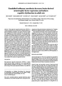
2.3. Involvement of GD1α Over-Expression in I 2.3. Involvement of G D1α Over-Expression in Interaction Processes of Breast Cancer Cell Lines with the BBB 2.3.Involvement Involvementof of GD1α D1α Over-Expression inOver-Expression InteractionProcesses Processes ofBreast BreastCancer CancerCell Cell Lineswith with theCell BBBLines with the BBB 2.3. Involvement of G D1α Over-Expression in Interaction Processes of Breast Cancer Cell Lines with the BBB 2.3. G Over-Expression in Interaction of the BBB 2.3. Involvement of GD1α in Interaction Processes of Lines Breast Cancer In order to specifically identify the eff In order to specifically identify the effect of ST6GALNAC5 over-expression on the adhesion and In order to specifically identify the effect of ST6GALNAC5 over-expression on the adhesion and In order to specifically identify the effect of ST6GALNAC5 over-expression on the adhesion and transmigration of breast In order to specifically identify the effectidentify of ST6GALNAC5 over-expression the adhesion and In order to specifically the effect of ST6GALNAC5 on over-expression on the adhesion and cancer cells, tw transmigration ofcells, breast cancer cells, two cell populations were generated, Clone #13 and a cell population, transmigration of of breast breast cancer cancer cells, two cell populations were generated, Clone #13 and transmigration of breast cancer two cell populations were generated, Clone #13 and polyclonal transmigration two cell populations were generated, Clone #13 and transmigration of cells, breast cancer cells, two cell populations were generated, Clone #13aaaandGFP-positive a polyclonal polyclonal GFP-positive cell population, in which ST6GALNAC5 cDNA was 10-fold andcompared 60-fold to control MDA polyclonal GFP-positive GFP-positive cell population, in which ST6GALNAC5 cDNA was 10-fold and 60-fold polyclonal GFP-positive cell population, in which ST6GALNAC5 cDNA was 10-fold and 60-fold over-expressed polyclonal cell population, in which ST6GALNAC5 cDNA was 10-fold and 60-fold GFP-positive cell population, in which ST6GALNAC5 cDNA was 10-fold and 60-fold over-expressed over-expressed compared to control MDA-MB 231 wt, respectively [9]. These two cell populations over-expressedcompared compared tocontrol controlMDA-MB MDA-MB231 231wt, wt,respectively respectively[9]. [9].These Thesetwo twocell cellpopulations populations over-expressed compared to control MDA-MB 231 wt, respectively [9]. These two cell populations were previously demonstrated to expres over-expressed comparedto to control MDA-MB 231 wt, respectively were previously were previouslyto demonstrated to express GD1α ganglioside by MALDI-TOF/TOF fragmentation were previously previouslydemonstrated demonstrated to express GD1α D1α ganglioside by MALDI-TOF/TOF MALDI-TOF/TOF fragmentation were previously demonstrated express G D1α ganglioside by MALDI-TOF/TOF fragmentation analysis (Figure S1). The were demonstrated express G ganglioside fragmentation toto express GD1α ganglioside by by MALDI-TOF/TOF fragmentation analysis (Figure S1). adhesion and tra analysis (Figure S1). The adhesion andcapacities transmigration capacities of these two cell populations were analysis(Figure (FigureS1). S1). The adhesion and transmigration capacities of these two cell populations were analysis (Figure S1). The adhesion and transmigration of these two cell populations were determined using the human BBB in vitro analysis The adhesion and transmigration capacities of these two cell populations were The adhesion and transmigration capacities of these two cell populations were determined using the determined using the human BBB in vitro model. As shown in Figure 4a, the results obtained were determinedusing using thehuman human BBB inmodel. vitromodel. model. Asshown shown inFigure Figure 4a,the theobtained resultsobtained obtained wereto those determined using the human BBB in vitro model. As shown in Figure 4a, the results obtained were thoseobtained obtained with BrM2 cells determined the in vitro As in 4a, results were human BBB inBBB vitro As shown in Figure 4a, the results were similar similar similar to with thoseBrM2 obtained BrM2 cells as adhesion theGFP+ Clonecell #13population and GFP+ similarto tothose thoseobtained obtained with BrM2 cellswith asadhesion adhesion ofthe the Clone#13 #13of and GFP+ cell population similar to those obtained cells as adhesion of the Clone #13 and were 40%cell andpopulation 50% decreased compared to similar with BrM2 cells as of Clone and GFP+ cell population were 40% andcompared 50% decreased compared towt, MDA-MB-231 respectively. A and 50% decrease were40% 40%and and50% 50% decreased compared toMDA-MB-231 MDA-MB-231 wt, respectively.wt, A55% 55% and50% 50%decrease decrease were 40% and 50% decreased to MDA-MB-231 respectively. A 55% and 50% decrease of55% transmigration rate was also observed were decreased compared to wt, respectively. A and of rate transmigration rate wasfor also observed forthe Clone #13cell and the GFP+ cell population, respectively oftransmigration transmigration rate wasalso alsoobserved observed for Clone #13and and the GFP+ cell population, respectively of transmigration was also observed Clone #13 and GFP+ population, respectively of rate was for Clone #13 the GFP+ cell population, respectively
Int. J. Mol. Sci. 2016, 17, 1309
6 of 13
number of adherent cells (%) number of adherent cells (%)
150 150
** **
* *
100 100
N.S N.S
50 50 0 0
MDA wt MDA wt
ST6 cl.13 ST6 cl.13
(a) (a)
ST6 GFP+ ST6 GFP+
number of transmigrated number of transmigrated cellscells (%) (%)
with BrM2 of the Clone #13 and GFP+ cell population were 40% and 50% decreased Int. J. Mol. Sci.cells 2016, as 17, adhesion 1309 6 of 12 compared to MDA-MB-231 wt, respectively. A 55% and 50% decrease of transmigration rate was also Int. J. Mol. Sci. 2016, 17, 1309 6 of 12 (Figure 4b). Following adhesion and transmigration assays, no increase in BBB permeability was observed for Clone #13 and the GFP+ cell population, respectively (Figure 4b). Following adhesion −1 and 0.92 ± 0.07 × 10⁻³ measured for Clone #13 and cellintransmigration populations (0.87 ±was 0.03no × increase 10−3 cm·min and transmigration assays, noGFP+ increase BBB permeability measured for #13 and GFP+ cell (Figure 4b). Following adhesion and assays, in Clone BBB permeability was ´3 cm¨min ´control 1 and 0.92 ´3 transmigration ´1 −1 −1 respectively) compared −3 cm·min to the condition before (Pe = 0.90 ± 0.04 populations (0.87 ˘ 0.03 ˆ 10 ˘ 0.07 ˆ 10 cm¨min respectively) compared measured for Clone #13 and GFP+ cell populations (0.87 ± 0.03 × 10 cm·min and 0.92 ± 0.07 × 10⁻³ × ´3 cm¨min´1 ). −3 −1). −1 respectively) 10 cm·min to cm·min the control condition before transmigration (Pecondition = 0.90 ˘ 0.04 ˆ 10 compared to the control before transmigration (Pe = 0.90 ± 0.04 × 10⁻³ cm·min−1). 150 150
*** ***
100 100
N.S N.S
50 50 0 0
MDA wt MDA wt
ST6 cl.13 ST6 cl.13
(b) (b)
ST6 GFP+ ST6 GFP+
Figure 4. Adhesion Adhesion (a); and and transmigration (b) of MDA-MB-231wt, MDA-MB-231wt, Clone #13 (ST6 cl. 13), and GFP+ Figure 4. Clone#13 #13(ST6 (ST6cl. cl.13), 13),and andGFP+ GFP+ Figure 4. Adhesion(a); (a); andtransmigration transmigration (b) (b) of of MDA-MB-231wt, Clone breast cancer cell population (ST6 GFP+) on the human Brain-Like endothelial Cells (BLECs) model. breast cancer cell population (ST6 GFP+) on the human Brain-Like endothelial Cells (BLECs) model. breast cancer cell population (ST6 GFP+) on the human Brain-Like endothelial Cells (BLECs) model. The number number of adherent or transmigrated MDA-MB-231 wt cells was set up 100% and equal to, The wt cells cellswas wasset setup uptoto to100% 100%and andequal equal The numberofofadherent adherentorortransmigrated transmigrated MDA-MB-231 MDA-MB-231 wt to,to, respectively, 533 and 117 cells. Results are the mean in triplicate, and representative of two or three respectively, 533 and and representative representativeofoftwo twoororthree three respectively, 533 and117 117cells. cells.Results Resultsare arethe the mean mean in in triplicate, triplicate, and independent experiments. N.S: not significant; * p < 0.01; ** p < 0.005; *** p < 0.001. independent experiments. ** pp