Dec 11, 1992 - (G. A. Hebert, C. G. Crowder, G. A. Hancock, W. R. Jarvis, and C. Thornsberry, J. Clin. ..... Kloos, W. E., and C. G. George. 1991. Identification of ...
JOUR±JAL
OF
Vol. 31, No. 3
CLINICAL MICROBIOLOGY, Mar. 1993, p. 490-493
0095-1137/93/030490-04$02.00/0 Copyright C 1993, American Society for Microbiology
Numerical Approach to Reference Identification of Staphylococcus, Stomatococcus, and Micrococcus spp. DWANE L. RHODEN,* GARY A. HANCOCK, AND J. MICHAEL MILLER Hospital Infections Program, Nosocomial Pathogens Laboratory Branch,
Centers for Disease Control and Prevention, Atlanta, Georgia 30333 Received 10 September 1992/Accepted 11 December 1992
A numerical-code system for the reference identification of Staphylococcus species, Stomatococcus mucilaginosus, and Micrococcus species was established by using a selected panel of conventional biochemicals. Results from 824 cultures (289 eye isolate cultures, 147 reference strains, and 388 known control strains) were used to generate a list of 354 identification code numbers. Each six-digit code number was based on results from 18 conventional biochemical reactions. Seven milliliters of purple agar base with 1% sterile carbohydrate solution added was poured into 60-mm-diameter agar plates. All biochemical tests were inoculated with 1 drop of a heavy broth suspension, incubated at 35°C, and read daily for 3 days. All reactions were read and interpreted by the method of Kloos et al. (G. A. Hebert, C. G. Crowder, G. A. Hancock, W. R. Jarvis, and C. Thornsberry, J. Clin. Microbiol. 26:1939-1949, 1988; W. E. Kloos and D. W. Lambe, Jr., P. 222-237, in A. Balows, W. J. Hansler, Jr., K. L. Herrmann, H. D. Isenberg, and H. J. Shadomy, ed., Manual of Clinical Microbiology, 5th ed., 1991). This modified reference identification method was 96 to 98% accurate and could have value in reference and public health laboratory settings. As the numbers and types of nosocomial infections caused by Staphylococcus aureus and coagulase-negative staphylococci increase, identification of these causative agents becomes more important clinically. Because Staphylococcus spp. remain an important component of commensal florae, assigning a pathogenic role to routine isolates of this genus is difficult. For years, laboratories have equated coagulasepositive gram-positive cocci with S. aureus and coagulasenegative cocci with Staphylococcus epidermidis, even though there are 27 recognized species of staphylococci. There are at least 13 human strains of coagulase-negative staphylococci, some of which are clearly pathogenic while others remain of questionable clinical significance (3). Although assigning a role to these agents is difficult, an accurate identification method would improve our understanding of the pathogenic potential of many of the coagulase-negative staphylococcal species and their epidemiology. Several automated, semiautomated, and commercially packaged methods for the identification of staphylococci are available. However, most have some sort of drawback, and their accuracy for rarely isolated species has been questioned (4, 7). The reference method for identification described by Kloos and Lambe (5) is considered by many to be the "gold standard" for final identification, but, like most reference methods, it requires large numbers of biochemicals along with a degree of experience in interpreting the reactions. Few laboratories can afford the luxury of the complete reference procedure. Abbreviated methods have been reported (6), but the need for a rational approach to definitive identification appears to be increasing. In this study, we combined selected reference biochemical tests with the ease and convenience of numerical coding to arrive at a cost-effective, abbreviated reference method for
*
definitive identification of members of the family Micrococcaceae.
MATERIALS AND METHODS Cultures tested. A total of 824 members of the Micrococfrom various sources (Table data base of 354 six-digit identification code numbers. Of these isolates, 388 were known strains and 41 were characterized by and obtained from W. E. Kloos (North Carolina State University at Raleigh). The remaining cultures were from collections at the Centers for Disease Control and Prevention (CDC). These included 289 frozen (-70°C in sheep blood) and fresh eye culture isolates and 147 reference strains. All test cultures were streaked onto Trypticase soy agar (BD Microbiology Systems, Cockeysville, Md.) containing 5% sheep blood and incubated overnight at 35°C. A single colony from each culture was streaked onto a second blood agar plate. Isolated colonies from the second subculture were then transferred to 5 ml of brain heart infusion broth (BD) to prepare a suspension equivalent to a 0.5 McFarland turbidity standard and used to inoculate primary biochemicals (Table 2). Additional colonies were transferred to 2 ml of 0.85% sterile saline to prepare a second suspension equivalent to a no. 3 McFarland turbidity standard and used to inoculate a STAPH-IDENT system (Analytab Products, Plainview, N.Y.). Biochemical identification. Nine 60-mm-diameter plates containing 7 ml of purple agar base with 1% sterile carbohydrate solution added (glucose, D-maltose, D-mannitol, D-Xylose, sucrose, D-mannose, D-ribose, a-lactose, or D-turanose), a urea agar slant, arginine, nitrate, and acetoin were each inoculated with 1 drop of the broth suspension. In addition to the primary biochemicals, all screening and confirmatory tests were inoculated at this time. The no. 3 McFarland saline suspension was used to inoculate a caceae representing 27 species 1) were used to establish a
Corresponding author.
490
VOL. 31, 1993
NUMERICAL APPROACH TO REFERENCE IDENTIFICATION
TABLE 1. Cultures used to establish data base CDC identfication CDC identification
Staphylococcus aureus Staphylococcus auricularis Staphylococcus capitis S. capitis subsp. ureolyticus Staphylococcus caprae Staphylococcus camnosus Staphylococcus caseolyticus Staphylococcus chromogenes Staphylococcus cohnii S. cohnii subsp. urealyticum Staphylococcus epidermidis Staphylococcus haemolyticus Staphylococcus hominis Staphylococcus hyicus Staphylococcus intermedius Staphylococcus kloosii Staphylococcus lentus Staphylococcus lugdunensis Staphylococcus saccharolyticus Staphylococcus saprophyticus Staphylococcus sciuri Staphylococcus schleifen Staphylococcus simulans Staphylococcus warneri Staphylococcus xylosus Stomatococcus mucilaginosus Micrococcus species
No. of isolates
% of
143 16 12 9 2 2 2 4 9 10 328 56 44 3 16 3 2 45 1 25 5 1 20 25 13 12 15
17.4 2.0 1.5 1.1 0.2 0.2 0.2 0.5 1.1 1.2 40.0 7.0 5.4 0.4 1.9 0.4 0.2 5.5 0.1 3.0 0.6 0.1 2.4 3.0 1.6 1.5 1.8
total
TABLE 2. Tests used to establish profile number and confirm identification of certain gram-positive cocci
No. of isolates with unique
profiles 17 15 3 5 2 1 2 4 4 2 93 36 29 1 11 2 2 21 1 7 5 1 8 21 6 12 10
STAPH-IDENT strip (API); the strip was incubated and the reactions were recorded after 5 h according to the manufacturer's instructions. Novobiocin susceptibility was determined by standard methods (1). These tests and the STAPHIDENT (API) tests for phosphatase, ,B-glucosidase, ,3-glucuronidase, and ,-galactosidase made up the 18 primary biochemical tests (Table 2). Conventional biochemical test cultures were incubated at 35°C for 72 h. Reactions were recorded daily, and the tests for nitrate and acetoin were done at the 72-h reading. Profile number. The primary biochemical tests (Fig. 1) were arranged into groups of three, and each member of the triad was assigned a value of 1, 2, or 4 in order. A positive reaction received the assigned value for that test, and the values for each triad were combined to generate a six-digit profile number. All results needed to obtain the identification profile were recorded. If the test profile was located in the profile data base, the identification was made. If the test profile was not matched, the results were evaluated by conventional biochemical means and the identification was then made (5). This new profile number was then added to the data base along with the accompanying identification. When an unusual identification or profile number was obtained, the results were arbitrated by an independent microbiologist in another laboratory. Table 1 lists the number of different profiles exhibited by each of the species tested. Screening tests. All isolates were subjected to six screening tests. Glucose utilization, catalase production, and the Gram strain tests were always done. In addition, three tests were used to establish the coagulase reaction. These included both tube and slide coagulase methods (Difco Laboratories, Detroit, Mich.) and latex agglutination (STAPHAUREX; Wellcome Diagnostics, Research Triangle Park, N.C.). All tests were carried out as directed by the manufacturer. A
491
Test
Primary D-Maltosea D-Trehalose D-Mannitol D-Xylose Sucrose D-Mannose D-Ribose a-Lactose D-Turanose Nitrate Urea agar slante Arginine dihydrolasec Acetoin (tube) Novobiocin susceptibility" Phosphated
lB-Glucosidased I-Glucuronidased
P-Galactosidased
Screening Glucose Catalase Coagulase (slide)e Coagulase (tube)e Latex agglutinationf Gram stain
Confirmatory Esculin Gelatin Omithine decarboxylasec Xylitol D-Cellobiose Raffinose ,-D-Fructose Galactose Arabinose D-Melezitose Furazolidone susceptibilityb a 1% carbohydrate solution (Sigma Chemical Co., St. Louis, Mo.) in 7.0 ml of purple agar base (BD). b BD.
Carr-Scarborough.
dSTAPH-INDENT strip (Analytab Products). Difco Laboratories. f STAPHAUREX kit (Wellcome Diagnostics).
positive result in any of the three tests was considered positive for identification purposes. Confirmatory tests. The following tests were used to resolve questions about rare or missing profiles. Resistance to a 100-jig furazolidone disc (BD) helped rule out Micrococcus species. Positive esculin and gelatin results were used to confirm Stomatococcus mucilaginosus. A positive ornithine decarboxylase test (Carr-Scarborough Microbiologicals, Inc., Decatur, Ga.) confirmed Staphylococcus lugdunensis. The confirmatory carbohydrate test plates (Table 2) were inoculated and the reactions were recorded, but the results were rarely used in the identification process. STAPH-INDENT system. STAPH-IDENT is a 10-test kit system designed to identify members of the family Micrococcaceae within 5 h of inoculation. The kit was inoculated
and incubated and the reactions were recorded according to the manufacturer's instructions. In this study, the results
492
Mal
I11
J. CLIN. MICROBIOL.
RHODEN ET AL.
1Tre
Xyl
Suc
Mno
1111
2
4
IMan
4
2
IL
1
L
|Rib 1[11
|Lac |Tur
2
4
Nit
Ure 1
2
Arg 4
Act 1
Nov
P04
2
4
1 L I L IL
Gco 1l11
1
GCU
2
Gal
4 _
L
Profile No.
ID:
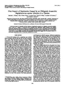
FIG. 1. Sample worksheet. Mal, maltose; Tre, trehalose; Man, mannitol; Xyl, xylose; Suc, sucrose; Mno, mannose; Rib, ribose; Lac, lactose; Tur, turanose; Nit, nitrate; Ure, urea agar slant; Arg, arginine; Act, acetoin; Nov, novobiocin susceptible; P04, phosphate; Gco, P-glucosidase; Gcu, 13-glucuronidase; Gal, 3-galactosidase. The last four reactions on the worksheet are taken from the API STAPH-IDENT.
from phosphatase, P-glucosidase, P-glucuronidase, and ,-galactosidase tests were used as part of the coding system. RESULTS AND DISCUSSION
A six-digit-code identification system was developed by using 824 isolates of Micrococcaceae. We found that the 18 biochemicals selected for the CDC Micrococcaceae profile system (MPS) were those that provided the best discrimination between species. Others were tried, but they added no more accuracy to the MPS. The MPS correctly identified more than 95% of the Staphylococcus, Micrococcus, and Stomatococcus strains tested. By using this system, the confusion experienced when large, complex identification charts are used is greatly reduced because of the limited number of tests involved in the MPS and because the reactions for each isolate are listed on separate worksheets. The profile numbers are distinct and reproducible. Therefore, phenotypic traits among cultures to be tracked within given institutions and situations may be easily documented and reviewed. The MPS allows laboratories to use a complex reference method with fewer biochemicals, thus providing a more cost-effective approach to the identification of Micrococcaceae in a reference setting. As shown in Table 1, many profile numbers are possible for some strains. The biotype (profile number) may be useful and discriminating as an epidemiologic tool for those strains that demonstrated many different profile numbers. We are assessing this possibility but are aware that phenotypes are usually less helpful in epidemiologic problems than genotypic analysis. While most of the S. epidernidis isolates were from eyes, isolates of this organism from other body sites showed no unique profile characteristics. Indeed, a wide variety of body sites is represented in the 535 noneye isolates tested, from which both coagulase-positive and coagulase-negative strains were collected. We found that organisms submitted for confirmation that had been previously identified by a commercial method were often misidentified. Our experience with STAPH-IDENT and its accuracy in identifying members of this group are the subject for a future publication. The primary purpose for using the commercial system in this study was to provide data on four tests not commonly done (Table 2). Conventional tube tests for these four biochemicals were not evaluated. For a specific genus and species to be identified, the profile number must match an existing number or must reflect no more than two aberrant tests. If three tests were aberrant, the organism was reported as "probable [genus and species]" or "Staphylococcus species, most closely resembling [genus and species]." In our experience, few errors have resulted when one or two tests were aberrant and the
resulting code number could be applied to either of two different species. When this rare incident occurred, we consulted the reference charts (2, 5) to determine which of the two tests would provide the higher percentage of reactivity. The final decision would be based on the higherpercentage reaction. During the early development phase of the MPS, selected isolates were submitted to a referee laboratory for confirmation or were hand delivered to the reference laboratory and tested by CDC personnel using the reference procedures of Kloos and Lambe (5). The MPS was accurate for more than 95% of the isolates tested. Although not used often, confirmatory test plates were inoculated along with the primary set of biochemicals. These additional tests would not be necessary in most reference laboratories unless a strain could not be clearly identified without them. For our purposes in building a data base for the MPS, we routinely inoculated the confirmatory test plates but the results were not a part of the profile numbers. Pouring, storing, and handling large numbers of small plates require a great deal of time. We recommend preparing a 2-month supply of plates and storing them sealed in plastic bags at 4°C. If stored unsealed, the plates should be held no more than 2 weeks. A more convenient multiwell tissue culture plate is being evaluated for use. Usually, the yellow color exhibited by a positive reaction on the purple agar base is clearly determined. There are few, if any, "half colors" or "almost yellow" reactions to interpret. We believe that the CDC MPS is a reliable alternative for laboratories which require reference identification of members of the Micrococcaceae. This method could provide public health and clinical laboratories an accurate, costeffective approach to the reference identification of these organisms. ACKNOWLEDGMENTS We thank Wesley Kloos for his assistance and advice during the evaluation of this study and Tamara Bannerman for her able technical expertise in arbitrating discrepant identifications.
REFERENCES 1. Almeida, R. J., and J. H. Jorgensen. 1982. Use of Mueller-Hinton agar to determine novobiocin susceptibility of coagulase-negative staphylococci. J. Clin. Microbiol. 16:1155-1156. 2. Hebert, G. A., C. G. Crowder, G. A. Hancock, W. R. Jarvis, and C. Thornsberry. 1988. Characteristics of coagulase-negative staphylococci that help differentiate these species and other members of the family Micrococcaceae. J. Clin. Microbiol. 26:1939-1949. 3. Horan, T., D. Culver, W. Jarvis, G. Emori, S. Banerjee, W. Martone, and C. Thornsberry. 1988. Pathogens causing nosocomial infections. Antimicrob. Newsl. 5:65-67. 4. Kloos, W. E., and C. G. George. 1991. Identification of Staphy-
VOL. 31, 1993
NUMERICAL APPROACH TO REFERENCE IDENTIFICATION
lococcus species and subspecies with the MicroScan Pos ID and Rapid Pos ID panel systems. J. Clin. Microbiol. 29:738-744. 5. Kloos, W. E., and D. W. Lambe, Jr. 1991. Staphylococcus, p. 222-237. In A. Balows, W. J. Hausler, Jr., K. L. Herrmann, H. D. Isenberg, and H. J. Shadomy (ed.), Manual of clinical microbiology, 5th ed. American Society for Microbiology, Washington, D.C.
493
6. Kloos, W. E., and K. H. Schleifer. 1975. Simplified scheme for routine identification of human Staphylococcus species. J. Clin. Microbiol. 1:82-88. 7. Kloos, W. E., and J. F. Wolfshohl. 1982. Identification of Staphylococcus species with the API STAPH-IDENT system. J. Clin. Microbiol. 16:509-516.