PHYSIOLOGY AND REPRODUCTION Steroid Hormones During Embryonic Development in Japanese Quail: Plasma, Gonadal, and Adrenal Levels1 M. A. Ottinger, S. Pitts, and M. A. Abdelnabi2 Department of Animal and Avian Sciences, University of Maryland, College Park, Maryland 20742 females consistently had overall higher levels of estradiol than males. Adrenal gland steroid content remained relatively high and did not change significantly with age. In contrast, steroid content of gonads followed patterns similar to those observed for plasma levels. These results provide evidence for steroid hormone production by the gonads of both sexes, as well as for distinct differences in the patterns observed in the adrenal gland and gonads. These results provide evidence for gonadal regulation of changes in circulating hormone levels. Further, these hormonal patterns were associated with the timing of steroid-induced sexual differentiation in the Japanese quail, suggesting that plasma gonadal steroids are critical in sexual differentiation.
ABSTRACT The purpose of this experiment was to measure plasma, gonad, and adrenal steroid hormones during embryonic and early posthatch development in Japanese quail. Blood plasma samples were collected from male and female Japanese quail embryos at 2-d intervals between Day 10 of incubation and Day 5 posthatch. Gonads and adrenal glands were collected from a separate set of embryos at the same ages. Concentrations of androgen (testosterone and 5α-dihydrotestosterone) and 17β-estradiol (E2) were determined by RIA. Plasma androgen changed significantly (P < 0.001) with age in males and females, and there were significant differences (P < 0.001) between sexes in the hormonal patterns. Males had higher plasma androgen than females; conversely,
(Key words: avian embryo, steroid hormone, gonad, adrenal, sexual differentiation) 2001 Poultry Science 80:795–799
ovaries from female embryos do appear to be steroidogenic (Abdelnabi et al., 2000). The differences in the embryonic gonadal activity for steroid production could be explained by sex differences in steroid enzymes (Imataka et al., 1988) or by gene expression at early developmental stages (Nomura et al., 1999). However, the source of the plasma steroid hormones during embryonic development is unclear. Therefore, the purpose of this experiment was to measure plasma, gonad, and adrenal gonadal steroid concentrations in male and female quail between Day 10 of incubation (E10) and Day 5 posthatch.
INTRODUCTION Gonadal steroid hormones are critical for sexual differentiation of endocrine and behavioral components of reproduction. In birds, estradiol appears to be central in the sexual differentiation of females in a number of species (Ottinger and Abdelnabi, 1997). Steroid treatment of quail embryos resulted in behavioral demasculinization of males, with the most potent effects observed with treatment at 12 to 14 d of incubation (Adkins, 1975, 1976; Adkins-Regan et al., 1982; Adkins-Regan, 1985). In the domestic chick, steroids are synthesized by the embryonic gonad and adrenals as early as 3.5 d of incubation (Woods and Erton, 1978), and these hormones are measurable in the circulation by 6.5 d of incubation (Woods et al., 1975, 1977; Tanabe et al., 1979, 1986; Woods and Brazzill, 1981). Plasma testosterone peaks late in embryonic development in male quail embryos (Ottinger and Bakst, 1981). However, gonadal histology does not show evidence of steroidogenesis in male embryos. Conversely, histologically,
MATERIALS AND METHODS Japanese quail (Coturnix japonica) from the University of Maryland quail colony were used as a source of fertilized eggs. Embryos were sampled at 2-d intervals between E10 and E17 and Day 5 posthatch. Blood samples were collected from the embryo by cutting a window in the shell above the air sac, exteriorizing the chorioallantoic vein, puncturing the vein, and collecting blood into a heparinized capillary tube (Ottinger and Bakst, 1981). Blood was centrifuged for 20 min at 2,500 × g; the plasma
2001 Poultry Science Association, Inc. Received for publication July 10, 2000. Accepted for publication February 5, 2001. 1 To whom correspondence should be addressed:
[email protected]. edu. 2 Department of Animal Sciences, University of Assiut, Assiut, Egypt.
Abbreviation Key: 5α-DHT = 5α-dihydrotestosterone; E2 = 17-β estradiol; E5 = Day 5 of embryonic incubation.
795
796
OTTINGER ET AL.
was frozen at −40 C until assay. The sex of the embryo was determined by examination of the gonads under a dissecting microscope. Plasma samples from embryos before hatch were pooled with equal volumes of plasma from each embryo to allow measurement of androgen in females (200 µL) and 17β-estradiol (E2) (100 µL) and androgen in males. E2 was measured in females previously (Abdelnabi et al., 2000). The number of samples used varied with the age of the embryos, with decreasing numbers of embryos pooled until single samples were analyzed from embryos sampled at hatch. There were at least five samples (pooled or single) for each age and sex. Hormones were measured by single antibody androgen RIA3 that had been previously validated for use with quail plasma (Ottinger and Mahlke, 1984). Assay sensitivity was 3 pg/tube; intraassay CV was 3 to 5%, and interassay CV was 5 to 7%. Antibody cross-reactivity was 100% with testosterone and 46% with 5α-dihydrotestosterone (5α-DHT). Previous experiments have shown that 5α-DHT levels are 25 to 30% of testosterone levels in quail and in zebra finches (Ottinger and Mahlke, 1984; Adkins-Regan et al., 1994). Adrenal and gonads were dissected out, weighed, and stored at −80 C until assay. The left ovary, both testes, and the adrenal glands were homogenized separately in phosphate buffer (pH 7.4) and then subjected to double extractions with ethyl ether. Next, the tissues were dried and reconstituted with diluent and then assayed for E2 and androgen.
Experimental Design and Analysis Samples were collected from two replicate hatches. Statistical analysis showed nonsignificant differences between replicates. Therefore, the results were combined to increase the sample size. A 2 × 2 × 8 factorial experimental design was used in this study. The factors were as follows: sex (male and female); gland (adrenal and gonad); embryonic days E10, E12, E14, E16; hatch; and Days 1, 3, and 5 posthatch. Data were analyzed using the general linear model procedure, followed by the least-squares means procedure, to compare among means. Number of samples for each age and sex are shown in Table 1.
RESULTS AND DISCUSSION In this study, levels of androgen (testosterone + 5αDHT) as well as E2 were measured in the plasma, testes, left ovaries, and adrenal glands from E10 through Day 5 posthatch, Table 1. Although there were three variables (sex, gland, and age) in this experiment, we limited our discussion to the main effects (overall) due to the small sample size (n) for some ages. The results showed that there were significant differences among levels of plasma androgen by age (P < 0.0001) and sex (P < 0.001). Males of
3
Amersham Corp., Arlington Heights, IL.
all ages had higher androgen concentrations than females with one exception. On the day of hatch, females had higher androgen (0.9 ± 0.2) than males (0.8 ± 0.1 ng/mL). Androgen concentration was the highest in females at E12 (1.3 ± 0.1 ng/ml) and lowest at E16 prehatch (0.4 ± 0.1) and on Day 5 posthatch (0.6 ± 0.1 ng/ml). By comparing levels of E2 in the females from our previous work with males, we found that E2 concentrations were significantly different between sexes (P < 0.0001) and also among ages (P < 0.0001) (Abdelnabi et al., 2000). E2 concentrations were higher in females than in males at all ages. Males had the highest concentration on E12 (94.7 ± 3.6 pg/mL) and the lowest concentration at Day 5 posthatch (26.8 ± 1.1 pg/mL). The results for adrenal and gonad wet weights and steroid hormone content in the gonads and adrenal glands are given in Table 1 and Figure 1. Statistical analysis conducted for relative weight of glands showed no differences. There were highly significant differences (P < 0.001) in steroid hormone content for the gonads and adrenal glands by age and sex. When considering the androgen concentration for gonads and adrenal glands, males had higher androgen levels at each developmental stage than females, in spite of similar androgen levels in the left ovary and testes. This result indicated that the contribution of the adrenal gland provided significantly more (P < 0.01) androgen in males (13.02 pg/mL) than in females (8.41 pg/mL). By contrast, females had somewhat higher E2 content in gonads and adrenal glands compared to males across developmental stages. Males and females showed significant differences (P < 0.001) across ages, with the highest amount produced from the gonads and adrenals for both sexes at E10 and the lowest levels at Days 3 and 5 posthatch in males and females. These data are similar to those reported by Tanabe and coworkers (1986) for chick embryos. Therefore, it appears that the relatively higher estrogen level in the female embryo may be consistent across galliformes. Gonadal hormones have been implicated as essential for sexual differentiation of the accessory sex organs and brain. The results from this and earlier studies provide information about endogenous levels of androgen and E2 as well as the relative amounts of these hormones in both sexes during the early embryonic and postnatal period of development in Japanese quail (Abdelnabi et al., 2000). The results show measurable amounts of steroid hormones in males and females starting at E10 until Day 5 posthatch, which indicates the capability of the gonads as well as the adrenal glands to synthesize androgens and estrogens. Females had higher overall levels of E2 than males, which agree with previous observations in the chick embryo (Woods and Brazzill, 1981). These high levels of E2 likely reflect the importance of this hormone in the process of sexual differentiation and later activation of female sexual behavior at puberty.
797
EMBRYONIC STEROID HORMONES IN JAPANESE QUAIL
In addition, the present study showed that males had higher plasma levels of androgen than females, with peaks at E10 and E12 (Figure 1A). This observation is of interest, especially if the testosterone is aromatized to E2 in the brain. As such, E2 would be expected to demasculinize male behavior, as shown by injection into the eggs before E14 (Adkins, 1979). Therefore, these data present a possible contradiction in that the avian male is regarded as the neutral sex. The apparent contradiction might be addressed in light of the data available about the male system during the critical period of sex differentiation. Available data have shown a high activity of 5β-reductase enzyme in the brain of the male embryos between E7 and E15 (Balthazart and Ottinger, 1984). This enzyme may protect males from being behaviorally demasculinized by metabolizing testosterone in the brain into 5β-dihydrotestosterone and not into E2. In addition, in mammalian embryos, α-feto protein, produced by the liver, has been shown to bind to the estrogen in the blood of the female embryo, which provides protection for the female brain from masculinization. Both mechanisms may be active in the avian male to protect hypothalamic areas from demasculinization. Finally, relative exposure to E2 and
testosterone may provide the critical signal, rather than the absolute concentration of the hormones. Our results showed nonsignificant differences in androgen content between the adrenal glands and testes. This finding supports the contention that the embryonic adrenal glands of quail have an important role in androgen production and secretion to raise overall levels, perhaps to achieve a threshold level of steroid hormone. Therefore, the gonadal steroids released by the adrenal combined with the gonadal contribution then provide sufficient hormone to prime the system, induce receptors, and activate the hypothalamus-pituitary-gonadal axis. The role of adrenal contribution in this study may explain the results found by Ottinger and Bakst (1981) in which it was observed that peripheral androgen concentrations in quail peaked at Day 2 prehatch, but the testicular tissue did not appear to be steroidogenic until about 16 d posthatch. Finally, it was observed that steroid content of testes and adrenal glands were higher in males than in females, reflecting a sex-specific effect on steroid production. Although females had significant (P < 0.01) and higher plasma E2 than males, the present study showed nonsig-
TABLE 1. Androgen and 17β-estradiol (E2) concentrations in the plasma, adrenal glands, and gonads of embryonic and posthatch Japanese quail.1 Plasma
Days of age
Sex2
10
F M
12
F M
14
F M
16
F M
hatch
F M
1
F M
3
F M
5
F M
Adrenals 3
4.4
Androgen (ng/ml)
Estradiol (pg/ml)
Androgen (pg/mg)
Estradiol (pg/mg)
Androgen (pg/mg)
Estradiol (pg/mg)
0.9 ± 0.1 (11) 1.8 ± 0.3 (8) 1.3 ± 0.1 (17) 2.2 ± 0.2 (11) 0.9 ± 0.2 (7) 1.1 ± 0.1 (11) 0.4 ± 0.1 (6) 1.1 ± 0.2 (5) 0.9 ± 0.2 (7) 0.8 ± 0.1 (11) 0.8 ± 0.1 (10) 1.7 ± 0.3 (7) 0.7 ± 0.1 (9) 1.4 ± 0.1 (8) 0.6 ± 0.1 (6) 0.8 ± 01 (22)
135.4 ± (10) 66.1 ± (5) 108.8 ± (16) 94.7 ± (7) 140.0 ± (10) 71.6 ± (6) 102.6 ± (10) 91.7 ± (6) 69.8 ± (16) 26.9 ± (5) 68.4 ± (11) 36.4 ± (8) 49.6 ± (10) 32.7 ± (10) 67.6 ± (16) 26.8 ± (17)
13.4 ± 4.1 (5) 19.8 ± 4.6 (3) 6.3 ± 1.4 (3) 15.7 ± 1.8 (41) 5.7 ± 1.4 (5-6) 8.9 ± 3.1 (8) 6.6 ± 1.7 (5) 12.8 ± 5.5 (6) 12.8 ± 2.9 (8) 15.5 ± 3.3 (11) 5.9 ± 1.4 (5) 4.8 ± 1.3 (6) 9.3 ± 1.7 (11) 20.6 ± 3.5 (9) 7.5 ± 1.3 (7) 5.7 ± 1.2 (11)
4.8 ± 1.1 (4) 1.9 ± 1.2 (3) 5.5 ± 1.0 (3) 5.3 ± 0.7 (8) 3.9 ± 0.9 (7) 5.5 ± 0.8 (6) 5.7 ± 2.4 (2) ND4 ( ) 0.8 ± 0.1 (7) 1.5 ± 0.4 (8) 1.1 ± 0.2 (9) 1.7 ± 3.3 (13) 1.7 ± 0.5 (13) 1.4 ± 0.3 (9) 1.4 ± 0.4 (11) 0.6 ± 0.1 (7)
28.1 ± 6.9 (3) 7.3 ± 2.8 (3) 11.7 ± 1.9 (20) 4.5 ± 0.6 (8) 7.2 ± 3.2 (6) 6.4 ± 2.3 (5) 8.3 ± 1.1 (20) 13.1 ± 2.7 (19) 13.9 ± 6.2 (8) 8.6 ± 2.2 (11) 5.4 ± 1.1 (6) 6.1 ± 3.2 (5) 9.5 ± 2.0 (16) 24.5 ± 5.8 (11) 6.2 ± 1.3 (9) 9.9 ± 2.7 (7)
26.1 ± 8.1 (5) 29.8 ± 1.0 (2) 8.9 ± 4.3 (7) 5.7 ± 0.6 (3) 3.3 ± 0.5 (7) 3.3 ± 0.7 (8) 3.2 ± 0.2 (2) ND4 ( ) 3.0 ± 1.0 (11) 2.9 ± 0.9 (14) 1.4 ± 0.5 (9) 1.8 ± 0.4 (7) 1.0 ± 0.2 (16) 1.0 ± 0.2 (12) 1.0 ± 0.2 (8) 1.8 ± 0.4 (8)
4.4 4.5 3.6 4.2 4.1 4.8 4.9 4.5 2.6 2.1 1.8 1.5 1.5 3.1 1.1
Values are presented as mean ± SE with number of samples in parentheses. F = female; M = male. 3 Levels of E2 in the female (from Abdelnabi et al., 2000). 4 ND = not detected. 1 2
Gonads
798
OTTINGER ET AL.
likely to reflect sexually differentiated hypothalamic responses. Moreover, these sex differences in plasma steroids are then responsible for differentiation of male and female behavior and accessory sex structures.
ACKNOWLEDGMENTS Special thanks are extended to M. Mobarak (Suiza Canal University, Egypt) for his assistance in this experiment and to Phyllis Bokman (Animal and Avian Science Department, University of Maryland) in the preparation of the manuscript.
REFERENCES
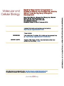
Figure 1. Ovary and testes weights (A) and adrenal gland weights (B) in embryonic and posthatch Japanese quail at various ages.
nificant differences in the content of the left ovaries and adrenal glands compared to E2 in the testes and adrenal glands in males, regardless of age. However, when E2 contents of testes and left ovaries were compared, we observed that ovaries contained more E2 than the testes. Sex differences in the amount of steroid production could be due to the differences in the steroidogenic enzymes (Imataka et al., 1988). Nakamaura et al. (1978) reported that 17α-hydroxylase activity, which is responsible for the production of testosterone, was detectable in the adrenals of the embryonic chicken but disappeared soon after hatch. Other studies have indicated that mRNA for steroidogenic enzymes required to convert cholesterol to androgens are present in the avian embryo before gonadal differentiation (Nomura et al., 1999). In contrast, p450 aromatase mRNA was detected in female embryos during E5 to E9 but not in male embryos (Nomura et al., 1999). In summary, the contributions from adrenal glands and gonads are required to establish plasma levels of androgen and E2 during embryonic and early postnatal development. However, the pattern of change in plasma steroid hormones appear to be dependent on the gonad’s production and release. Because the hypothalamus-pituitary-gonadal axis appears functional later in embryonic development (Li et al., 1991), steroid feedback to the hypothalamus is likely to impact on gonadal steroid production. As a result, sex-specific patterns in plasma steroids are
Abdelnabi, M. A., M. R. Bakst, J. E. Woods, and M .A. Ottinger, 2000. Plasma 17β estradiol levels and ovarian interstitial cell structure in embryonic and posthatch Japanese quail. Poultry Sci. 79:564–567. Adkins, E. K., 1975. Hormonal basis of sexual differentiation in the Japanese quail. J. Comp. Physiol. Psychol. 89:61–71. Adkins, E. K., 1976. The effects of antiestrogen CI-628 on sexual behavior activated by androgen or estrogen in quail. Horm. Behav. 7:417–429. Adkins, E. K., 1979. Effects of embryonic treatment with estradiol or testosterone on sexual differentiation of the quail brain. Critical period and dose response relationships. Neuroendocrinology 29:178–185. Adkins-Regan, E. K., 1985. Exposure of embryos to an aromatization inhibitor increase copulatory behavior of male quail. Behav. Proc. 2:153–158. Adkins-Regan, E., V. Manukhani, C. Siewert, and R. Thompson, 1994. Sexual differentiation of brain and behavior in the Zebra finch: Critical periods for effects of early estrogen treatment. J. Neurobiol. 25:865–877. Adkins-Regan, E., P. Pickett, and D. Koutnik, 1982. Sexual differentiation in quail: Conversion of androgen to estrogen mediates testosterone-induced demasculinization of copulation but not other male characteristics. Horm. Behav. 16:259–278. Balthazart, J., and M. A. Ottinger, 1984. 5β-reductase activity in the brain and cloacal gland of male and female embryos in the Japanese quail (Coturnix coturnix Japonica). J. Endocrinol. 102:77–81. Imataka, H., K. Suzuki, H. Inano, K. Kohmoto, and B. Tamaok, 1988. Sexual differences of steroidogenic enzymes in embryonic gonads of the chicken (Gallus domesticus). Gen. Comp. Endocrinol. 69:153–162. Li, Q., G. B. Alston-Mills, and M. A. Ottinger, 1991. Avian LHRH-I during embryonic development: Measurement by competitive ELISA with a monoclonal antibody. Gen. Comp. Endocrinol. 82:444–450. Nakamaura, T., Y. Tanabe, and H. Hirano, 1978. Evidence of in vitro formation of cortisol by the adrenal gland of embryonic and young chickens (Gallus domesticus). Gen. Comp. Endocrinol., 35:302–308. Nomura, O., O. Nakabayashi, K. Nishimori, H. Yasue, and S. Mizuno, 1999. Expression of five steroidogenic genes including aromatase gene at early developmental stages of chicken male and female embryos. J. Steroid Biochem. Mol. Biol. 71:103–109. Ottinger, M. A., and M. A. Abdelnabi, 1997. Neuroendocrine system and avian sexual differentiation. Am. Zool. 37:514– 523. Ottinger, M. A. and M. R. Bakst, 1981. Peripheral androgen concentrations and testicular morphology in embryonic and young male Japanese quail. Gen. Endocrinol. Comp. Endocrinol. 43:170–177.
EMBRYONIC STEROID HORMONES IN JAPANESE QUAIL Ottinger, M. A., and K. Mahlke 1984. Androgen concentrations in testicular and peripheral blood in the Japanese quail. Poultry Sci. 63:1851–1854. Tanabe, Y., T. Nakamura, K. Fujioka, and O. Doi, 1979. Production and secretion of sex steroid hormones by the testes, the ovary, and the adrenal glands of embryonic and young chickens (Gallus domesticus). Gen. Comp. Endocrinol. 39:26–33. Tanabe, Y., N. Saito, and T. Nakamura. 1986. Ontogenetic steroidogenesis by testes, ovary, and adrenals of embryonic and postembryonic chickens (Gallus domesticus). Gen. Comp. Endocrinol. 63:456–463.
799
Woods, J. E., and D. M. Brazzill, 1981. Plasma 17β-estradiol levels in the chick embryo. Gen. Comp. Endocrinol. 44:37–43. Woods, J. E., and L. H. Erton, 1978. The synthesis of estrogens in the gonads of the chick embryo. Gen. Comp. Endocrinol. 36:360–370. Woods, J. E., E. S. Podczaski, L. H. Erton, F. E. Rutherford, and C. F. McCarter, 1977. Establishment of the adenohypophysialtesticular axis in the chick embryo. Gen. Comp. Endocrinol. 32:390–394. Woods, J. E., R. M. Simpson and P. L. Moore, 1975. Plasma testosterone levels in the chick embryo. Gen. Comp. Endocrinol. 27:543–547.