International Journal of Pharmacological Research ISSN: 2277-3312
www.ssjournals.com Journal DOI:10.7439/ijpr
Steven Johnson syndrome and toxic epidermal necrolysis: A review Sriram Anne*, Sreya Kosanam, and Lakshmi Prasanthi N Hindu college of Pharmacy, Amaravathi Road, Guntur-522006, India.
Corresponding author* Sriram Anne, Hindu College of Pharmacy, Amaravathi Road, Guntur-522006, India. E-mail:
[email protected]
Abstract Toxic epidermal necrolysis (TEN) and Stevens Johnson Syndrome (SJS) are severe adverse cutaneous drug reactions that predominantly involve the skin and mucous membranes. They are characterized by mucocutaneous tenderness and typically hemorrhagic erosions, erythema and more or less severe epidermal detachment presenting as blisters and areas of denuded skin. Drugs are assumed or identified as the main cause of SJS/TEN in most cases, but Mycoplasma pneumoniae and Herpes simplex virus infections are well documented causes alongside rare cases in which the etiology remains unknown. Several drugs are at "high" risk of inducing TEN/SJS including: Allopurinol, Trimethoprim-sulfamethoxazole and other sulfonamide-antibiotics, aminopenicillins, cephalosporins, quinolones, carbamazepine, phenytoin, phenobarbital and NSAID's of the oxicam-type. Differential diagnosis includes linear IgA dermatosis and paraneoplastic pemphigus, pemphigus vulgaris and bullous pemphigoid, acute generalized exanthematous pustulosis (AGEP), disseminated fixed bullous drug eruption and staphyloccocal scalded skin syndrome (SSSS). Due to the high risk of mortality, management of patients with SJS/TEN requires rapid diagnosis, identification and interruption of the culprit drug, specialized supportive care ideally in an intensive care unit, and consideration of immunomodulating agents such as high-dose intravenous immunoglobulin therapy. Keywords: Toxic epidermal necrolysis, Stevens Johnson Syndrome, skin and mucous membranes.
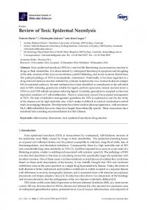
1. Introduction Stevens-Johnson syndrome (SJS) is an immune-complex–mediated hypersensitivity complex that typically involves the skin and the mucous membranes1. Stevens-Johnson syndrome was first described in 1922 as an extraordinary, generalized epidermal eruption. It is accompanied by fever, inflammation of the buccal mucosa, and severe purulent conjunctivitis. Incidence of this disorder is unknown, but it is thought to be very low. The etiology of this disorder is multiple, including drugs, infectious agents, and idiopathic causes. The mortality rate mainly depends on the age and health of the patient, and rates can range from 30 to 100 percent. Individuals at opposite ends of the age spectrum, i.e., the very young and the old, are usually fatal cases. Death is commonly due to infectious complications 2-5. Toxic epidermal necrolysis (TEN) is usually drug-related6,7. Drugs are an important cause of Stevens–Johnson syndrome, but infections or a combination of infections and drugs has also been implicated. In case reports and studies, more than 100 drugs have been implicated as causes of Stevens–Johnson syndrome or toxic epidermal necrolysis8-13. A limited number of drugs, including sulfonamides, anticonvulsant agents, and allopurinol, are the most consistently associated with the conditions; whether nonsteroidal anti-inflammatory drugs (NSAIDs), analgesic agents, and nonsulfonamide antibiotics are associated with them is controversial. The relative risk associated with the use of specific drugs has never been quantified. When there is very extensive skin detachment typical pattern of toxic epidermal necrolysis. And a poor prognosis (death rates of 30 to 40 percent), the condition is usually called toxic epidermal necrolysis. Milder forms are known as Stevens–Johnson syndrome Typical Pattern of Stevens–Johnson Syndrome or overlapping Stevens– Johnson syndrome and toxic epidermal necrolysis14. The simplest classification breaks the disease down as follows1 : Stevens-Johnson syndrome: A minor form of toxic epidermal necrolysis, with less than 10% body surface area (BSA) detachment Overlapping Stevens-Johnson syndrome/toxic epidermal necrolysis: Detachment of 10-30% of the BSA. Toxic epidermal necrolysis: Detachment of more than 30% of the BSA. Both SJS and TEN are debatably included in the same spectrum as Erythema Multiforme (EM). This mucocutaneous condition has similarities in clinical presentation to SJS/TEN but has some distinct differences 11,14 as mentioned in table-1 and figure-1.
IJPR Volume 4 Issue 4 (2014)
158
Sriram Anne
Review Article Table-1: Similarities in clinical presentation Characteristics EM SJS SJS-TEN overlap %BSA involved in detachment 90% Spots No Yes Yes Atypical targets Raised Flat Flat Mortality Rare 10% 30% Common cause Infection Medication Medication Recurrent Yes (30%) No No Sequelae Rare Common Common
TEN >30% >905 Yes Flat 50% Medication No Common
Figure: 1: Difference in severity of disease
2. Etiology Various etiologic factors have been implicated as causes of Stevens-Johnson syndrome. Drugs most commonly are blamed. The 4 etiologic categories are as follows: Infectious Drug-induced Malignancy-related Idiopathic 2.1. Infectious causes Viral diseases that have been reported to cause Stevens-Johnson syndrome include the following: Herpes simplex virus (possibly; remains a debated issue) AIDS Coxsackie viral infections Influenza Hepatitis Mumps In children, Epstein-Barr virus and enteroviruses have been identified. More than half of the patients with Stevens-Johnson syndrome report a recent upper respiratory tract infection. Bacterial etiologies include the following: Group A beta-hemolytic streptococci Diphtheria Brucellosis Lymphogranuloma venereum Mycobacteria Mycoplasma pneumonia15,16 Rickettsial infections Tularemia Typhoid IJPR Volume 4 Issue 4 (2014)
159
Sriram Anne
Review Article
2.2. Drug induced Antibiotics are the most common cause of Stevens-Johnson syndrome, followed by analgesics, cough and cold medication, NSAIDs, psychoepileptics, and antigout drugs. Of antibiotics, penicillins and sulfa drugs are prominent; ciprofloxacin has also been reported17. The following anticonvulsants have been implicated: Phenytoin Carbamazepine oxcarbazepine (Trileptal) Valproic acid Lamotrigine Barbiturates Mockenhapupt et al stressed that most anticonvulsant-induced SJS occurs in the first 60 days of use18. Antiretroviral drugs implicated in Stevens-Johnson syndrome include nevirapine and possibly other non-nucleoside reverse transcriptase inhibitors.19 Stevens-Johnson syndrome has also been reported in patients taking the following drugs: Modafinil (Provigil) Allopurinol20 Mirtazapine21 TNF-alpha antagonists (eg, infliximab, etanercept, adalimumab)22 Cocaine Sertraline Pantoprazole Tramadol 2.3. Genetic factors: Carriage of the following human leukocyte antigens has been associated with increased risk: HLA-B*1502 HLA-B*5801 HLA-B*44 HLA-A29 HLA-B12 HLA-DR7 HLA-A2 HLA-B*5801 HLA-A*0206 HLA-DQB1*0601 Certain of these HLA alleles are associated with an increased probability of developing Stevens-Johnson syndrome upon exposure to specific drugs. HLA-B*5801 confers a risk of allopurinol-related reactions.23 Pretreatment screening is not readily available.24 Whites with HLA-B*44 appear to be more susceptible to develop Stevens-Johnson syndrome. HLA-A29, HLA-B12, and HLA-DR7 are frequently associated with sulfonamide-induced Stevens-Johnson syndrome, while HLA-A2 and HLA-B12 are often encountered in Stevens-Johnson syndrome induced by nonsteroidal anti-inflammatory drugs (NSAIDs). HLA-A*0206 and HLA-DQB1*0601 allele have been shown to be was strongly associated with Stevens-Johnson syndrome with ocular disease. 25,26 Nevertheless, whether the presence of those genes constitutes a predisposition to Stevens-Johnson syndrome or whether those genes are in linkage disequilibrium with more relevant adjacent genes is unknown. 27
3. Signs and symptoms Typical prodromal symptoms of Stevens-Johnson syndrome are as follows: Cough productive of a thick, purulent sputum Headache Malaise Arthralgia Patients may complain of a burning rash that begins symmetrically on the face and the upper part of the torso. The cutaneous lesions are characterized as follows: The rash can begin as macules that develop into papules, vesicles, bullae, urticarial plaques, or confluent erythema The typical lesion has the appearance of a target; this is considered pathognomonic In contrast to the typical lesions of erythema multiforme, these lesions have only 2 zones of color The lesion’s core may be vesicular, purpuric, or necrotic; that zone is surrounded by macular erythema Lesions may become bullous and later rupture, leaving denuded skin; the skin becomes susceptible to secondary infection
IJPR Volume 4 Issue 4 (2014)
160
Sriram Anne
Review Article
Urticarial lesions typically are not pruritic Infection may be responsible for the scarring associated with morbidity Although lesions may occur anywhere, the palms, soles, dorsum of the hands, and extensor surfaces are most commonly affected The rash may be confined to any one area of the body, most often the trunk Signs of mucosal involvement can include the following: Erythema Edema Sloughing Blistering Ulceration Necrosis The following ocular signs may be noted on slit-lamp examination: Eyelids: Trichiasis, distichiasis, meibomian gland dysfunction, blepharitis Conjunctiva: Papillae, follicles, keratinization, subepithelial fibrosis, conjunctival shrinkage, foreshortening of fornices, symblepharon, ankyloblepharon Cornea: Superficial punctate keratitis, epithelial defect, stromal ulcer, neovascularization, keratinization, limbitis, conjunctivalization, stromal opacity, perforation.
4. Pathophysiology The pathogenesis of SJS/TEN is not fully understood but is believed to be immune-mediated, as re-challenging an individual with the same drug can result in rapid recurrence of SJS/TEN 29,29. An idiosyncratic, delayed hypersensitivity reaction has been implicated in the pathophysiology of StevensJohnson syndrome. Certain population groups appear more susceptible to develop Stevens-Johnson syndrome than the general population. Slow acetylators, patients who are immunocompromised (especially those infected with HIV 30,31 ), and patients with brain tumors undergoing radiotherapy with concomitant antiepileptics are among those at most risk. Slow acetylators are people whose liver cannot completely detoxify reactive drug metabolites. These drug metabolites may have direct toxic effects or may act as haptens that interact with host tissues, rendering them antigenic. 32,33 Antigen presentation and production of tumor necrosis factor (TNF)–alpha by the local tissue dendrocytes results in the recruitment and augmentation of T-lymphocyte proliferation and enhances the cytotoxicity of the other immune effector cells.34A "killer effector molecule" has been identified that may play a role in the activation of cytotoxic lymphocytes.35 The activated CD8+ lymphocytes, in turn, can induce epidermal cell apoptosis via several mechanisms, which include the release of granzyme B and perforin. Both SJS and TEN are characterised by the detachment of epidermis from the papillary dermis at the epidermaldermal junction, manifesting as a papulomacular rash and bullae as a result of keratinocyte apoptosis36,37. Keratinocyte apoptosis mediated by cytotoxic T-lymphocytes (CD8) in SJS and TENS is modulated by plasma TNF-alpha and interferon-gamma, which are increased in patients with SJS and TEN38,39. This process is currently hypothesised to occur through 3 possible pathways: Fas-Fas ligand interaction; perforin/granzyme B; and granulysin-mediated.38,40-44
5. Diagnosis The diagnosis relies on the one hand on clinical symptoms and on the other hand on histological features. Typical clinical signs initially include areas of erythematous and livid macules on the skin, on which a positive Nikolsky sign can be induced by mechanical pressure on the skin, followed within minutes to hours by the onset of epidermal detachment characterized by the development of blisters. To distinguish SJS, SJS-TEN and TEN the surface area of the detachment is the main discriminating factor. Histological work up of immediate cryosections or conventional formalin-fixed sections of the skin revealing wide spread necrotic epidermis involving all layers confirms the diagnosis. In order to rule out autoimmune blistering diseases, direct immune fluorescence staining should be additionally performed and no immunoglobulin and/or complement deposition in the epidermis and/or the epidermal-dermal zone should be detected. 5.1. Differential diagnosis Major differential diagnosis of SJS/TEN are autoimmune blistering diseases, including linear IgA dermatosis and paraneoplastic pemphigus but also pemphigus vulgaris and bullous pemphigoid, acute generalized exanthematous pustulosis (AGEP), disseminated fixed bullous drug eruption and staphyloccocal scalded skin syndrome (SSSS). SSSS was one of the most important differential diagnoses in the past, but the incidence is currently very low with 0.09 and 0.13 cases per one million inhabitants per year45.
6. Treatment 6.1. Prompt withdrawal of culprit drug(s) Prompt withdrawal of causative drugs should be a priority when blisters or erosions appear in the course of a drug eruption. Garcia-Doval et al. have shown that the earlier the causative drug is withdrawn, the better the prognosis, and that patients exposed to causative drugs with long half-lives have an increased risk of dying46. In order to identify the culprit IJPR Volume 4 Issue 4 (2014)
161
Sriram Anne
Review Article
drug(s) it is important to consider the chronology of administration of the drug and the reported ability of the drug to induce SJS/TEN. The chronology of administration of a culprit drug, or time between first administration and development of SJS/TEN, is between 1 and 4 weeks in the majority of cases. The reported ability or likelihood of a drug be the cause of SJS/TEN can be found in Pubmed/Medline or other appropriate sources such as the Litt's drug eruption reference manual47. 6.2. Supportive Care A critical element of supportive care is the management of fluid and electrolyte requirements. Intravenous fluid should be given to maintain urine output of 50 - 80 mL per hour with 0.5% NaCl supplemented with 20 mEq of KCl. Appropriate early and aggressive replacement therapy is required in case of hyponatraemia, hypokalaemia or hypophosphataemia which quite frequently occur. Wounds should be treated conservatively, without skin debridement which is often performed in burn units, as blistered skin acts as a natural biological dressing which likely favors reepithelialization. Non-adhesive wound dressings are used where required, and topical sulfa containing medications should be avoided48. 6.3. Drug therapy 6.3.1. Systemic steroids A recent retrospective monocenter study suggests that a short course "pulse" of high dose corticosteroids (dexamethasone) may be of benefit 49. 6.3.2. High-dose intravenous immunoglobulin’s As a consequence of the discovery of the anti-Fas potential of pooled human intravenous immunoglobulins (IVIG) in vitro50, IVIG have been tested for the treatment of TEN, and their effect reported in different non-controlled studies. To date, numerous case reports and 12 non-controlled clinical studies containing 10 or more patients have analyzed the therapeutic effect of IVIG in TEN. All except one study51 confirm the known excellent tolerability and a low toxic potential of IVIG when used with appropriate precaution in patients with potential risk factors (renal insufficiency, cardiac insufficiency, IgA deficiency, thrombo-embolic risk)52. The concomitant administration of corticosteroids or immunosuppressive agents remains controversial. IVIG has also been applied in a few children with SJS/TEN, and two non-controlled studies suggest a possible benefit53,54. 6.3.3. Ciclosporin (CsA) Patients treated with CsA had significantly shorter time to complete re-epithelialisation, and fewer patients with multi-organ failure and death were observed55. A small case series with three TEN patients treated initially with high-doses of intravenous dexamethasone followed by CsA showed a stop in disease progression within 72 hrs56. Other single case reports also reported a positive effect of the use of CsA in TEN 57,58. Recently, Valeyrie-Allanore L conducted an open, phase II trial to determine the safety and possible benefit of ciclosporin59. 6.4. Treatment for ocular manifestations Treatment of acute ocular manifestations usually begins with aggressive lubrication of the ocular surface. As inflammation and cicatricial changes ensue, most ophthalmologists use topical steroids, antibiotics, and symblepharon lysis. In case of exposure keratopathy, tarsorrhaphy may be required. Maintenance of ocular integrity can be achieved through the use of amniotic membrane grafting, adhesive glues, lamellar grafts, and penetrating keratoplasty, either in the acute phase or in subsequent follow-up care. Visual rehabilitation in patients with visual impairment can be considered once the eye has been quiet for at least 3 months. 6.5. Topical treatment Although the blisters are fragile, they should be left in place or only be punctured. Erosions can be treated with chlorhexidine, octenisept or polyhexanide solutions and impregnated nonadhesive mesh gauze. The latter is important if environmental factors, such as high room temperature or alternating pressure mattress, lead to skin dryness. Silver sulfadiazine should be avoided, at least if the causative drug was cotrimoxazole or another anti-infective sulfonamide. Some burn care specialists debride the skin under general anesthesia and apply allografts or other types of coverage. However, this rather aggressive procedure is not tolerated well by many elderly patients with underlying diseases 60. Furthermore, hypertrophic scars may occur if debridement is carried out extensively and if allografts are fixed with staples directly into the skin. For affected mucosal surfaces, specialized care is critical. The severity of the mucosal involvement is often not in line with the amount of skin detachment and overlooked mucosal lesions can lead to life-long problems. A multidisciplinary approach is needed and in case of urethral involvement urologists should be involved. Appropriately placed wet dressings or sitz baths may help to avoid adhesions or strictures of genital erosions in girls and women. Disinfectant mouth wash should be used to treat oral erosions and mild ointment, such as dexpanthenol, should be applied on erosions and bloody crusts of the lips. In the case of eye-involvement, regular ophthalmologic consultation is crucial. Specialized lid care is needed on a daily basis and anti-inflammatory eye drops should be given several times per day. Severe blepharitis may lead to entropion with trichiasis (in growing eye lashes) causing further corneal damage. Various specialized approaches to ocular involvement have been suggested, such as stem cell generation of replacement cells, amniotic membrane transplantation and scleral lenses, but are not yet widely accepted61,62. Nevertheless, experienced ophthalmologists should be involved in the care of all patients with SJS/TEN, even those that do not present with eye-involvement right away, since it may occur with some delay. IJPR Volume 4 Issue 4 (2014)
162
Sriram Anne
Review Article
7. Conclusion SJS and toxic epidermal necrolysis (TEN) are considered as one disease entity of different severity. SJS/TEN is mainly caused by drugs, infections and probably other risk factors not yet identified. The pathogenesis of SJS/TEN has not been completely solved, but specific genetic predispositions, which vary among ethnic groups and differ between certain causing drugs, were identified. Since to date no treatment has been identified to be capable of halting the progression of skin detachment, supportive management is crucial to improve the patient’s state, probably more than specific immunomodulating treatments. Despite all therapeutic efforts, mortality is high and increases with disease severity, patients’ age and underlying medical conditions. Survivors may suffer from long-term sequelae such as strictures of mucous membranes including severe eye problems. Therefore, interdisciplinary care and follow-up of patients with SJS/TEN is important.
References 1. French LE. Toxic epidermal necrolysis and Stevens Johnson syndrome: our current understanding. Allergol Int. Mar 2006; 55(1):9-16. 2. Huff JC, Weston WL, Tonnesen MG. Erythema multiforme: Critical review of characteristics, diagnostic criteria, and causes. J Am Acad Dermatol 1983; 8:763-775. 3. Ackerman AB, Penneys NS, Clark WH. Erythema multiforme exudativum: Distinctive pathological process. Br J Dermatol 1971; 84:554-566. 4. Yetiv JZ, Bianchine JR, Owen JA. Etiologic factors of the Stevens-Johnson syndrome. South Med J 1980; 73:599-602. 5. Bianchine JR, Macaraeg PV, Lasagn L, et al. Drugs as etiologic factors in the Stevens-Johnson syndrome. Am J Med 1968; 44:390-405. 6. Lyell A. Toxic epidermal necrolysis (the scalded skin syndrome): a reappraisal. Br J Dermatol 1979;100:69-86 7. Roujeau JC, Stern RS. Severe cutaneous adverse reactions to drugs. N Engl J Med1994; 331:1272-1285 8. Stern RS, Chan HL. Usefulness of case report literature in determining drugs responsible for toxic epidermal necrolysis. J Am Acad Dermatol1989; 21:317-322. 9. Ruiz-Maldonado R. Acute disseminated epidermal necrosis types 1, 2, and 3: study of sixty cases. J Am Acad Dermatol 1985; 13:623-635. 10. Guillaume J-C, Roujeau J-C, Revuz J, Penso D, Touraine R. The culprit drugs in 87 cases of toxic epidermal necrolysis (Lyell's syndrome). Arch Dermatol 1987; 123:1166-1170. 11. Roujeau J-C, Guillaume J-C, Fabre J-P, Penso D, Flechet ML, Girre JP. Toxic epidermal necrolysis (Lyell syndrome): incidence and drug etiology in France, 1981-1985. Arch Dermatol1990; 126:37-42. 12. Schopf E, Stuhmer A, Rzany B, Victor N, Zentgraf R, Kapp JF. Toxic epidermal necrolysis and Stevens-Johnson syndrome: an epidemiologic study from West Germany. Arch Dermatol 1991; 127:839-842. 13. Correia O, Chosidow O, Saiag P, Bastuji-Garin S, Revuz J, Roujeau J-C. Evolving pattern of drug-induced toxic epidermal necrolysis. Dermatology 1993; 186:32-37. 14. Bastuji-Garin S, Rzany B, Stern RS, Shear NH, Naldi L, Roujeau J-C. Clinical classification of cases of toxic epidermal necrolysis, Stevens-Johnson syndrome, and erythema multiforme. Arch Dermatol 1993; 129:92-96. 15. Hillebrand-Haverkort ME, Budding AE, bij de Vaate LA, van Agtmael MA. Mycoplasma pneumoniae infection with incomplete Stevens-Johnson syndrome. Lancet Infect Dis. Oct 2008; 8(10):586-7. 16. Sendi P, Graber P, Lepère F, Schiller P, Zimmerli W. Mycoplasma pneumoniae infection complicated by severe mucocutaneous lesions. Lancet Infect Dis. Apr 2008; 8(4):268. 17. Hällgren J, Tengvall-Linder M, Persson M, Wahlgren CF. Stevens-Johnson syndrome associated with ciprofloxacin: a review of adverse cutaneous events reported in Sweden as associated with this drug. J Am Acad Dermatol. Nov 2003; 49(5 Suppl):S267-9. 18. Mockenhaupt M, Messenheimer J, Tennis P, Schlingmann J. Risk of Stevens-Johnson syndrome and toxic epidermal necrolysis in new users of antiepileptics. Neurology. Apr 12 2005; 64(7):1134-8. 19. Metry DW, Lahart CJ, Farmer KL, Hebert AA. Stevens-Johnson syndrome caused by the antiretroviral drug nevirapine. J Am Acad Dermatol. Feb 2001; 44(2 Suppl):354-7. 20. Halevy S, Ghislain PD, Mockenhaupt M, et al. Allopurinol is the most common cause of Stevens-Johnson syndrome and toxic epidermal necrolysis in Europe and Israel. J Am Acad Dermatol. Jan 2008; 58(1):25-32. 21. Belkahia A, Hillaire-Buys D, Dereure O, Guillot B, Raison-Peyron N. Stevens-Johnson syndrome due to mirtazapine first case. Allergy. Oct 2009; 64(10):1554. 22. Salama M, Lawrance IC. Stevens-Johnson syndrome complicating adalimumab therapy in Crohn's disease. World J Gastroenterol. Sep 21 2009; 15(35):4449-52. 23. Kardaun SH, Jonkman MF. Dexamethasone pulse therapy for Stevens-Johnson syndrome/toxic epidermal necrolysis. Acta Derm Venereol. 2007; 87(2):144-8. 24. Fernando SL, Broadfoot AJ. Prevention of severe cutaneous adverse drug reactions: the emerging value of pharmacogenetic screening. CMAJ. Mar 23 2010; 182(5):476-80. 25. Hynes AY, Kafkala C, Daoud YJ, Foster CS. Controversy in the use of high-dose systemic steroids in the acute care of patients with Stevens-Johnson syndrome. Int Ophthalmol Clin. Fall 2005; 45(4):25-48. IJPR Volume 4 Issue 4 (2014)
163
Sriram Anne
Review Article
26. Khalili B, Bahna SL. Pathogenesis and recent therapeutic trends in Stevens-Johnson syndrome and toxic epidermal necrolysis. Ann Allergy Asthma Immunol. Sep 2006; 97(3):272-80; quiz 281-3, 320. 27. Meth MJ, Sperber KE. Phenotypic diversity in delayed drug hypersensitivity: an immunologic explanation. Mt Sinai J Med. Sep 2006; 73(5):769-76. 28. Halevi A, Ben-Amitai D, Garty BZ. Toxic epidermal necrolysis associated with acetaminophen ingestion. Ann Pharmacother.2000; 34:32–34. doi: 10.1345/aph.19064. 29. Schmidt D, Kluge W. Fatal toxic epidermal necrolysis following reexposure to phenytoin: a case report. Epilepsia. 1983; 24:440–443. doi: 10.1111/j.1528-1157.1983.tb04914.x. 30. Rotunda A, Hirsch RJ, Scheinfeld N, Weinberg JM. Severe cutaneous reactions associated with the use of human immunodeficiency virus medications. Acta Derm Venereol. 2003; 83(1):1-9. 31. Gruchalla RS. 10. Drug allergy. J Allergy Clin Immunol. Feb 2003; 111(2 Suppl):S548-59. 32. Ahmed AR, Dahl MV. Consensus statement on the use of intravenous immunoglobulin therapy in the treatment of autoimmune mucocutaneous blistering diseases. Arch Dermatol. Aug 2003; 139(8):1051-9. 33. Assier-Bonnet H, Aractingi S, Cadranel J, Wechsler J, Mayaud C, Saiag P. Stevens-Johnson syndrome induced by cyclophosphamide: report of two cases. Br J Dermatol. Nov 1996; 135(5):864-6. 34. De Rojas MV, Dart JK, Saw VP. The natural history of Stevens Johnson syndrome: patterns of chronic ocular disease and the role of systemic immunosuppressive therapy. Br J Ophthalmol. Aug 2007; 91(8):1048-53. 35. Morel E, Escamochero S, Cabañas R, Díaz R, Fiandor A, Bellon T. CD94/NKG2C is a killer effector molecule in patients with Stevens-Johnson syndrome and toxic epidermal necrolysis. J Allergy Clin Immunol. Mar 2010; 125(3):703-10, 710.e1-710.e8. 36. Paul C, Wolkenstein PC, Adle H, et al. Apoptosis as a mechanism of keratinocyte death in toxic epidermal necrolysis. Br J Dermatol. 1996; 134:710-714. 37. Le Cleach L, Delaire S, Boumsell L, et al. Blister fluid T lymphocytes during toxic epidermal necrolysis are functional cytotoxic cells which express human natural killer (NK) inhibitory receptors. Clin Exp Immunol. 2000; 119:225-230. 38. Schwartz RA, McDonough PH, Lee BW. Toxic epidermal necrolysis. Part I: introduction, history, classification, clinical features, systemic manifestations, etiology, and immunopathogenesis. J Am Acad Dermatol. 2013; 69:173. 39. Caproni M, Torchia D, Schincaglia E, et al. Expression of cytokines and chemokine receptors in the cutaneous lesions of erythema multiforme and Stevens-Johnson syndrome/toxic epidermal necrolysis. Br. J Dermatol. 2006; 155:722728. 40. Mockenhaupt M. The current understanding of Stevens-Johnson syndrome and toxic epidermal necrolysis. Expert Rev Clin Immunol. 2011; 7:803-813. 41. Schwartz RA, McDonough PH, Lee BW. Toxic epidermal necrolysis. Part II: prognosis, sequelae, diagnosis, differential diagnosis, prevention, and treatment. J Am Acad Dermatol. 2013; 69:187. 42. Viard I, Wehrli P, Bullani R, et al. Inhibition of toxic epidermal necrolysis by blockade of CD95 with human intravenous immunoglobulin. Science. 1998; 282:490-493. 43. Chung WH, Hung SI, Yang JY, et al. Granulysin is a key mediator for disseminated keratinocyte death in StevensJohnson syndrome and toxic epidermal necrolysis. Nat Med. 2008; 14:1343-1350. 44. Abe R, Shimizu T, Shibaki A, et al. Toxic epidermal necrolysis and Stevens-Johnson syndrome are induced by soluble Fas ligand. Am J pathol 2003; 162:1515-1520. 45. Mockenhaupt M, Messenheimer J, Tennis P, Schlingmann J. Risk of Stevens-Johnson syndrome and toxic epidermal necrolysis in new users of antiepileptics. Neurology.2005; 64:1134–1138. 46. Garcia-Doval I, LeCleach L, Bocquet H, Otero XL, Roujeau JC: Toxic epidermal necrolysis and Stevens-Johnson syndrome: does early withdrawal of causative drugs decrease the risk of death? Arch Dermatol 2000, 136:323-327. 47. Litt JZ: Litt's Drug Eruptions and Reactions Manual.16 th edition. 2010. 48. Thomas Harr and Lars E French. Toxic epidermal necrolysis and Stevens-Johnson syndrome. Orphanet Journal of Rare Diseases 2010; 5. 49. Kardaun SH, Jonkman MF: Dexamethasone pulse therapy for Stevens-Johnson syndrome/toxic epidermal necrolysis. Acta Derm Venereol 2007, 87:144-148. 50. Viard I, Wehrli P, Bullani R, Schneider P, Holler N, Salomon D, Hunziker T, Saurat JH, Tschopp J, French LE: Inhibition of toxic epidermal necrolysis by blockade of CD95 with human intravenous immunoglobulin. Science 1998, 282:490-493. 51. Bachot N, Revuz J, Roujeau JC: Intravenous immunoglobulin treatment for Stevens-Johnson syndrome and toxic epidermal necrolysis: a prospective noncomparative study showing no benefit on mortality or progression. Arch Dermatol 2003, 139:33-36. 52. Prins C, Gelfand EW, French LE: Intravenous immunoglobulin: properties, mode of action and practical use in dermatology. Acta Derm Venereol 2007, 87:206-218. 53. Mockenhaupt M, Messenheimer J, Tennis P, Schlingmann J: Risk of Stevens-Johnson syndrome and toxic epidermal necrolysis in new users of antiepileptics. Neurology 2005, 64:1134-1138. 54. Tennis P, Stern RS: Risk of serious cutaneous disorders after initiation of use of phenytoin, carbamazepine, or sodium valproate: a record linkage study. Neurology 1997, 49:542-546. IJPR Volume 4 Issue 4 (2014)
164
Sriram Anne
Review Article
55. Arevalo JM, Lorente JA, Gonzalez-Herrada C, Jimenez-Reyes J: Treatment of toxic epidermal necrolysis with cyclosporin A. J Trauma 2000, 48:473-478. 56. Rai R, Srinivas CR: Suprapharmacologic doses of intravenous dexamethasone followed by cyclosporine in the treatment of toxic epidermal necrolysis. Indian J Dermatol Venereol Leprol 2008; 74:263-265. 57. Hashim N, Bandara D, Tan E, Ilchyshyn A: Early cyclosporine treatment of incipient toxic epidermal necrolysis induced by concomitant use of lamotrigine and sodium valproate. Acta Derm Venereol 2004, 84:90-91. 58. Robak E, Robak T, Gora-Tybor J, Chojnowski K, Strzelecka B, Waszczykowska E, Sysa-Jedrzejowska A: Toxic epidermal necrolysis in a patient with severe aplastic anemia treated with cyclosporin A and G-CSF. J Med 2001, 32:31-39. 59. Valeyrie-Allanore L, Wolkenstein P, Brochard L, Ortonne N, Maitre B, Revuz J, Bagot M, Roujeau J: Open trial of ciclosporin treatment for Stevens-Johnson syndrome and toxic epidermal necrolysis. Br J Dermatol 2010: 163(4):847853. 60. Struck MF, Hilbert P, Mockenhaupt M, Reichelt B, Steen M. Severe cutaneous adverse reactions: emergency approach to non-burn epidermolytic syndromes. Intensive Care Med. 2010; 36(1), 22–32. 61. Yip LW, Thong BY, Lim J et al. Ocular manifestations and complications of Stevens–Johnson syndrome and toxic epidermal necrolysis: an Asian series. Allergy 2007; 62, 527–531. 62. Gregory DG. The ophthalmologic management of acute Stevens–Johnson syndrome. Ocul. Surf. 2008; 6: 87–95.
IJPR Volume 4 Issue 4 (2014)
165