Rom J Morphol Embryol 2013, 54(2):377–383
RJME
ORIGINAL PAPER
Romanian Journal of Morphology & Embryology http://www.rjme.ro/
Structural chromosomal anomalies detected by prenatal genetic diagnosis: our experience SIMONA FARCAŞ1), C. D. CRIŞAN2), NICOLETA ANDREESCU1), MONICA STOIAN1), A. G. M. MOTOC3) 1)
2)
Department of Microscopic Morphology, Division of Genetics
Department of Obstetrics and Gynecology
3)
Department of Anatomy and Embryology
“Victor Babeş” University of Medicine and Pharmacy, Timisoara
Abstract
The prenatal diagnosis is currently widely spread and facilitates the acquiring of important genetic information about the fetus by a rate extremely accelerate and considered without precedent. In this paper, we like to present our experience concerning the genetic diagnosis and counseling offered for pregnancies in which a structural chromosomal aberration was found. The study group is formed by 528 prenatal samples of amniotic fluid and chorionic villi, received by our laboratory from 2006 through October 2012 for cytogenetic diagnosis. The appropriate genetic investigation was selected based on the indications for prenatal diagnosis. The cases with structural chromosomal anomalies and polymorphic variants were analyzed as regard to the maternal age, gestational age, referral indications and type of chromosomal anomaly found. A total number of 21 structural chromosomal anomalies and polymorphic variants were identified in the study group. Out of 21 structural chromosomal anomalies and polymorphic variants, six deletions and microdeletions, four situations with abnormal long “p” arm of acrocentric chromosomes, two duplications, two reciprocal translocations, two inversions, two additions, one Robertsonian translocation associating trisomy 13, one 9q heteromorphism and one complex chromosome rearrangement were noticed. To the best of our knowledge, this is the first Romanian study in which the diagnostic strategies and the management of the prenatal cases with structural rearrangements are presented. The data provided about the diagnosis strategy and the management of the prenatal cases with structural chromosomal anomalies represents a useful tool in genetic counseling of pregnancies diagnosed with rare structural chromosomal anomalies. Keywords: genetic prenatal diagnosis, structural chromosomal anomalies, genetic counseling.
Introduction The prenatal diagnosis is currently widely spread and facilitates the acquiring of important genetic information about the fetus by a rate extremely accelerate and considered without precedent [1]. Since 70s when it was first used, until now new techniques for screening and diagnosis were introduced. Using the new screening tests that can select the pregnancies at risk, many syndromes can be identified before birth by performing prenatal diagnosis. Currently, new techniques such as microarray CGH are used in clinical studies and different strategies were elaborated for rapid and efficient prenatal diagnosis [2–4]. In prenatal diagnosis, the new molecular techniques can offer a rapid result for a selected range of chromosomal aberrations, but the cytogenetic analysis still remains the gold standard. By using conventional cytogenetic studies, not only the major aneuploidies can be identified, but also the structural rearrangements [5]. The conventional cytogenetic investigations, even with a poor banding resolution, are required for the validations of the results after the identification of a specific chromosomal anomaly by molecular analyses. Whether for the numerical chromosomal anomalies ISSN (print) 1220–0522
there are many techniques available, the structural chromosomal aberrations, rarely found, can raise different problems for a correct diagnosis and an accurate genetic counseling. Regarding the structural chromosomal anomalies the most important aspect is about the viability of the fetus, depending on the chromosome(s) involved, the type of anomaly and the size of the defect [6]. For these cases, in some situations, additional investigations should be performed, as a more comprehensive molecular characterization is essential. The genetic investigations must be associated with ultrasound evaluation in order to have a complete tableau of the case [7]. The next questions raised are about the risk for a future pregnancy and the couple’s reproductive alternatives available in order to have a healthy child. In this paper, we like to present our experience concerning the genetic diagnosis and counseling offered for pregnancies in which a structural chromosomal aberration was found. Materials and Methods The study group is formed by the prenatal samples of amniotic fluid and chorionic villi, received by our laboratory from 2006 through October 2012 for ISSN (on-line) 2066–8279
Simona Farcaş et al.
378
cytogenetic diagnosis. A total number of 528 cases were referred by the clinicians from the “Dr. Dumitru Popescu” Obstetrics and Gynecology Hospital, Timişoara, Romania, for genetic investigations. The indications for prenatal diagnosis included: advanced maternal age (≥35-yearold at the expected date of confinement), abnormal biochemical screening markers in maternal serum (double test or triple test), abnormal ultrasound findings, previous history of a fetus/child with chromosomal abnormalities or congenital anomalies, family history of chromosomal abnormalities or congenital anomalies, patient’s anxiety and twin pregnancies. The appropriate genetic investigation was selected based on the indications for prenatal diagnosis. For the majority of cases the cytogenetic investigation was considered sufficient. The FISH analysis was performed for the cases referred with suspicion of microdeletion/ microduplication and as an additional investigation for the cases where structural chromosomal aberrations were found. The chorionic villi samples were released from maternal deciduas and blood clots. The samples were divided into fragments of 3–5 mg, and an amount of approximately 15 mg villi were exposed for 15 minutes to collagenase digestion at 370C. Two long-term monolayer cultures in Chang medium (Irvine Scientific), from two
different groups, were established in a 5% CO2 incubator at 370C. The cultures were harvested after 10 days and then used for karyotyping by standard cytogenetic techniques or FISH analysis. For amniotic cells, the cultures were established in two different groups of Gibco AmnioMAX-C100 Basal Medium supplemented with Gibco AmnioMAX-C100 Supplement (Invitrogen). The flask method was used for cultures set in a 5% CO2 incubator at 370C. Approximately after 10 days, the cultures were harvested and the standard chromosome preparation or FISH technique was done. For FISH analysis, the probes used were acquired from Abbott (Downers Grove, IL, USA). Denaturation and hybridization of the DNA specimen and the probe was performed according to the manufacturer’s instructions. The slides were observed under a fluorescence microscope using appropriate filters. A minimum of 200 nuclei/ metaphases were scored for each probe. Results A total number of 21 structural chromosomal anomalies and polymorphic variants were identified in the study group. The results are summarized in the Table 1.
Table 1 – The gravidae data and the results of the cytogenetic and molecular analysis of the prenatal cases with structural chromosomal anomalies Case Maternal age No. [years] 1. 26 2.
26
3.
29
4.
36
5.
26
6.
33
7.
24
8.
23
9. 10. 11.
38 32 27
12.
34
13.
31
14.
34
15.
29
16.
33
17.
36
18.
30
19.
30
20.
20
21.
26
Gestational Indications for prenatal FISH analysis Fetus karyotype Parental karyotype age [weeks] genetic diagnosis for fetus 12 Ultrasound markers 46,XX,del11qter del11qter 46,XX,t(10;11)(p14;q21) Biochemical abnormal del9qter, 11 46,XX,add9qter 46,XX,t(9;10)(q34;p11.2) screening dup10pter 10 Ultrasound markers 46,XX,t(4;15)(p16;q22) 46,XX,t(4;15)(p16;q22) Advanced maternal age, 11 46,XX,add12q 46,XX,t(3;12)(q27;q24.3) ultrasound markers Biochemical abnormal 18 46,XX,dup(22)(q12.2q13.1) screening Biochemical abnormal 16 46,XX,9qh+ screening Biochemical abnormal 18 46,XY,del(4)(p16pter) del4pter screening Biochemical abnormal 18 46,XY,inv(9)(p13;q13) 46,XY,inv(9)(p13;q13) screening 18 Advanced maternal age 46,XY,t(4;6)(q23;q15) Normal 18 Ultrasound markers del22q11 20 Ultrasound markers del15q11-13 47,XY,t(4;21)(q13.3q22.3), 17 Ultrasound markers +mar 18 Ultrasound markers del22q11 Normal Biochemical abnormal 18 46,XY,dup(16) screening, ultrasound markers 17 Ultrasound markers del22q11 Biochemical abnormal 17 46,XY,13pstk+ 46,XY,13pstk+ screening Advanced maternal age, 17 46,XX,21pstk+ 46,XX,21pstk+ familial history of congenital malformations Biochemical abnormal 15 46,XY,trob(13;14) 45,XY,trob(13;14) screening Biochemical abnormal 16 46,XX,13pstk+ 46,XX,13pstk+ screening Biochemical abnormal 18 46,XY,inv(9)(p11q13) 46,XX,inv(9)(p11q13) screening Biochemical abnormal 18 46,XX,14pstk+ 46,XX,14pstk+ screening
Structural chromosomal anomalies detected by prenatal genetic diagnosis: our experience
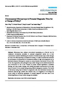
Additionally FISH analysis was performed in some cases in order to correctly characterize the cytogenetic anomalies found. The results of these analyses are also summarized Table 1. For 13 cases, parental karyotype was done and the results were confirmed by FISH analysis as can be seen in Table 1. The cases with structural chromosomal anomalies and polymorphic variants were analyzed as regard to the maternal age, gestational age, referral indications and type of chromosomal anomaly found. In the majority of cases, the maternal age was under 35-year-old (85.71%) and only 14.29% of the gravidae had an advanced maternal age. For 80.95% of the cases the gestational age was between 15th and 20th week of amenorrhea and for these cases, amniocentesis was done. For four (19.05%) cases the gestational age varied between 10 and 12 weeks of amenorrhea and for these cases chorionic villi biopsy was performed. The indications for prenatal genetic diagnosis in the group of the gravidae were: abnormal biochemical screening (double test or triple test) in nine (42.85%) cases, fetal malformations observed at ultrasound evaluations in eight (38.09%) cases, advanced maternal age one (4.76%) case, the association of two indications was found in three (14.28%) cases, one case with abnormal biochemical and ultrasound markers, one case with advanced maternal age and family history of congenital malformations and one case with advanced maternal age and fetal malformations identified by ultrasound (Figure 1).
Figure 1 – Indications of the prenatal diagnosis for the cases with structural chromosomal anomalies.
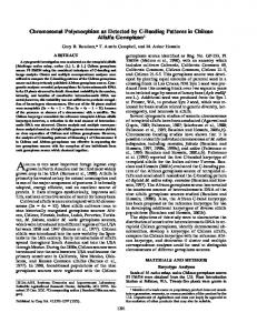
The fetal malformations identified through ultrasound evaluation comprised heart anomalies, choroid plexus cysts, pulmonary atresia, single umbilical artery and thickened nuchal fold (>3 mm). Out of 21 structural chromosomal anomalies and polymorphic variants, six deletions and microdeletions, four situations with abnormal long “p” arm of acrocentric chromosomes, two duplications, two reciprocal translocations, two inversions, two additions, one Robertsonian translocation associating trisomy 13, one 9q heteromorphism and one complex chromosome rearrangement were noticed. We have classified the karyotypes in three groups (Figure 2): unbalanced chromosome aberrations (deletions/microdeletions, duplications, additions, the complex chromosomal rearrangement and the Robertsonian translocation associated with trisomy 13) balanced chromosomal rearrangements (inversions, enlargement of the heterochromatic region) and apparently balanced
379
anomalies (translocation and long “p” arm of acrocentric chromosomes).
Figure 2 – Classification of structural chromosomal anomalies found through prenatal diagnosis.
Discussion Genetic counseling has improved the pregnancy management and the use of an appropriate diagnosis method followed by a risk assessment [8]. In our country, as in all European countries, the screening programs used for identification of fetal anomalies in the first or second trimester of the pregnancy lead to a better selection of the pregnancies at risk of having conception products with chromosomal aberrations [9]. More efficient for detection of the most frequent aneuploidies, these screening programs also facilitated the identification of the rare structural anomalies. Currently there is only little information about the frequency and consequences of the rare chromosomal defects and most of the data are from selected studies that cannot offer a real incidence [9]. Structural unbalanced chromosomal aberrations lead to phenotype anomalies that can vary from minor dysmorphism to sever malformations affecting the vital prognosis of the fetus. For these cases, the management of the pregnancy implies a comprehensive ultrasound evaluation followed by genetic counseling in order to inform the parents about the pregnancy risk, the possible fetal development and the consequences of the genetic defect identified. Depending on the severity of the fetal malformations, the parents can choose to terminate the pregnancy or to have the child. For the cases with unbalanced chromosomal defects, the parents were informed about the genetic defect found and the consequences for the fetus were discussed for each case. In the majority of the cases, the parents option was to elective terminate the pregnancy. The parents were informed about the importance of testing their genetic material in order to establish if the fetal chromosomal anomaly is inherited or de novo. Out of the group of fetuses with unbalanced chromosomal aberrations the parental karyotype was done for five cases, the rest of the couples refused further investigations or did not present for blood sampling. In one case with 22q11 microdeletion detected in the fetus (Figure 3), both parents karyotype did not show any structural aberration. Only one case of Robertsonian translocation was found in the study group, involving chromosomes 13
380
Simona Farcaş et al.
and 14, associated with trisomy 13. For this case, an important aspect is that the pregnancy was obtained after in vitro fertilization. An interesting finding was that the father carried the same Robertsonian translocation. This finding emphasizes the importance of performing cytogenetic investigation to all couples before undergoing in vitro fertilization, so these situations could be identified and preimplantation genetic diagnosis be offered to those in need.
Figure 5 – Mother’s karyotype showing a reciprocal translocation 46,XX,t(3;12)(q27;q24.3). Figure 3 – FISH analysis of the fetus showing the microdeletion 22q11.2 (green signal for both regions 22q13 and only one red signal corresponding to the chromosomal region 22q11.2).
In another case, where a deletion 11q was found in fetus, maternal karyotype showed a balanced translocation between chromosomes 10 and 11. For the fetus and mother, FISH analysis was performed and the cytogenetic results were confirmed. By extending the family investigation, two other members from the mother side (her mother and her brother) were identified to be carriers of the same chromosomal aberration. Another case was of a fetus were an addition on the chromosome 12q was found (Figure 4). The maternal karyotype revealed a reciprocal translocation between chromosomes 3 and 12 (Figure 5). This finding allowed us to conclude that the additional genetic material on the chromosome 12 was the terminal region of the chromosome 3.
Figure 4 – The fetal karyotype showing an addition on the chromosome 12.
The last case where parental karyotype was performed was the one with an addition on the chromosome 9q (Figure 6). The FISH analysis using telomeric 9q probe (Spectrum Orange) revealed a terminal deletion of the long arm of chromosome 9 (Figure 7). We supposed that a deletion of the 9qter and a partial trisomy were associated in this case. The maternal karyotype in this case revealed a balanced translocation between chromosomes 9 and 10 (Figure 8). Additional FISH analysis using probes for the short arm of the chromosome 9 – TelVysion 9p SpectrumGreen and for the long arm of the chromosome 9 – TelVysion 9q SpectrumOrange was performed for the mother. The result of the FISH analysis of the mother confirmed the translocation of the 9qter and did not revealed any deletion (Figure 9). After this finding, we concluded that the addition found in fetus karyotype could be due to a partial trisomy 10p. This case was the only one from the group with unbalanced chromosomal anomalies of the fetus where the parents decided to continue the pregnancy. The delivery was at term but the plurimalformed baby had an infaust evolution.
Figure 6 – The image shows an addition in the chromosome 9q.
Structural chromosomal anomalies detected by prenatal genetic diagnosis: our experience
Figure 7 – The picture present the deletion of the telomeric region 9q (only one hybridization signal for 9qTEL probe).
Figure 8 – The image presents the maternal karyotype with a balanced translocation (9;10).
Figure 9 – Picture of FISH analysis of the pregnant woman showing a translocation of the 9qter and no deletions of the telomeric regions 9p and 9q.
381
These cases, in which one of the parents was carrier of a balanced chromosomal rearrangement, underwent genetic counseling in order to be informed about the recurrence risk, but also about their reproductive options. The couples were informed that the risk estimation for a future pregnancy is mainly empirical. The couples ascertained after having a an affected baby with an unbalanced karyotype have a higher risk (20–22%) as compared with the couples identified with balanced translocations after recurrent miscarriages (2–5%) [10]. An option for these cases is to undergo preimplantation genetic diagnosis that can reduce to risk of having an affected child. For this procedure, in vitro fertilization is required and the couples have been informed about the associated risk. The inversions of the chromosome 9 were considered as balanced chromosomal rearrangements based on the previous research that established that these are polymorphic variants that do not produce phenotypic effect [11]. In the two cases of inversions of the chromosome 9 we recommended cytogenetic investigations of the parents. In both cases, a similar inversion was found, one case with a paternal, the second with a maternal inversion of the chromosome 9. The enlargement of heterochromatic region of the chromosome 9 was included in the balanced chromosomal anomalies taking in consideration Liehr T et al. report that concluded that partial trisomies or polysomies of the centromeric heterochromatin of this chromosome are not associated with clinical effects [12]. For the apparently balanced structural anomalies, special care measures can be necessary. Usually additional tests are required in order to correctly characterize the anomaly. A useful strategy in these cases is to have the parents karyotype in order to find if the defect is inherited or is a de novo anomaly. With the new molecular techniques, now is possible to establish if the chromosomal aberrations considered balanced can be associated with submicroscopic deletions/duplications. These are especially necessary for de novo chromosomal rearrangements. The variations of the length of the “p” arm of the acrocentric chromosomes were considered as chromosomal polymorphisms without any clinical relevance, but recent studies suggested that in prenatal diagnosis additional investigations should be performed [13]. In the literature, several cases are reported in which the variations in size of the short arm of acrocentric chromosomes were due to partial trisomies of different chromosomes [14, 15]. In the cases where an abnormal “p” arm length of chromosomes 13 (Figure 10), 14 or 21 was noticed a special attention was offered in order to make sure that the morphological modification represents a normal variant and not a structural aberration with phenotypic relevance. For these four situations, we performed additionally cytogenetic investigations of the parents. For all the cases a similar chromosomal polymorphism was establish in one of the parents (Figure 11). Based on these results the cases were considered as polymorphic variants without clinical relevance and the pregnancies were continued.
382
Simona Farcaş et al.
Figure 10 – The picture showing the fetus karyotype 46,XX,13pstk+.
Figure 11 – The maternal karyotype showing a long “p” arm of the chromosome 13: 46,XX,13pstk+.
Reciprocal translocations were included in the apparently balanced group of chromosomal anomalies. Apparently, balanced translocations can be associated with microdeletions/microduplications detectable by FISH or array-CGH [16, 17]. First, for these cases we recommended that cytogenetic investigations of the parents should be done in order to establish if the defect is de novo or inherited. The first prenatal case where a reciprocal translocation was found had the following karyotype: 46,XY,t(4;6) (q23;q15). Advanced maternal age was the indication for amniocentesis and prenatal genetic diagnosis. For this case, parental karyotype was required and an ultrasound examination was indicated. In the parents karyotype no chromosomal anomalies were found which allowed us to conclude that it was a de novo chromosomal anomaly. For this case, FISH analysis was
not available at that time. At ultrasound examination, a defect of the abdominal region was observed. The parents decided to continue the pregnancy regardless of the possible consequences and a plurimalformed baby was born. As previous reported, the phenotypic anomalies in cases with balanced chromosomal rearrangements detected by conventional cytogenetics are due to gain/ loss of genetic material at the breakpoints of the chromosomes involved in translocation [18]. In the second case where a reciprocal translocation – 46,XX,t(4;15)(p16;q22) – was found, at the cytogenetic investigations of the parents, a similar translocation was found in the maternal karyotype. FISH analysis did not reveal loss or gain of chromosomal material at the breaking points. In this case, the pregnancy was continued and the baby was delivered at term, showing no dysmorphic features. For this study, the incidence of the structural chromosomal aberrations was of 3.97% showing a slightly lower value when compared with another recent study done on a selected Romanian pregnant women group where an incidence of 4.5% was found [19]. From this data, we consider that the frequency of those chromosomal aberrations justifies the use of cytogenetic characterization and the additional molecular and ultrasound tests in order to correctly evaluate the possible cryptic genetic anomalies as well as the clinical consequences. Currently the prenatal genetic diagnosis can benefit from the advances of the molecular techniques that are very useful for the accurate characterization of the chromosomal anomalies. The genetic counseling, corroborating the cytogenetic information with the ultrasound abnormal findings, where present, and the results of the molecular investigations are essential for the pregnancy management. To the best of our knowledge, this is the first Romanian study in which the diagnostic strategies and the management of the prenatal cases with structural rearrangements are presented. Further studies done on larger cohorts are necessary because they can improve the outcome of the pregnancies showing chromosomal rearrangement as well as the knowledge about next reproductive options in the couples that had a fetus with a genetic anomaly. Conclusions The data provided about the diagnosis strategy and the management of the cases with structural chromosomal anomalies detected through prenatal genetic diagnosis represents a useful tool in genetic counseling of pregnancies diagnosed with rare structural chromosomal anomalies. We have showed that the identification of fetal chromosomal defects has an important impact for the pregnancy course, but also for the parents and even for other family members. An accurate characterization of the fetal chromosomal defects has implications in the couple decision regarding the continuing of the pregnancy or elective abortion and brings important information for the future reproductive options in order to give birth to a healthy baby.
Structural chromosomal anomalies detected by prenatal genetic diagnosis: our experience
References [1] Hui L, Bianchi DW, Recent advances in the prenatal interrogation of the human fetal genome, Trends Genet, 2012, 29(2):84–91. [2] Park SJ, Jung EH, Ryu RS, Kang HW, Ko JM, Kim HJ, Cheon CK, Hwang SH, Kang HY, Clinical implementation of whole-genome array CGH as a first-tier test in 5080 pre and postnatal cases, Mol Cytogenet, 2011, 4:12. [3] Fiorentino F, Caiazzo F, Napolitano S, Spizzichino L, Bono S, Sessa M, Nuccitelli A, Biricik A, Gordon A, Rizzo G, Baldi M, Introducing array comparative genomic hybridization into routine prenatal diagnosis practice: a prospective study on over 1000 consecutive clinical cases, Prenat Diagn, 2011, 31(13):1270–1282. [4] Lee CN, Lin SY, Lin CH, Shih JC, Lin TH, Su YN, Clinical utility of array comparative genomic hybridisation for prenatal diagnosis: a cohort study of 3171 pregnancies, BJOG, 2012, 119(5):614–625. [5] Shaffer LG, Dabell MP, Fisher AJ, Coppinger J, Bandholz AM, Ellison JW, Ravnan JB, Torchia BS, Ballif BC, Rosenfeld JA, Experience with microarray-based comparative genomic hybridization for prenatal diagnosis in over 5000 pregnancies, Prenat Diagn, 2012, 32(10):976–985. [6] Clancy S, Shaw KM, DNA deletion and duplication and the associated genetic disorders, Nature Education, 2008, 1(1), http://www.nature.com/scitable/topicpage/dna-deletion-andduplication-and-the-associated-331. [7] Gagnon A, Wilson RD, Allen VM, Audibert F, Blight C, Brock JA, Désilets VA, Johnson JA, Langlois S, MurphyKaulbeck L, Wyatt P; Society of Obstetricians and Gynaecologists of Canada, Evaluation of prenatally diagnosed structural congenital anomalies, J Obstet Gynaecol Can, 2009, 31(9):875–881, 882–889. [8] Forabosco A, Percesepe A, Santucci S, Incidence of nonage-dependent chromosomal abnormalities: a populationbased study on 88965 amniocenteses, Eur J Hum Genet, 2009, 17(7):897–903. [9] Wapner R, Thom E, Simpson JL, Pergament E, Silver R, Filkins K, Platt L, Mahoney M, Johnson A, Hogge WA, Wilson RD, Mohide P, Hershey D, Krantz D, Zachary J, Snijders R, Greene N, Sabbagha R, MacGregor S, Hill L, Gagnon A, Hallahan T, Jackson L; First Trimester Maternal Serum Biochemistry and Fetal Nuchal Translucency Screening (BUN) Study Group, First-trimester screening for trisomies 21 and 18, N Engl J Med, 2003, 349(15):1405–1413. [10] Franssen MTM, Korevaar JC, van der Veen F, Leschot NJ, Bossuyt PM, Goddijn M, Reproductive outcome after chromosome analysis in couples with two or more miscarriages: case-control study, BMJ, 2006, 332(7544): 759–763. [11] McKinlay Gardner RJ, Sutherland GR, Chromosome abnormalities and genetic counseling, Oxford University Press, 2004.
383
[12] Liehr T, Mrasek K, Weise A, Dufke A, Rodríguez L, Martínez Guardia N, Sanchís A, Vermeesch JR, Ramel C, Polityko A, Haas OA, Anderson J, Claussen U, von Eggeling F, Starke H, Small supernumerary marker chromosomes – progress towards a genotype–phenotype correlation, Cytogenet Genome Res, 2006, 112(1–2): 23–34. [13] Kowalczyk M, Srebniak M, Tomaszewska A, Chromosome abnormalities without phenotypic consequences, J Appl Genet, 2007, 48(2):157–166. [14] Benzacken B, Monier-Gavelle F, Siffroi JP, Agbo P, Chalvon A, Wolf JP, Acrocentric chromosome polymorphisms: beware of cryptic translocations, Prenat Diagn, 2001, 21(2): 96–98. [15] Strake H, Mrasek K, Liehr T, Three cases with enlarged acrocentric p-arms and two cases with cryptic partial trisomies, J Histochem Cytochem, 2005, 53(3):359–360. [16] De Gregori M, Ciccone R, Magini P, Pramparo T, Gimelli S, Messa J, Novara F, Vetro A, Rossi E, Maraschio P, Bonaglia MC, Anichini C, Ferrero GB, Silengo M, Fazzi E, Zatterale A, Fischetto R, Previderé C, Belli S, Turci A, Calabrese G, Bernardi F, Meneghelli E, Riegel M, Rocchi M, Guerneri S, Lalatta F, Zelante L, Romano C, Fichera M, Mattina T, Arrigo G, Zollino M, Giglio S, Lonardo F, Bonfante A, Ferlini A, Cifuentes F, Van Esch H, Backx L, Schinzel A, Vermeesch JR, Zuffardi O, Cryptic deletions are a common finding in “balanced” reciprocal and complex chromosome rearrangements: a study of 59 patients, J Med Genet, 2007, 44(12):750–762. [17] Baptista J, Mercer C, Prigmore E, Gribble SM, Carter NP, Maloney V, Thomas NS, Jacobs PA, Crolla JA, Breakpoint mapping and array CGH in translocations: comparison of a phenotypically normal and an abnormal cohort, Am J Hum Genet, 2008, 82(4):927–936. [18] Giardino D, Corti C, Ballarati L, Colombo D, Sala E, Villa N, Piombo G, Pierluigi M, Faravelli F, Guerneri S, Coviello D, Lalatta F, Cavallari U, Bellotti D, Barlati S, Croci G, Franchi F, Savin E, Nocera G, Amico FP, Granata P, Casalone R, Nutini L, Lisi E, Torricelli F, Giussani U, Facchinetti B, Guanti G, Di Giacomo M, Susca FP, Pecile V, Romitti L, Cardarelli L, Racalbuto E, Police MA, Chiodo F, Rodeschini O, Falcone P, Donti E, Grimoldi MG, Martinoli E, Stioui S, Caufin D, Lauricella SA, Tanzariello SA, Voglino G, Lenzini E, Besozzi M, Larizza L, Dalprà L, De novo balanced chromosome rearrangements in prenatal diagnosis, Prenat Diagn, 2009, 29(3):257–265. [19] Neagos D, Cretu R, Sfetea RC, Bohiltea LC, The importance of screening and prenatal diagnosis in the identification of the numerical chromosomal abnormalities, Maedica (Buchar), 2011, 6(3):179–184.
Corresponding author Nicoleta Andreescu, Assistant Professor, MD, PhD, Department of Microscopic Morphology, Division of Genetics, “Victor Babeş” University of Medicine and Pharmacy, 2 Eftimie Murgu Square, 300041 Timişoara, Romania; Phone +40256–204 250, e-mail:
[email protected]
Received: November 30th, 2012 Accepted: May 25th, 2013