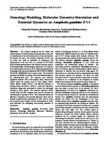
International Journal of Computational Bioinformatics and In Silico Modeling Vol. 3, No. 5 (2014): 483-490 Research Article Open Access
ISSN: 2320-0634
Structural Modeling and Molecular Dynamics Simulation Studies of Camel Milk Kappa Casein Protein Abdul Wadood1*, Huma1, Zahid ullah1, Muhammad Riaz1, Sulaiman Shams1, Mehreen Gufran1, Amir Fahira1, Muhammad Naeem1, Sahib Gul1 and Ayaz Ahmad2 Computational Medicinal Chemistry Laboratory, Department of Biochemistry, Abdul Wali Khan University Mardan, Mardan23200, Pakistan. 2 Department of Biotechnology, Abdul Wali Khan University Mardan, Mardan-23200, Pakistan. 1
*Corresponding author: Abdul Wadood; e-mail:
[email protected] Received: 15 August 2014
Accepted: 31 August 2014
Online: 01 September 2014
ABSTRACT Kappa casein protein belong to phosphoprotein’s casein family which contribute 80% of milk protein comprise of αS1, αS2, β-casein and κ-casein. Kappa casein gene (CNS3) is highly conserved in mammalian species and show variation containing 1997 base pairs. Kappa-casein involved in vital physiological processes. Along with the stabilizing coagulum by interfering with lactation cycle, it has many antimicrobial activities against Escherichia, Helicobacter pylori, Listeria, Salmonella and Staphylococcus, yeast and filamentous fungi. To date the X-ray structure of Kappa casein is not available. The sequence under accession no of P79139 was retrieved from uniprot and 3D structure was built with template ATP-dependent RNA helicase p47 (PDB ID 1XTI.A) showing 43.4% sequence identity using MOE2010.11. The alignment of target and template protein was done using MUSCLE server. The structure was refined by subjecting to MOE-DYNAMIC tool and final structure was used for protein-protein interaction with Helicobacter pylori (data not shown).The consistency of modeled structure was confirmed by getting acceptable scoring from ERRAT and Rampage owing to high accuracy of MOE for modeling and dynamics simulation. Toward the inhibitory activity this modeled structure may unclose the mechanism of eradication of helicobacter pylori from gastric epithelial cell.
Keywords: Homology modeling, Md simulation, Casein and Inhibitor INTRODUCTION Kappa casein (κ-casein) is a protein which is post transcriptionally modified by addition of one or more phosphate molecules. It belong to phosphoproteins’s casein family divided into four groups αS1, αS2, βcasein and κ-casein [1] that contribute 80% of milk protein fraction. Among which the most abundant proteins are αs1-casein and β-casein at 10-12 mg/ml and 10 mg/ml respectively while αs2-casein and κcasein are present at 3.7mg/ml and 3.4 mg/ml respectively [2]. Casein is secreted by mammary gland cells, genes of which are resides in chromosome 6 arranged in following orders: α-s1 casein, β-casein, αs2 casein and κ-casein [3]. Kappa casein gene (CNS3) is highly conserved in mammalian species and show
variation containing 1997 base pairs. Kappa-casein involved in vital physiological processes. The efficiency of digestion upsurge by fragmented kappa casein into insoluble peptide (para kappa-casein) and a soluble hydrophilic glycopeptide in the gastro intestinal tract (GIT) prevents infant hypersensitivity to proteins and the activity of several pathogens was inhibited [4]. For example, human milk kappa casein inhibits Helicobacter pylori adhesion to sections of human gastric mucosa [5]. Stabilizing the coagulum was powered by Kappa-casein and it also has major Influences on milk composition and processing properties by associating with lactation cycle [6]. κcasein peptide has been found to have antimicrobial activity against wide range of pathogenic organisms
http://bioinfo.aizeonpublishers.net/content/2014/5/bioinfo483-490.pdf
483
Abdul Wadood et al. / Int J Comput Bioinfo In Silico Model. 2014, 3(5): 483-490
such as Escherichia, Helicobacter pylori, Listeria, Salmonella and Staphylococcus, yeast and filamentous fungi [7]. Secondary structures have been found to be of great importance in antimicrobial effect [8]. In the absence of experimentally three dimensional Xray structure, a homology model could provide a rational opportunity to obtain three dimension (3D) model [9, 10]. The application of homology model such as mechanism investigation and structure based molecular mechanism has been recognized. This approach gives a good model with template sharing more than 25% identity. The template used in this study was the X-ray structure of ATP-dependent RNA helicase p47 (PDB ID 1XTI.A) showing 43.4% sequence identity from homo sapiens. This template has two domains. The N-terminal domain consists of seven βsheets surrounded by eight α-helix and C-terminal domain contains seven β-sheets surrounded by five αhelix. Our model showed similarity with N-terminal domain. The present study deals with the prediction of first 3-D model of kappa casein (Camelusdromedarius) based on comparative homology modeling using MOE 2010.11
MATERIALS AND METHODS Retrieval of sequence The primary sequence of Kappa casein (accession number: P79139) of Camelus dromedarius was retrieved in FASTA format from the Universal Protein Resource (UniProt) (http://www.uniprot.org/) and was saved in text file with name Kappa casein in local directory. The retrieved sequence of Kappa casein was described in silico using Expasy-ProtParam tool [11]. Secondary structure prediction Secondary structure of our protein under observation was predicted using different methods. The protein sequence was submitted to the following server and was analyzed for secondary structure. Servers used in this task were double prediction method (DPM) [12], Discrimination of protein secondary structure class (DSC) [13], PHD[14], Predator [15], SIMPA96 [16], SOPM [17], Self-optimized prediction method with alignment (SOPMA) [18], Sec. Cons [19]. Template selection Similarity search was performed using PDB Search Tools suited in MOE 2010-11 against PDB databank of MOE. The sequence was pasted in Sequence Query and run the Search keeping parameters like Gap start -12, Gap Extend -2, E-value cutoff 10, E-value Accep 0.5, Z iterations 100 and Z cutoff 6 using identity Matrix. Xray crystallographic structure of Probable ATPdependent RNA helicase p47 (PDB ID 1XTI.A) was chosen as template sharing highest sequence identity of 43.4% with the target protein and was loaded to MOE Sequence Editor. This template consists of two domains. Our sequence showed similarity with Nterminal domain.
Target-template alignment Sequence alignment of target protein kappa casein and template protein (PDB ID 1XTI.A) was performed by using MUSCLE [20] server (http://www.ebi.ac.uk/ Tools/msa/muscle/). In parameters set the Output format to ClustalW. Prediction of intrinsic disorder In order to identify segments of disordered and higher flexibility, the tools DisEMBL[21], Globplot [22] and Regional order neural network (RONN) [23] were used. Protein disordered prediction is important in determining protein function and protein folding pathway [24, 25]. Homology modeling The model was generated using MOE Homology tool. Chain 1 is our target sequence and chain 2 is template sequence. In model refining tool Intermediate was set to Medium, Final model to Medium,using scoring function Generalized Born/Volume Integral (GB/VI). The Force field was set to Amber99 with Solvation RField. Total of 10 models was generated, and the final model was loaded to MOE main window. The sequences were aligned before starting homology modeling by using MOE-Align application. Dynamic simulation The model generated by MOE Homology tool was subjected to MOE Dynamic tool in order to refine it. The energy of protein was minimized by using MOE Force field AMBER99 by concerning calculation toward solvation energy with Born implisit solvation. The energy was minimized to RMS Gradient 0.05. The overall charge of protein was optimized with partial charge option in MOE 2010-11. The other parameter was set to default values that were ensemble at NVT and algorithm at NPA for creating ensemble trajectory. The acceleration, velocity and position were saved after each 0.5 picoseconds (ps). Before starting MD simulation, addition of water molecule by Water Soak option with soak mode BOX and layer width 5 was followed by energy minimization. The system was heated from 0K to 300K in 20 picoseconds (heat time) followed by production time of 200 picoseconds. The system was cooled back to 0K in 20 picoseconds. Validation of modeled structure The stereo chemical quality of the generated model was initially evaluated by MOE package using Rampage .Then it was subjected to ERRAT [26] server to check the overall quality.
RESULTS AND DISCUSSION The sequence of camel milk kappa casein was obtained from Universal Protein Resource (UniProt) under the accession no of P79139and the obtained sequence was submitted to Expasy-ProtParam server to study it physiochemical properties (http://web.expasy.org/ protparam/). The protein has 182 amino acids with molecular weight of 20417.5 and estimated theoretical isoelectric point (pI) of 8.55. The Extension coefficient
http://bioinfo.aizeonpublishers.net/content/2014/5/bioinfo483-490.pdf
484
Abdul Wadood et al. / Int J Comput Bioinfo In Silico Model. 2014, 3(5): 483-490
of protein is 10555 indicates that at a definite wavelength how much light was absorbs by a protein [27]. It is used by spectrophotometer in purification of protein. The instability index (II) is 44.72 that displaying protein stability [28]. Instability index smaller than 40 for a protein is expected as stable. Protein may be unstable with Instability index more than 40. The hydrophilic nature is predicted by The Grand average of hydropathy (GRAVY) index value of 0.150[29]. The Aliphatic index is 90.49 indicating the relative volume of aliphatic side chains (leucine, valine,
isoleucine, and alanine). The thermostability of globular proteins is calculated by Aliphatic index [30]. Secondary structure prediction Prediction of secondary structure ensures whether amino acids lie in strand, helix or coil. The sequence was submitted to different servers and results were obtained as shown in Table1.The inspection of the table show that random coil dominate over extended strand followed by alpha helix.
Table 1. Secondary structure prediction calculated by different tools. Secondary Structure Alpha helix 310 helix Pi helix Beta bridge Extended strand Beta turn Bend region Random coil Ambigous states Other states
DPM
DSC
Sec.Cons
SOPMA
PHD
Predator
SIMPA96
SOPMA
10.99% 0% 0% 0% 35.16% 1.10% 0% 52.75% 0% 0%
11.54% 0% 0% 0% 16.48% 0% 0% 71.98% 0% 0%
10.99% 0% 0% 0% 17.03% 0% 0% 68.68% 3.30% 0%
16.48% 0% 0% 0% 15.93% 1.10% 0% 66.48% 0% 0%
1.65% 0% 0% 0% 25.27% 0% 0% 73.08% 0% 0%
18.68% 0% 0% 0% 15.38% 0% 0% 65.93% 0% 0%
15.85% 0% 0% 0% 13.66% 0% 0% 69.95% 0% 0.55%
17.03% 0% 0% 0% 18.68% 4.95% 0% 59.34% 0% 0%
Prediction of intrinsic disorder Protein disorder can be defined as the region without regular secondary structure and of high degree of flexibility in protein chain [31]. Recently it has been assured that functional segments of polypeptide are mostly present outside of globular domain specially the region of intrinsic disordered [31]. Globular domains are regions of high ordered and contain regular
secondary structure. The cellular and structural aspects of these regions are unknown but it has been thought about it that it become ordered when bound to another molecule [32, 33]. The sequence was submitted to different online server having free access and the result of different server is given in Table 2 and was shown in Figure 1.
Table 2. Intrinsic disorders predicted by different server. Server
Disordered
GLOBPROT
by
4-42, 72-77, 201-205, 209-214, 223-228 166-182
DISEMBL RONN
Disordered REM465
Disordered by loop/coil definition
Disordered by HOTLOOP definition
15-29,40-84, 152,161-174
16-25, 97-111, 116132, 139-149, 160-182
91-
21 - 33, 98 - 176
Figure 1. Protein intrinsic disorders predicted by different server (A) DisEMBL (B) Globprot (C) PRODOS. http://bioinfo.aizeonpublishers.net/content/2014/5/bioinfo483-490.pdf
485
Abdul Wadood et al. / Int J Comput Bioinfo In Silico Model. 2014, 3(5): 483-490
Figure 2. Sequence alignment between kappa casein and template 1XTI.A N-Terminal.
Target-template alignment The alignment of query sequence to template sequence was done by using MUSCLE [20] server. The alignment was done in order to generation a consequent model. The alignment was shown in Figure 2. Homology modeling To model 3D structure Molecular operating environment software (MOE 2010-11) was used which implement comparative protein structure modeling based on the following facts. 1) Initial partial geometry of target sequence was copied by MOE from template structure where residues identity was conserved. 2) By using a specialized logic insertions and deletions are treated for those residues having no assigned backbone coordinates [34].
3) Loops are modeled first in random order. A list of possible candidates were analyzed by using contact energy function chosen based on Boltzmann weighted averaging [35, 36]. Model was generated by keeping parameters like Model Scoring to Generalized Born/Volume Integral (GB/VI) methodology [37], Force fields to Amber99 supported in MOE recommended for protein homology applications [38]. After modeling, energy of structure was minimized to 0.05 Gradient by using Force field AMBER99 which is parameterized for proteins and nucleic acids and is not suitable for small molecules. The generated structure (Figure 3A) was saved in PDB format with suitable name. The generated model was superimposed on template structure with root mean square deviation (RMSD) of 1.242 angstroms showing a close homology (Figure 3B) using chimera version 1.7.
Figure 3. (A) Homology model for kappa casein (B) Superposition of target (magenta) and template (cyan).
http://bioinfo.aizeonpublishers.net/content/2014/5/bioinfo483-490.pdf
486
Abdul Wadood et al. / Int J Comput Bioinfo In Silico Model. 2014, 3(5): 483-490
Figure 4. Potential energy plots during MD simulation at 300K.
Figure 5. RMSD of alpha carbon verses time graph show equality after 350 picoseconds.
Dynamics simulation Molecular dynamics simulation, which solve the classical equation of motion, results in conformational trajectories providing configurationally information to calculate thermodynamic properties of the system and explore the conformational space available to the system. The generated structure was further refined by subjecting to molecular dynamic simulation tool implemented into MOE-2010-11 [39]. The structure was saturated with partial charges followed by minimizing up to 0.05 RMS by using force field AMBER99 implemented in MOE-2010-11 [40, 41]. NVT ensemble was used as the temperature; volume and number of atom were held constant. The NPA algorithm was used as it is the most accurate and sensitive method in generating true ensemble trajectories [42, 43]. The Nose-Poincare-Anderson (NPA) method generate theoretically correct NVE, NVT, NPH and NPT ensembles (N, V, T, H, E, P) shows Number of atoms, volume, temperature, enthalpy, energy and pressure respectively). In heating phase the temperature was raised to 300K as is the normal human body temperature followed by production phase of 200 picoseconds in which temperature kept constant. After 150 picoseconds the system reaches the equilibrium indicating the validity of our protein at human body temperature. The system was then cooled for 20 picoseconds to get stable bonds energies. If it is not done, additionally energy minimization should be done.
There are many ways to analyses the MD simulation. In this study we were interested in potential energy plot of protein conformation with respect to time. Conformation mention the three dimensional arrangement of protein which can be change without fluctuating covalent bonds [44]. The potential energy plot verses time was given in Figure 4. From the analysis of trajectories as a result of MD simulation, After 350 picoseconds Root Mean Square Deviation (RMSD) of alpha carbon does not showed any fluctuation showing that system reached the equilibrium (Figure 5). The RMSD graph of backbone atoms as a function of time was also shown (Figure 6). The super position of initial and final structure after MD simulation was shown in Figure7 having 0.5 RSMD value, owing to greater stability of protein at 300 K and also showing the highly accurate modeling of MOE software. Validation of modeled structure The geometry of model was evaluated with Ramachandran plot calculation using Molecular operating environment (MOE 2010.11). Ramachandran plot revealed that 81.1% of residues are in favored region, 13.3% are in additionally allowed region and 5.6% are in outlier region (Figure 8C). The TYR63, GLN64, GLN92, ILE102 and THR139 were out of energetically favorable region. Ramachandran plot shows that total of 94% residues are in favored region
http://bioinfo.aizeonpublishers.net/content/2014/5/bioinfo483-490.pdf
487
Abdul Wadood et al. / Int J Comput Bioinfo In Silico Model. 2014, 3(5): 483-490
showing the high reliability of structure. The analyses of bond length of main chain backbone were also calculated in MOE 2010.11 revealing good quality of protein (Figure 8A). In Figure 8B the X-axis represents the sequential arrangement of amino acids and Y-axis shows the Z-Scores of the particular backbone bond length. Z score cutoff of 4 are drawn in red colour, above which the residue are in outlier region. Bond length calculation was presented in three separated plots. First plot is for the bond length of carbonyl carbon (pC) of previous amino acid with nitrogen (N)
and nitrogen with alpha carbon (Cα) of next amino acid (pC-N and N-Cα). Second plot is for bond length of alpha carbon with carbonyl carbon and side chain carbon (CB) with alpha carbon (Cα-C and CB-Cα). Third plot is for bond length of carbonyl oxygen and carbon (C=O) and carbon of side chain eith alpha carbon. The errate score for our model is 92.529. Errat analyses the statistics of non-bonded interaction between different atoms and a score of normally 50 is acceptable. The score for our model showed the great accuracy (Figure 9).
Figure 6. RMSD graph of backbone atoms verses Time graph show equality after 350 picoseconds.
Figure 7. Superposition of initial and final structure.
Figure 8. (A) Z-score of bond length (B) Bond length definition (C) Ramachandran plots.
Figure 9. Overall Quality factor for kappa casein. http://bioinfo.aizeonpublishers.net/content/2014/5/bioinfo483-490.pdf
488
Abdul Wadood et al. / Int J Comput Bioinfo In Silico Model. 2014, 3(5): 483-490
CONCLUSION Protein structure enables the understanding of mechanism of protein’s function. Solving the protein structure by experimental method is difficult process as it cannot meet the demand resulted from the exponential growth of protein sequences. Homology modeling is very important in drug design or in assessment of function to a protein as it gives insight into the interaction of protein with other proteins or with ligands. We modeled for the first time, the Kappa casein protein from camel milk which has a role in inhibition of Helicobacter pylori to gastric cell. MOE2010.11 was used for modeling purpose giving a good quality model .the quality of model was check by Errat2 giving 92.529 quality factor, normally 50 quality factor is accepted. Furthermore, the model was refined by molecular dynamics simulation using MOE. The predicted three dimensional homology model might be used to unclose the mechanism of eradication of helicobacter pylori from gastric epithelial cell.
REFERENCES 1.
2. 3.
4.
5.
6.
7.
8.
9.
10. 11.
12.
13.
14. 15.
16.
Lucey JA, Johnson ME (2003). Horne DS: Invited review: Perspectives on the basis of the rheology and texture properties of cheese. J. Dairy Sci. 86:27252743. Martin P, Grosclaude F: Improvement of milk protein quality by gene technology. Livestock Prod Sci 1993,35:95115. Lien S, Rogne S (1993) Bovine casein haplotypes number, frequencies and applicability as genetic marker. Anim. Genet. 24:373376. Ageitos JM, Vallejo JA, Poza M, Villa TG (2006) Fluorescein thiocarbamoyl-kappa-casein assay for the specific testing of milk-clotting proteases. J Dairy Sci. 89:37703777. Stromqvist M, Falk P, Bergstrom S , Hansson L, Lonnerdal B, Normark, S, Hernell O (1995) Human milk kappa-casein and inhibition of Helicobacter pylori adhesion to human gastric mucosa. J. Pediatr. Gastroenterol. Nut. 21:288296. Gangaraja DR, Swathi SB, Govindaiaha MG, Nagaraja CS, Byregowdac SM, Jayashankara MR (2008) Molecular characterization of kappa-casein gene in buffaloes. Science Asia. 34:435439. Haque E, Chand R (2008) Antihypertensive and antimicrobial bioactive peptides from milk proteins. Euro. Food Res. Tech. 227:715. Vajiheh F (2012) Milk Proteins-derived antibacterial peptides as novel functional food ingredients. Annal. Biol. Resh. 3:25202526. Hilbert M, Böhm G, Jaenicke R (1993) Structural relationships of homologous proteins as a fundamental principle in homology modeling. Proteins. 17:138151. Sali A (1995) Modelling mutations and homologous proteins.Curr. Op. Struc. Biol. 6:437451. Gasteiger E, Hoogland C, Gattiker A, Duvaud S, Wilkins MR, Appel RD, Bairoch A: Protein Identification and Analysis Tools on the ExPASy Server;(In) John M. Walker (ed): The Proteomics Protocols Handbook, Humana Press2005, 571607. Deleage G, Roux B (1987) An algorithm for protein secondary structure prediction based on class prediction. Protein Eng.1:289294. King RD, Sternberg MJ (1996) Identification and application of the concepts important for accurate and reliable protein secondary structure prediction. Protein Sci. 5:2298310. Rost B, Sander C (1993) Prediction of protein secondary structure at better than 70% accuracy. J. MolBiol. 232:58499. Frishman D, Argos P (1996) Incorporation of non-local interactions in protein secondary structure prediction from the amino acid sequence. Protein Eng. 9:133142. Levin JM, Robson B, GarnierJ (1986) An algorithm for secondary structure determination in proteins based on sequence similarity. FEBS Lett. 205:303308.
http://bioinfo.aizeonpublishers.net/content/2014/5/bioinfo483-490.pdf
17. Geourjon C, DeleageG: SOPM (1994) A self-optimized method for protein secondary structure prediction. Protein Eng.7:157164. 18. Geourjon C, Deleage G (1995) SOPMA: Significant improvements in protein secondary structure prediction by consensus prediction from multiple alignments. Comput. Appl. Biosci. 11:681684. 19. Deleage G, Blanchet C, Geourjon C (1997) Protein structure prediction. Implications for the biologist. Biochimie. 79:681686. 20. Edgar RC: MUSCLE (2004) multiple sequence alignment with high accuracy and high throughput. Nucleic Acids Res. 32:17921797. 21. Linding R, Jensen LJ, Diella F, Bork P, Gibson TJ, Russell RB (2003) Protein disorder prediction: implications for structural proteomics. Structure. 11:14531459. 22. Linding R, Russell RB, Neduva V, Gibson TJ (2003) GlobPlot: Exploring protein sequences for globularity and disorder. Nucleic Acids Res.31:37013708. 23. Yang ZR, Thomson R, McNeil P, Esnouf RM (2005) RONN: the bio-basis function neural network technique applied to the detection of natively disordered regions in proteins. Bioinformatics. 21:33693376. 24. Plaxco KW, Gross M (2001) Unfolded, yes, but random? Nat. Struct. Biol. 8:659660. 25. Verkhivker GM, Bouzida D, Gehlhaar DK, Rejto PA, Freer ST, Rose PW (2003) Simulating disorder-order transitions in molecular recognition of unstructured proteins: where folding meets binding.Proc. Natl. Acad. Sci. U.S.A.100:51485153. 26. Colovos C, Yeates TO (1993) Verification of protein structures: patterns of nonbonded atomic interactions. Protein Sci. 2:15111519. 27. Gill SC, Hippel PH (1989) Calculation of protein extinction coefficients from amino acid sequence data. Anal. Biochem. 182:319326. 28. Guruprasad K, Reddy BV, Pandit MW (1990) Correlation between stability of a protein and its dipeptide composition: a novel approach for predicting in vivo stability of a protein from its primary sequence. Protein Eng. 4:155161. 29. Kyte J, Doolittle RF (1982) A simple method for displaying the hydropathic character of a protein. J. Mol. Biol. 157:105132. 30. Ikai AJ (1980) Thermostability and aliphatic index of globular proteins. J. Biochem. 88:18951898. 31. Wright P, Dyson H (1999) Intrinsically unstructured proteins: reassessing the protein structure-function paradigm. J. Mol. Biol. 293:321331. 32. Uversky V (2002) Natively unfolded proteins: a point where biology waits for physics. Protein Sci. 11:739756. 33. Dunker AK, Lawson JD, et al. (2001) intrinsically disordered protein. J. Mol. Graph Model. 19:2659. 34. Needleman SB, Wunsch CD (1970) A general method applicable to the search for similarities in the amino acid sequence of two proteins. J. Mol. Biol. 48:443453. 35. Sippl MJ (1993) Boltzmann’s principle, knowledge-based mean fields and protein folding. An approach to the computational determination of protein structures. J. Comp. Aid. Mol. 7:473501. 36. Sippl MJ (1993) Recognition of errors in three-dimensional structures of proteins. Proteins. J. Comp. Aid. Mol. 17:355362. 37. Labute P (2008) The Generalized Born / Volume Integral (GB/VI) Implicit Solvent Model: Estimation of the Free Energy of Hydration Using London Dispersion Instead of Atomic Surface Area. J. Comp. Chem. 29:16931698. 38. Summa CS, Levitt M (2007) Near-native Structure Refinement Using InVacuo Energy Minimization. PNAS. 104:31773182. 39. Montreal QC: Molecular Operating Environment (MOE), 2011.10; Chemical Computing Group Inc. 1010 Sherbooke St. West, Suite #910, Canada, H3A 2R7, 2011. 40. Weiner SJ, Kollman PA, Nguyen DT, Case DA (1986) An All Atom Force Field for Simulations of Proteins and Nucleic Acids. J. Comp. Chem. 7:230252. 41. Cornell WD, Cieplak P et al. (1995) A second generation force field for the simulation of proteins and nucleic acids; J. Am. Chem. Soc. 177:51795197.
489
Abdul Wadood et al. / Int J Comput Bioinfo In Silico Model. 2014, 3(5): 483-490 42. Sturgeon JB, Laird BB: Symplectic Algorithm for Constant Pressure Molecular Dynamics Using a Nosé-Poincaré Thermostat. University of Kansas Technical Paper 2002. 43. Bond SD,Benedict JL, Laird BB (1999) The Nosé-Poincaré Method for Constant Temperature Molecular Dynamics. J. Comp. Phys. 151:114134. 44. Bhagavan NV: Medical Biochemistry. 4th Edn. Academic Press, San Diego, ISBN-10: 0120954400, pp: 1016.
© 2014; AIZEON Publishers; All Rights Reserved This is an Open Access article distributed under the terms of the Creative Commons Attribution License which permits unrestricted use, distribution, and reproduction in any medium, provided the original work is properly cited.
*****
http://bioinfo.aizeonpublishers.net/content/2014/5/bioinfo483-490.pdf
490