Jan 25, 2016 - Dimethyl sulfate, hydrazine, piperidine, and cesium chloride were purchased from Aldrich, East- man Kodak, Fischer, and Schwarz/Mann, ...
Vol. 261, No. 3, Issue of January 25, pp. 1329-1338,1986 Printed in U.S.A.
THEJOURNAL OF BIOLOGICAL CHEMISTRY 0 1986 by The American Society of Biological Chemists, Inc.
Structure and Expressionof the Gene Locus Encoding the Phosphatidylglycerophosphate Synthase of Escherichia coli* (Received for publication, July 22, 1985)
Alampallam S. Gopalakrishnan, Yang-Chang ChenS, MegTemkin, and William Dowhang From the DeDartment of Biochemistrv and Molecular Biology, University of Texas Medical School a t Houston, Houston, Texas 77225
This paper presents definitive results which estab- mutants at the pgsB locus (minute 4) which result in the lishes a direct gene-protein product relationship be- complete cessation (in a partial pgsA mutant) of both acidic tween thepgsA gene and the phosphatidylglycerophos- phospholipid synthesis and the synthesis of residual phosphaphate synthase of Escherichia coli. The predicted pro- tidylglycero-P synthase activity at the restrictive temperature tein sequence derived from the determined DNA se- (Nishijima et al., 1981). Introduction of the cloned pgsA’ quence of pgsA is in close agreement with the amino locus into a pgsA pgsB double mutant results in the overproacid composition and partially determined amino acid duction of synthase activity, the restorationof phospholipid sequence of the purified enzyme. The purified synthase synthesis, and the loss of thetemperaturesensitivity for has the same apparent molecular mass as the gene growth. Introduction of the cloned pgsB+ locus corrects the productmade by a plasmid-directed transcription- temperature-sensitive phenotype and restores phospholipid translation system. The plasmid-borne copy of the synthesis but results in restoration of only mutant synthase pgsA gene is also capable of expressing enzymatically active synthase in vitro. The DNA sequence analysis levels. Strains carryingonly the pgsB mutation have no phehas established the exact linear relationship between notype except the accumulation of one early precursor of the uurC, pgsA, and gly W loci and revealed that these lipopolysaccharide biosynthesis (Nishijima et al., 1981; Nishof this three genes are transcribed in the same direction. The ijima and Raetz,1981); it is now clear that the product terminal coding regions of these three genes also share gene locus is a tetraacyldisaccharide l-phosphate synthetase common sequences with transcriptional regulatory ele- (Bulawa and Raetz, 1984; Ray et al., 1984), which catalyzes an early step in lipopolysaccharide biosynthesis. Therefore, ments for the adjacent genes. the relationshipbetween the pgsA and pgsB loci still remains unclear, and more importantly the evidence that the pgsA locus is the structural gene for thephosphatidylglycero-P The phosphatidylglycerophosphate synthase (CDP-1,2-di- synthase is incomplete. acyl-sn-glycerol-3-P phosphatidyltransferase, EC 2.7.8.7)of EXPERIMENTALPROCEDURES Escherichia coli catalyzes the committed step to the synthesis of the acidic phospholipids of this organism. This enzyme, Materials-All chemicals were reagent grade or better.Radiochemlike most of the enzymes of phospholipid metabolism, is an icals and scintillation supplies were obtained from ICN Pharmaceuintegral membrane protein found tightly associated with the ticals, Amersham/Searle Corp., or New England Nuclear. Sepharose cytoplasmic membrane of E. coli (Raetz, 1978). The purified dB, blue dextran 2000, PBE 94 chromatofocusing resin, and Poly96 were purchased from Pharmacia. Agarose, acrylamide, enzyme from E. coli B also displays many of the propertiesof buffer N,N’-methylenebisacrylamide, ammoniumpersulfate, Coomassie a membrane protein and requires a detergent-lipid substrate Blue R-250, and sodium dodecyl sulfate (SDS’) were purchased from mixed micelle for maximum activity i n vitro (Harabayashi et Bio-Rad. Restriction endonucleases, T4 polynucleotide kinase, DNA polymerase (Klenow fragment), T4 DNA polymerase, RNA polymal., 1976). The apparent structural gene, pgsA at minute 42 of the E. erase, and nuclease S1 were obtained from New England Biolabs, coli genetic map (Nishijima and Raetz, 1979), has beencloned Bethesda Research Laboratories, or Boehringer Mannheim. A nicktranslation reagent kit was purchased from and used as directed by and when carried on multicopy number plasmids results in BethesdaResearchLaboratories.CNBr was a product of Pierce the expression of activity in direct proportion togene dosage Chemical Co., and DEAE membranes and nitrocellulose sheets were (Ohta et al., 1981). The cloned gene corrects the mutation at suppliedby Schleicherand Schuell. Dimethylsulfate, hydrazine, the pgsA locus (Ohta et al., 1981), the major phenotypes of piperidine, and cesium chloride were purchased from Aldrich, Eastwhich are the lack of measurable in vitro enzymatic activity man Kodak, Fischer, and Schwarz/Mann, respectively. CMP-morand a partial reduction in the acidic phospholipid content of pholidate, protein standards for gel electrophoresis, ethidium brodimethyl sulfoxide, RNase A, and TritonX-100 were purchased the membrane (Nishijimaet al., 1981); these mutants display mide, fromSigma. Bacterial growthmedium was obtained from Difco. no known growth phenotypes. Formamide obtained from Aldrich was deionized prior to use with The genetic and biochemical understanding of these mu- the mixed bed resin AG 501-X8 from Bio-Rad. Renex 690 (nonyltants is complicated by the isolationof temperature-sensitive phenoxypolyethoxyethanol) was obtained from ICI. BacterialStrains,
Plasmids, and Growth Conditiom-Bacterial
* This work was supported in partby United States Public Health strains JA200(pgsA+ recA), AD100 @gsA+ recA), and AD10 (pgsAI0 Service Grant GM 20478 from the National Institutes of General Medical Sciences. The costs of publication of this article were defrayed in part by the payment of page charges. This article must therefore be hereby marked “advertisement” in accordance with 18 U.S.C. Section 1734 solely to indicate this fact. $ Present address: Abbott Laboratories, North Chicago, IL 60064. § T o whom correspondence should beaddressed.
recA) have been described previously (Ohta et al., 1981). Plasmids used in this work are listed in Table I. All pgsA-containing plasmids designated with an “L” were derived from pPGlL (see Fig. 1). The gene product(phosphatidylglycero-Psynthase) coded by the gene
’
The abbreviations used are: SDS,sodium dodecyl sulfate; bp, base pair; DTT, ditbiothreitol.
1329
Sequence of pgsA Gene
1330 TABLE I Plasmids used in this work
striction enzyme digestion, DNA ligation, and bacterial transformation have been previously described (Ohta et al., 1981; Maniatus et al., 1982).Isolation, 3'- or 5'-labeling, strand separation, and sequencmarkers Relevant Plasmid ing of DNA fragments were performed as described by Maxam and Ohta et al., 1981 pPGl pgsA+ uurC+ glyW+ Tet' Gilbert (1980). Specific DNA fragments of plasmids generated by pPGlL pgsA+ uurC+ glyW+ IS1 Tet' Ohta et al., 1981 restriction endonucleases were isolated from preparative agarose gels Tucker et al., 1982 pPGL2019 pgsA+ glyW+ Amp' Kana using the DNA band-capture procedure of Lizardi et al. (1984). mRNA Tucker et al., 1982 pPGL3008 pgsA+ Tet' Amp" was synthesized in vitro using the appropriate plasmid DNA and E. This work pPGL2134 pgsA+ Amp' Tet" coli RNA polymerase as described byAoyama et al. (1983). The Tet' Amp' Ohta et al., 1981 pBR322 reaction mixture was precipitated by ethanol afterphenol extraction. AmD' Kan' DKC~ Rao and Rogers. 1979 The mRNA was used for nuclease S1 mapping after hybridization to the appropriate 32P-labeledDNA fragment as described by Sharp et al. (1980). After the DNA-RNA hybrid was subjected to nuclease S1 H treatment, the labeled product was sized relative to the sequencing 2 ladder generated from a DNA sequence analysis of the original labeled DNA fragment. Southern hybridization analysis was carried out essentially as described by Maniatus et al. (1982)except the blocking and hybridization solutions were made as suggested by Johnson et al. (1984) using 0.05% nonfat dry milk. Computer Programs-All computer analysis was done using an uvrC vgsA olyW Apple I1 plus computer. Programs for storage, retrieval, and printing FIG. 1. DNA segment containing the pgsA gene locus. The segment lacking the IS1insert was derived from pPGl andcodes for ofDNA and protein sequences and for generation of restriction enzyme maps originated in our laboratory with the aid of Dr. Terry the native gene product (PGSN). The segment containing an IS1 insert at theposition indicated was derived from pPGlL (Ohtaet al., Landers. Analysis of the protein secondary structure and hydropho1981; Tuckeret al., 1982) and codes for an altered gene product bicity was done using modifications of the programs supplied to us by Sam Circillo and Robert Henkens of Duke University. (PGSL). kb, kilobase pair. Protein Analysis-The purified enzyme was usually in a dilute (pgsA-N) originating from pPG1 is designated PGSN and thatcoded solution containing high concentrations of salt, Triton X-100, and by the gene @gsA-L) originating from pPGlL is designated PGSL. phospholipid; all of these interfered with studies of the chemistry of All plasmids designated gly W+contain the gly W gene sequence, but the protein. Samples were lyophilized, and the residue was extracted it is not known whether the sequence is expressed (Tucker et al., with 90% acetone to remove residual salt and the detergent. The 1982). pPGL2019 carries the smaller BglII-EcoRI fragment (Fig. I) residue was suspended in chloroform and then partitioned against in the kanamycin-resistance gene (Kan) of pKC7. pPGL3008 contains water. After removing the chloroform layer which contained the the BglII-PstI fragment in the ampicillin-resistance gene (Amp) of phospholipid and residual detergent, the aqueous layer including any insoluble material was lyophilized.The residue was insoluble in both pBR322, and pPGL2134 carries this same fragment in the tetracycline-resistance gene (Tet) of pBR322. Growth of bacterial cells has 88% formic acid and 5% SDS even in the latter case after boiling in been previously described (Ohta et al., 1981). the presence of mercaptide. The sample could be dissolved in anhyProtein Synthesis-Protein was synthesized in vitro using a plas- drose trifluoroacetic acid which was used to transfer the sample. mid DNA-directed, coupled transcription-translation system. JA200 To determine the amino acid composition using a Beckman model and AD10 (pgsA-) were used for the preparation of the 30,000 X g 119 amino acid analyzer, the above samples were subjected to hysupernatants (S30) as described by Zubay (1973); such preparations drolysis in 6 N HCl for 24, 48, and 72 h in vacuo prior to analysis. had an AZG0of 75/ml. Synthesis of protein in vitro was carried out in Serine and threonine content was determined by extrapolation to a final volume of 50 p1 containing 30 mM potassium acetate, 15 mM zero time. magnesium acetate, all four ribonucleotides each at 2 mM, 0.1 mM In order to specifically cleave the protein at the methionine resiCAMP, E. coli tRNA at 0.2 mg/ml, 5 mM phosphoenolpyruvate, dues, 10 nmolof protein in anhydrous trifluoroacetic acid was diluted pyruvate kinase at 0.01 mg/ml, 1 mM dithiothreitol, amino acids dropwise into 4 volumes of 88% formic acid containing a 1000-fold (except methionine) at 10 p~ each, 10 pCi of [35S]methionine,plasmid excess of CNBr. The reaction was allowed to proceed for 36 h in the DNA, and 30 pl of E. coli S30 extract. If the enzymatic activity of dark at room temperature. After addition of an excess of water, the phosphatidylglycero-P synthase rather than incorporation of radiolabel was to be measured, the labeled methionine was replaced with product was isolated by repeated lyophilization from water. The . protein residue was taken up in anhydrous trifluoroacetic acid and subjected unlabeled methionine at aconcentration of100 p ~ The synthesis mixtures were incubated at 37 "C for either 1 h or the to amino acid sequence analysis by automated Edman degradation indicated time and the reaction stopped by cooling on ice or by the using the Beckman model 890DAutomatic Sequencer with the 0.1 M addition of RNase A. Samples for SDS-polyacrylamide gel electro- Quadrol program. The 2-anilino-5-thiazolinone derivatives of the amino acids were converted manually to thecorresponding 3-phenylphoresis were precipitated with 10% trichloroacetic acid and then processed as described below. For measurement of enzyme activity, 2-thiohydantoins by heating for 10 min at 80 "C with 0.2 ml of 1 N 10 pl of the RNase A-treated sample was incubated for 2 h at 30 "C HCI containing 0.1% ethanethiol. The 3-phenyl-2-thiohydantoin in a final volume of 50 p1 containing 100 mM Tris-HC1, pH 7.5, 0.5 amino acids were separated, identified, and quantified by high permM sn-[U-'4C]glycero-P, 100 mM MgCl,, and 1%Triton X-100 with formance liquid chromatography using a Water-system, including a or without 0.1 mM CDP-diacylglycerol. The formation of phosphati- Model 660 solvent programmer, a Model 440 absorbance detector, a dylglycero-P dependent on CDP-diacylglycerol was quantified as Wisp 710B, and a Data Module. Amino acid analysis was used to previously described (Hirabayashi et al., 1976). verify that all of the peptides had been solubilized by trifluoroacetic SDS-Polyacrylamide Gel Analysis-Samples from protein synthe- acid and to know the extent of the CNBr reaction. The sequence sis experiments were heated to 100 "C for 5 min in the presence of analysis and amino acid analysis were performed for US by Dr. Finn electrophoresis buffer containing 1%SDS and 10 mM DTT and then Wold's laboratory. subjected to electrophoresis (Laemmli, 1970). The gels were stained Miscellaneous Procedures-Phosphatidylglycero-P synthase was using Coomassie Blue R-250 to visualize the protein standards. Prior assayed as previously described (Hirabayashi etal., 1976), and 1 unit to being dried under vacuum and exposed to x-ray film, the gels were of activity was defined as theamount of enzyme which forms 1 pmol subjected to thefollowing series of incubations: dimethyl sulfoxide (1 h); 7.5% Omnifluor in dimethyl sulfoxide (1h); water (2 times for 30 of product/min at 37 "C. Protein was determined by the method of min). Samples of isolated enzyme were concentrated, if necessary, by Lowry et al. (1951).Protein was transferred from SDS-polyacrylamide precipitation as described above and subjected to the same protocol gels to nitrocellulose filters for enzyme assay as previously described except proteins were visualized by the silver staining method (Merril (Poole et al., 1984). Synthesis of CDP-diacylglycerol (Agranoff and Suomi, 1963), CDP-diacylglycerol-Sepharose4B (Dutt andDowhan, et al., 1981). NucleicAcid Manipulations-Methods for plasmid (Ohta et al., 1985),and blue dextran-Sepharose 4B (Dutt and Dowhan, 1985)were 1981) and bacterial (Saito and Miura, 1963) DNA preparation, re- previously described.
Sequence of pgsA Gene RESULTS ANDDISCUSSION
1331
12345
678910
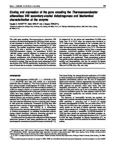
Enzyme Purification-The following buffers were used during the purification: Buffer A composed of 50 mM Tris-HC1, p H 7.0,lO mM MgC12, and 2 mM DTT; Buffer B composed of Buffer A plus 4% Triton X-100; Buffer C composedof Buffer 66K -m A plus 0.1% Triton X-100. The results for the purification of 45Kthe synthase fromJA200/pPGL2134 (PGSL) are summarized in Table 11. All procedures were carried out a t 4 “C unless otherwise indicated. Cell paste was suspended in Buffer A at a ratio of 1 g of cells, wet weight/2.5 ml of buffer and the mixture passed through a French pressure cell to break the 20.1K, cells. The membrane fractionwas collected by centrifugation 18.4Kat 105,000 x g for 2 h. The supernatant was discarded and 14.3Kp the membrane fraction suspended in 2.5 ml of Buffer B for each 1 g of starting cells, wet weight. The membrane suspension was stirred for aminimum of 2 h before being centrifuged FIG. 2. SDS-gel electrophoresis of purified PGSL. Sample at 105,000 X g for 2 h. The pellet was discarded and the supernatant was adsorbed preparation and electrophoresis was carried out as described under to a CDP-diacylglycerol-Sepharose affinity resin by stirring “Experimental Procedures” using a 7.5-15% gradient acrylamide gel. Lane I , standard proteins lysozyme, @-lactoglobulin, soybeantrypsin the membrane extract with resin overnight in the ratio of inhibitor I, ovalbumin, bovine serum albumin. Lanes 2-9, approxiapproximately 4000 units of enzyme/ml of settled resin vol- mately equivalent numbers of units of PGSL from sequential fracume. The resin was then poured into a chromatography col- tions across the activity peak from step 6, Table 11. Lane 10, pof the gel is also present in umn of convenient dimensions for batchwise washing of the lactoglobulin. The material at the bottom resin; generally, between55 and95% of the enzymatic activity the column elution buffer. was retained by the resin. The resin waswashed with 10 column volumes of Buffer C containing 3.5 M NaC1. During previously reported (22,000) for the enzyme from E. coli B these washing steps about 10-20% of the enzymatic activity (Hirabayashi et al., 1976). The apparent molecular mass of the major protein component in this preparation was 19,000 was lost. Thecolumn was thensaturatedwith BufferC containing 0.8 N hydroxylamine-HCl, p H 7.0, using about 4 daltons which is considerably lower than the previously recolumn volumes of this buffer. The column was allowed to ported value of 24,000 daltons (Hirabayashi etal., 1976). This stand overnight and then eluted with the same buffer. The discrepancy is largely due to differences in pretreatment of thesampleandtheSDS gel system employed sincethis eluant was dialyzed against several changes of Buffer C. The dialyzed preparation was adjusted to pH9.4 using 0.25 protein, like several other membrane proteins (Bridger and M ethanolamine and applied to a PBE 94 chromatofocusing Walker, 1976; Rubin and Tzagoloff, 1973; Spatz and Strittcolumn (approximately 7,000-10,000 units of enzyme/ml of mater, 1973), displays different apparent molecular weights packed resin volume) equilibrated with25 mM ethanolamine/ relative to molecular weight standards depending on the preacetate, pH9.4, containing 1% Triton X-100 and 2 mM DTT. treatment conditions and the gel system employed (data not plasmid source of the The column was washed with 2 column volumes of equilibra- shown). Thedifference is not due to the tion buffer and then developed using a 1:lO dilution of Poly- enzyme. The enzyme was also purifiedin small amountsfrom buffer 96 (adjusted to pH6.0 with acetic acid) containing1% JA200 (PGSN) lacking a plasmid borne copy of pgsA. Since Triton X-100 and 2 mM DTT. The peak fractions of enzy- the startingspecific activity of the enzyme was about 20-fold lower, the total amount of protein isolated was considerably matic activity routinely hada p H of 9.1. The peak fractions from the chromatofocusing column were lower, and themajor species, although corresponding exactly pooled and applied to a blue dextran-Sepharose column (3000 in mobility to the above 19,000-dalton species (not shown), units of enzyme/ml of settled volume) which wasequilibrated only represented about 80% of the total protein in the prepwith Buffer C. The column was washed sequentially with 2 aration. The mostdefinitive evidence that the19,000-dalton species column volumes each of Buffer C, Buffer C containing 1 M is the phosphatidylglycero-Psynthaseis shown in Fig. 3. NaC1, and Buffer C without MgC12 but containing 0.6 mM CDP-diacylglycerol and allowed to incubate overnight. The Purified enzyme from JA200 (PGSN) and JA200/pPGL2134 column was then developed with the lipid-containing buffer (PGSL) was first subjected to SDS-gel electrophoresis and thentransferred by electroelution to nitrocellulosefilters. until the enzymatic activitywas eluted. Although the overall yield was low, a highly purified prep- Each lane of the nitrocellulose filter was cut into 0.25-cm aration (Fig. 2) was obtained with a specific activity near that strips and assayed for enzymatic activity using a 1-h incubation and 10-foldhigher specific activity of substrate than usual. Enzymatic activity co-migrates with the 19,000-dalton TABLEI1 protein isolated from both strains. The total amount of enPurification of PGSL zymatic activity recovered was less than 1%of that applied Total Total Specific Step to the gel, but it is clear that the 19,000-dalton species is volume protein activity associated with the enzymaticactivity. ml mg unitslmg % DNA Sequence of the pgsARegion-The originalclone 645 40640 33 100 1. Cell-free extract covering the pgsA region was carried in a pSC101-based 500 27000 2. Membrane fraction 55 100 plasmid (pPG1) which is normally carried a t less than 10 350 6300 120 57 3. Triton extract copies/cell andcannot be amplified to higher levels after 355 15 4. Affinity column 64 5. Chromatofocusing 5 treatment of the hostcells with chloramphenicol. Subcloning 28 2 6. Blue dextran 22000 3 regions of this plasmid which carry the pgsA locus into high Based on startingwith 200 g of cell paste from JA200/pPGL2134. copy number plasmids which can be amplified has proved
Sequence of pgsA Gene
1332
Y
z
h
g~
2500
15000 14000 13000 12000 11000
N
Y
3
2000 1500
0
n
Y
0
.e
=
1200
Q)
800
2
E, 2 W
ze.
1000
2 W
D
,500
p
n
400
inverted repeat at its ends which are flanked on either side by a direct repair of the target DNA sequence; both of these features along with the partial sequencing of the insert as indicated in Figs. 4 and 5 are consistent with the insert being IS1 (Johnsrud,1979; Ohtsubo and Ohtsubo, 1978).The 11bp between the 2 Hinfl sites at the right end of the IS1 insert (see Fig. 4) were not actually sequenced by us. This region has been sequenced by Tucker and Murgola (1985) and is as indicated in Fig. 5. The composite sequence for the pgsA-N gene as derived from pPG1 andpPGL2019 is shown in Fig. 6. An open reading frame begins 82 bp beyond the BglII site and codes for a protein (PGSN, the native synthase) containing 216 amino acids (24,800 daltons). If the DNA sequence of pPGL2019 is used then a protein (PGSL) of 211 amino acids wouldbe coded for as shown in Fig. 5; only the first 203 amino acids of each proteinare homologous. Computer analysis by the method described by Mulligan et al. (1984) of this DNA sequence shows a strong RNA polymerase binding site upstream from the open reading frame. This promoter which has a66% homology with the consensus promoter falls within the top 25 strongest out of 110 promoters analyzed by Mulligan et al. (1984). Between the promoter and the initiation codon there is no apparent homology with a classical ribosomal binding site (Storm0et al., 1982).Although it is unusual for genes expressed in E. coli to lack such a site, there is precedence for translation of mRNAs lacking a binding site (Walz et al., 1978). The low level of the phosphatidylglyceroP synthase found in cells (about 1,400 molecules/cell;Larson and Dowhan, 1976) may be due tothe lack of astrong ribosomal binding site. Another contributing factor to low levels of the enzyme may be codon usage. Minor tRNA species are used at a frequency of less than 1% for translation of many abundantproteins in E. coli (Ikemura, 1981). The following minor tRNA species are used at a significantly higher frequency for translation of pgsA: Arg (CGC + AGA) at 2.3%; Thr (ACA ACG) at 1.9%; Pro (CCU) at 1.4%. The combination of a relatively strong promoter, an apparent weak ribosomal binding site, and significant usage of rare codons may result in a high amount of untranslated message and a low steady state level of gene product. Additional confirmation of the sequence at the 5‘-end of the gene comes from the work of Sancar et al. (1984) who reported the sequence of 150 bp beyond the BglII site which is near the 3‘ terminus of the uurC gene; the only discrepancy is that they reported base pair 35 of our sequence to be a “G” instead of an “A” which would have no effect on our conclusions. It is interesting to note that the -35 regionof the promoter ofpgsA lieswithin the coding region of uurC. Sancar et al. (1984) identified three potential transcription termination sites which are located after the initiation site for the pgsA mRNA. It is not clear if these sequence overlaps have any effect on the expression of either gene. The 3’-end of the pgsA-N gene shows some sequence homology with a p-independent transcription terminator; the TGA translation stopcodon is in thecenter of a GC-rich dyad symmetry which is characteristic of transcription terminators (Rosenberg and Court, 1979). However, this sequence is followed by an AT- rather than T-rich region which has been reported for only a small number of p-independent terminators (Horowitz and Platt, 1982). On the other hand, p-dependent terminators also appear to have a hairpin loop structure, but they are not followed by a T-rich region (Farnham and Platt, 1980). Near this terminus and within the coding region of pgsA there is a strong promoter (79% homology on the above discussed scale) which is indicated in Fig. 6. A
1 Y
30
25
20
15
10
5
1
Sample Number
-
PGSN
- STD
- PGSL Oirection of electrophoresis FIG. 3. Comparison of mobility of enzymatic activity and the major protein species. PGSL from Table 11, step 6, and PGSN from a similar purification using JA200 were subjected to electrophoresis and transferred to a nitrocellulose filter as described under “Experimental Procedures.” The positions of the proteins in each lane were determined by staining the nitrocellulose filter with 0.1% Amino Black in 10% acetic acid/45% methanol and are indicated on the reconstruction of the nitrocellulose filter. The standards (STD) used were lysozyme, soybean trypsin inhibitor I, and bovine serum albumin (see Fig. 2). The filter was cut and assayed for phosphatidylglycero-P synthase activity and thecounts/min in product plotted versus sample number. The apparent difference in mobility of the two forms of the enzyme is not significant since the lanes which were assayed were not next to each other.
unsuccessful (Tucker et al., 1982;Ohta et al., 1981).Therefore, the majority of the sequence work wasdone using a subcloned fragment from a spontaneously generated derivative of pPG1. This latterplasmid (pPG1L) was previously shown to contain an insert of approximately 750 bp near or in the pgsA locus whichallowed this region to be maintained in high copy number plasmids (Tucker et al., 1982; Ohta et al., 1981). Plasmid pPGL2019 containsa 2.2-kilobase pair fragment (BglII to EcoRI in Fig. 1)derived from pPGlL which complements pgsA cells and contains glyW and the complete IS1 element. The sequencing strategy used and theregions of pPGl and pPGL2019 sequenced are shown in Fig. 4. Except for one 10bp region the complete DNA sequence of the pgsA gene was determined by either sequencing both DNA strands or by sequencing the same strand in both directions. The sequence of the region from 445 to 455 bp could only be determined on one strand and in one direction due to apparent secondary structural anomalies. The evidence that pPGl and pPGL2019 differ only by an IS1 element inserted near the HinfI at a position 683 bp from the BglII is as follows. Previously reported restriction enzyme digestion results for these two plasmids were consistent with pPGL2019 containing an extra750 bp between pgsA and gly W (Tucker et al., 1982). DNA sequencing (Fig. 5) verified this conclusion and showed a sequence inserted at the position indicated in Fig. 4. The insert has a nearly perfect 23-bp
+
Sequence of pgsA Gene
::
0
(u
I
z
z
0
0
I
I
1
I
0
0
N
0
0 0 P
I
1
0 0
0 0
0
(D
h
I
1
I
Y)
1333 0
8
0 0
m
0
0 c
I
I
I
1
0 0
BASE PAIRS
._ c___o
0
-
0 0
0
N
5
I
I
!
FIG. 4. Sequencing strategy used for the pgsA gene. The restriction enzyme sites (MspI,O, TuqI, O; BglII,
. ;BstEII, 0;H i d , A;HueIII, A) a t which fragments were end labeled (either 3' or 5') are indicated along with the direction of sequencing. The DNA strand labeled and sequenced in each case is that strand closest to the arrows. The position of pgsA-N, pgsA-L, and glyW are also shown. The position of IS1 in pPGL2019 and its point of insertion into pPGlwhich resulted in the formationof pPGlL is diagrammed. Important structuralregions are carboxyl-terminal coding region of indicated as follows: 0, coding region of pgsA common to both plasmids; [ZB, PGSL derived from IS1;,I carboxyl-terminal coding region of PGSN which appears in bothpPGl andpPGL2019. Kn indicates the positionof the kanamycin-resistancegene in pPGL2019.
region of pgsA is in common with potential transcriptional signals of an adjacent gene. The IS1 insert which stabilizes the surrounding DNA regions in high copy number plasmids 2w ai0 pRy20ID like pPGL2019 is found in the carboxyl-terminal coding re.-1gion of pgsA, but also in the potential promoterfor gly W. At aw Si0 present it is notknown whether the insertionof IS1 prevents PPDL Z O I D err m-.b If& Tee CIY. coc expression of multiple plasmid-borne copies of gly W or alters is, -1."" "* ",., ". cc.0*, ccc.",C.CTthe gene product of pgsA in a way which may not affect its in FIG. 5. DNA and protein sequence change caused by IS1 insertion. The base pair numbering in the top line corresponds to vitro activity but may change its in uiuo properties allowing bold type indicates its overproduction. the numbering inFig. 6. The DNA sequence in the Structure of Genome Near thepgsA Locus-Since the DNA the 9-bp region of p P G l which was duplicated and flanks the IS1 in pPGL2019. The DNA sequence in pPGL2019 shown in small type cloned into pPG1 was originally derived from F'150 (Ohta et at each endof IS1. The sequence covered al., 1981) and multiple copies of the region covered by F'150 indicates the inverted repeat by a line at the 3'-end of IS1 is the sequence determined only by are known to be unstable in E. coli (Low, 1972),the relationTucker andMurgola (1958).The proteinsequence derived frompPG1 (PGSN) and pPGL2019 (PGSL) are also shown with the amino acids ship of the cloned DNA togenomic DNA was determined. E. coli genomic DNA was isolated from AD100 and JA200 and numbered. digested to completion with EcoRI and BglII. Such digests reading frame begins 51 bp after the end of pgsA which is were analyzed by Southern hybridization using pPGl radiohomologous with the knownsequence of the Gly 3 tRNA labeled by nick translation; pPGl digested by the same en(Squires and Carbon, 1971) which is coded by glyW. Tucker zymes was used as the size standard. pPGl digested as above and Murgola(1985)have reported the sequence beginning should yield three DNA fragments homologous with E. coli within the right end of the IS1 and extending to the EcoRI DNA (Ohta etal., 1981), twoof which are diagrammed in Fig. site shown in Fig. 1; our sequence beginning with the pointof 1. The Southern hybridization analysis verified that the E. insertion of IS1 into pPGl and extending through the glyW coli DNA inserted into pPGl had the same restriction pattern gene is in agreement with their sequence. Again the coding as thehomologous genomic DNA from both strains (data not PPDI
."
P o '
880
7:O
CTO~U.Llr1\TCBrr~.5~C1TSPCOCCAAOllLOT~O111(IC11CCCLrCW1CLYlC~CICTO* L...
"~I"...LIl.....L"...lI"."O*.".*.
.*- ."
."
ClO*11..T~,CC,,D,S,C.,50.~,~IDl.D(.YI...'...l~..0T(i,*,~"~0O,0 L,.
I.
AAC A r A AAA C I C
"
"-rn..
L
Ip.
Y . I
1 "L..
"L
cur..
L
*.
C 1 1 CZA A W ACA I T 0 CAA P
I
&
".
LTC CL.
~
1
I
I
100 500
I
110
520
120 540
580
S60
Loo ~~
~
CAGATGGTGGCGTTGGCPITGGCTGCTGTGGCGTCCQA~CATTTGGGTTGAGTPICGCCGGT~TTGCACTTTTCTTTGTGGCTGCGGTACTGPICTCTGTGGTCAATGTTQC~T~~T~ G1n~Va1Al~LeuPIlaTrpLeuLeuTrpArgProAsnI1eTrpValGluTyrAlaG1yI1eA1aLcuPhePheVa1A1aPIlaVa1LeuThrlruTrpSer~L.u01nTyrLmuBrr I
I
I
140
150
160
620
640
li0
720
700
6EO
660
GCTGCGCGGCAGATTTGCT~G~TCAGTGPI~C~TTTCGGC~TPIATTTTCPI~CAAA~~PITC~AAAGTGGTG~A~AATATC~~~ATC~CGCCA
Plla~laArgGlnI1eCysLeul1eSerPI~p~rgPheGlyValIlePh~SerLysArgScrLysV~lV~lLysPIsnI1eV~lPIspSerScrArgG1nV~lSerAr~Gln~rg~l I
I
180 740
so 760
CGGCE;GEAC;GATfEEEAGncGnTnnTnnnnrcnnoTonTGAA ArgArgHis
860
880
2 :o
260 E00
780
;E,
900
820
E40
CGOO~TA;iCTCAGTTQG;AGAGCACGAECTTGCCAAG;iTCGGGGT~OAQTTC~~ OlYW 920
940
960
CTCGTTTCCCGC~GTTTMAAQAC~TCOBWTCAAGCB~TQTCTQBCTG~~BCCTQA~~TTT8GCBCGTT~C~ffiCGGTTATGTAGCGGATTBC~TCC~TCTA
FIG. 6. DNA sequence and derived protein sequence of thepgsA-N locus. The sequence (5’ to 3’)begins at the BglII site shown in Fig. 4 and extends to beyond the glyW gene (flanked by the arrows near the 3’-end of the sequence). The potential promoters for pgsA and glyW are boxed in the sequences 5’ to each of the genes, respectively. The open reading frame beginningat base pair 82 is translated into thepredicted protein sequence of PGSN. The point of initiation of the mRNA for pgsA is indicated by the arrow near the 5’-end of the gene. The stop codon, TGA, marked at the 5’-endof the sequenceis for the uurC gene (Sancar et al., 1984). Adyad symmetry is indicated by the broken lines flanking base pair 730. The IS1 insertion occurred after base pair 690.
shown). Therefore, DNA in pPG1 is a true representative of pPGL3008 (data not shown) which carries only the pgsA-L genomic DNA. gene of E. coli. Therefore pPG1, pPGL3008, and pPGL2019 Initiation Point of mRNA Synthesis-mRNA generated in code for a protein of the same apparent size which displays vitro using pPGL2019 was hybridized to a 222-bp DNA frag- the same mobility when subjected to SDS-gel electrophoresis ment extending from theBglII to TaqI site (see Fig. 4) which as purified phosphatidylglycero-P synthase (PGSL). Thereis was 5’-labeled at theTaqI end. This fragment should containno indication that a larger precursor of PGSL or PGSN is 145 bp of the codingsequence for the gene and 77 bp of made by these plasmids. Further evidence that the pgsA locus codes for the phossequence priortotheMet-initiation codon, i.e. sequence complimentary to base pairs5-226 (Fig. 6). Fig. 7 shows that phatidylglycero-P synthase was obtained by measuring the the fragment protected fromnuclease SI terminates with the formation of enzymatic activity invitro as a function of time sequence 3’-ATTAGA-5’ which is complimentary to the se- and pPGL2019 concentrationusing an S30 made from AD10 quence 5’-TAATCT-3’ (base pairs48-56 in Fig. 6). There are (pgsA-); this extract had less than 0.1% residual synthase several minor species remaining after S1 digestion so that the activity. The formation of enzymatic activity(Fig. 9) was not precise mRNA initiation point remains uncertain; however, only found to be dependent on time and DNA concentration, the majority of E. coli mRNAs initiate with an A 6-8 bp but also on the presence during protein synthesis of a nonionic following the -10 region of the promoter (Hawley and Mc- detergent (Renex690); addition of CDP-diacylglycerol during Clure, 1983) so that the mRNAfor pgsA most likely initiates protein synthesis gave similar results, but addition of other phospholipids was not tried. Similar results were obtained where indicated in Fig. 6. Transcription and Translationof pgsA-The protein prod- using pPGL3008. Since the assay for enzymatic activity is ucts fromacoupled transcription-translation system were always carried out in the presence of a nonionic detergent, analyzed bySDS-gel electrophoresis (Fig. 8). Among the the requirement for detergent (presumably supplying a hydroproducts of pPGl (PGSN) andpPGL2019 (PGSL) only one phobic domain) for the synthesis of active enzyme must be protein is common to both, and this product has a mobility either during synthesisor very shortly after synthesis of the identical to that of purified PGSL. Thispolypeptide is missing polypeptide chain. The presenceor absence of detergent had from pBR322 which like pPGL2019 has a functional Amp no effect on the level of protein synthesized as judged by gene; thedoublet above 29,000 daltonsismost likely the analysis of labeled products by SDS-gel electrophoresis as product of this gene. Similarresults were obtained with described above (data not shown). These observations are
Sequence of pgsA Gene
1335
3
c 1
2
G
G+A
C+T
C
o
12
24
DNA CONCENTRATION
so
u)
eo
o
30
eo
120
180
PROTEIN SYNTHESIS TIME
(p~lrnl)
2io
(Mi)
FIG.9. Formation of phosphatidylglycero-Psynthase activity in vitro. After in vitrosynthesis of protein directed by pPGL2019 5‘
FIG.7. Determination of initiation site of the pgsA transcript. mRNA was synthesized in vitro from pPGL2019 and hybridized with a BglII-TuqI fragment (5’4abeled at the TuqI site). After treatment with nuclease S1 for 10 (lane 1 ) or 20 min (lane 2), the sizes of the protected DNA fragments were determined by comparing their mobilities relative to a sequencing ladder from the original labeled DNA fragment (5’4abeled a t the TuqI end and strand separated). The urrou, indicates the position in the sequencing ladder which corresponds to the major labeled fragment in lanes 1 and 2.
using an S30 made from AD10 (pgsA-), the sample was assayed for enzymatic activity. A , protein synthesis was allowed to proceed for 3 h a t various plasmid DNA concentrations in the presence of 0.1% Renex 690 prior to assaying for CDP-diacylglycerol-dependentincorporation of radiolabeled glycero-P into lipid by each reaction mixture. B, protein synthesiswas carried out either with (24 pg/ml) or without DNA and with (0.1%) or without Renex 690 for the times indicated. After the synthesis was stopped, the presence of synthase activity was determined as inA.
TABLE I11 Predicted and actual amino acid compositions The predicted amino acid compositions for PGSL and PGSN are based on the respective gene sequences. The actual number of residues in PGSL was determined by using the determined mole % of each amino acid times the predicted number of amino acids in PGSL (198 since cysteine and tryptophan were not determined). Amino acid
Actual number (PGSL)
Predicted
PGSL
PGSN
Number of residues=
-
29K
-
PGSL
18.5K -14K
ASP Asn Asp + Asn Thr Ser Glu Gln Glu + Gln Pro GlY Ala CYS Val Met Ile Leu TYr Phe LYS His Arg Trp
7
5 12.4 (12) 11.2 (11) 11.1(11) 13.2 8.7 12.2 28.7
(13) (9) (12) (29)
18.2 (18) 6.0 (6) 12.8 (13) 26.2 (26) 5.3 (5) 14.3 (14) 5.7 (6) 2.6 (3) 8.5 (9)
12 11 14 5 5 10 7 7 27 2 23 6 17
25 4 15 7 2 11
6 4 10 11 15 6 7 13 7 6 25 2 24 7 17 23 4 15 7 3 16 11 216
FIG.8. Protein products of the pgsA gene locus. The [35S] methionine-labeled products of an in vitro transcription-translation is in agreementwith the predicted system directed by the indicated plasmids were separated by SDS-gel compositionofPGSL electrophoresis. The position of each marker protein was determined composition as derived from the gene sequence (Table 111); by staining the acrylamide gel prior to autoradiography. insufficient pure PGSN was availableto carry out an accurate determination of its composition. The major differences beconsistent with the membrane location of this protein and tween the predicted sequences for the two proteins results from the insertion ofIS1 intopgsA-N. PGSN when compared further substantiatesthe direct relationship between gene and product. to PGSL has agreater number of residues (molecular mass of 24,800 versus 23,800 daltons, respectively), a significant inAminoAcidComposition and Sequence-The amino acid
Sequence of pgsA Gene
1336
crease in the number of basic residues (calculated isoelectric onine which after its removal by CNBr hydrolysis results in point of 8.9 uersus 8.5, respectively), and an additional me- the cyclization of the new amino-terminal residue. The detecthionine. Although the predictedcomposition of the two pro- tion of additional amino acids at cycles 2, 3, 5, and 8 can be teins are probably too close to be easily distinguished by explained by carry over from the previous cycle. This sequence amino acid analysis, the actual composition of PGSL favors data provides very strong verification of the predicted protein the predicted values from the pgsA-L gene for Asp + Asn, sequence for peptides 2 through 6 (see the positions of the Ala, Met, and Arg. The actual composition suggests that the methionine residues in Fig. 6). The evidence isconsistent with a blocked amino-terminal residue which makes thelargprotein still retains its N-terminal methionine. The predicted est peptide of the protein (77 out of 211 residues) unavailable molecular mass of the proteins is not consistent with the for easy sequence analysis. The predicted aminoacid comporesults of SDS-polyacrylamide gel electrophoresis as discussed elsewhere. The calculated isoelectric point is consistent with sition of a protein lacking this peptidewould be very different for PGSL. Theposition of the promoter at from that determined the elutionof the proteins from chromatofocusing columns for this gene and the start of the mRNA are also consistent a basic pH. Further confirmationof the proteinsequence was obtained with the presence of the blocked peptide. Fig. 10 summarizes other structural features of the proteins by direct sequence analysis. The amino-terminal end of the protein appears to be blocked sincenoaminoacids were (i.e. PGSN and PGSL) based on the amino acid sequence. released when intact PGSL was subjected to automated Ed- Making predictions about protein structure based on amino man degradation. Verification of the sequence of peptides acid sequence is even more tenuous for membrane-associated generated by CNBr cleavage at the methionine residues of proteins than for soluble proteins because structural data on PGSL was done asfollows. After cleavage of the protein(90% such proteins isso limited andmost model systems arebased on soluble protein structural information. On the other hand, as judged by residual methionine), the unresolved peptides certain interesting analogies can be drawn between proteins were subjected to 10 cycles of Edman degradation. Table IV by analyzing thestructural implications of the sequence. shows a very close correlation between the amino acidswhich Analysis of the hydrophobicity of PGSN asdescribed by Kyte should have been related at each cycle and those which were and Doolittle (1982), indicates extensive hydrophobic actually detected. No amino acids from the amino-terminal stretches within the sequence; PGSL differs from PGSN in peptide were detected which indicates that either the amino- that the very hydrophilic carboxyl-terminal end of PGSN is terminal residue in the protein pyroglutamic is acid-generated changed by theIS1insertto a very hydrophobic region. from glutamine (residue 2) or the residue is a blocked methi- Although this appears not to affect the activityof the protein
in vivo as measured in uitro, it may have affectsonthe function or regulation of this enzyme. The grand hydrophoSequence of CNBr-generated peptides from PGSL bicity of PGSN is 0.59 and of PGSL is 0.81; the major The predicted peptides are numbered sequentially beginning at the difference is due again to changes in a short region at the N terminus with the predicted aminoacids for each sequencingcycle (see Fig. 6) listed in column A. The major amino acids detected after carboxyl end. These values are well within the range detereach cycle are listed in columnB. Additional amino acids detected in mined for other membrane proteins by Kyte and Doolittle significant amounts over background levels, but well below the level (1982). Stretches of sequence on the order of 19 residues in of the major amino acids, are listed in the row labeled “Extra.” The length having anaverage hydrophobicity of 1.6 or greater are analysis systemwas not set up to detect tryptophan, serine, threonine, predicted by Kyteand Doolittle (1982) tobemembranelysine, or arginine; “X” indicates no amino acidoverbackground associated or membrane-spanning. Residues 14-28 (2.7), 33levels was detected.Althoughaccuratequantitation was also not possible, in cycles where 2 residues of the same amino acid should 44 (2.8), 1-44 (1.8), 62-82 (1.8), 132-142 (1.6), and 154-172 have beenreleased, a significantly higher level of this aminoacid was (2.0) are sufficiently hydrophobic to be membraneassociated. Analysis of the sequence for the location of 0-turns as dedetected. scribed by Rose (1978) indicates that such a turn may exist Cycle at almost every transition between a hydrophobic and a 1 2 3 4 5 Peptide hydrophilic protein. The amino-terminal region ~ _ _ _ _ stretch of the ~ ~ ~ A B A B A B A B A B of the protein hasconsiderable predicted structuralhomology withleadersequences of secreted proteins (Pearlman and Asn X Ile X Pro X 1 Gln X Phe X 2 Val Val Leu Leu Val Val Thr X Glu Glu Halvorson, 1983) and with helical hairpins of membraneGlu Glu Ile Ile 3 Ile Ile Ala Ala Arg X interacting domains (Engelman and Steitz,1981) which may 4 Ala Ala Glu Glu Leu Leu Gly Gly Lys X be responsible for the membrane association of the proteins. 5 Val Val Ala Ala Leu Leu Ala Ala Trp X Leader sequences begin with a positively charged aminoacid, 6 Leu Leu Gln Gln Tyr Tyr Leu Leu Ser X are followed by a hydrophobic domain lacking charged amino Extra X Val Ala X Ala acids which shows a preference for @-sheet rather than aCycle helical structure, and end with either a cluster of charged 6 7 8 9 10 Peptide acids or a &turn. Engelman and Steitz (1981) suggest ~ _ _ _ _ _ _ _ _ _ _ _ amino ~ A B A B A B a A B A B that a protein can interact with and remain associated with a membrane through two adjacent hydrophobic helices of at Thr X Leu X 1 Thr X Leu X Leu X least 11 residues in length which form a hairpin structure. 2 His His Tyr Tyr His His Ser X Trp X Ala Ala Leu Leu 3 Ile Ile Ile Ile Ser X After insertion, both the amino and carboxyl ends of the 4 Arg X Ser X Ser X Val Val Ala Ala protein would remain on the cytoplasmic side of the memPro Pro 5 Leu Leu Leu Leu Trp X Arg X brane. Residue 13 of PGSN is an arginine which precedes two Gln Gln Ile Ileb 6 Ala Ala Ala Ala Arg X hydrophobic domainsseparated by a 0-turn.The second Extra X X Leu X domain is followed by both a series of 0-turns and charged a In cycle 8 all of the amino acids detected in cycle 7 were present amino acids. From Chou and Fasman (1974) analysis this in significant amounts. In cycle 10 isoleucine was detected inlow but significant amounts region of the protein is predicted to be largely @-sheet in structure; since this region contains 4 proline residues, it is consistent with being preceded by glutamine in thesequence. TABLEIV
1337
Sequence of pgsA Gene
FIG. 10. Computer predicted struct u r a l f e a t u r e sof the protein.The hydropathy index (positive values indicate hydrophobicregions) as a function of linear proteinsequence was calculated as described by Kyte and Doolittle (1982) by averaging 7 residues at a time. The results are for PGSN with PGSL in the insert. Secondary structural features of the protein were predicted by using the approach outlined by Chou and Fasman (1974) and Rose (1978). Structural features are denoted asfollows: -, @-sheet; -, random coli; 4a-helix; -+,&turn. The approximate position of Asp and Glu (-) and Lys and Arg (+) are indicated along thesequence.
4.0
2.0
5.0
1.o
2.0
0
1.o
- 1.0
X
P
0
-1.0
-2.0
-3.0 -4.0
0
50
100
150
200
SEQUENCE NUMBER
R., Sato, H., and Ikehara, M. (1983) Nucleic Acids Res. 11, 5855unlikely to be in an a-helix. Based on completehydrogen 5864 bonding Engelman andSteitz’s (1981) model favors a-helical Bridger, J., and Walker, I. D. (1976) Biochemistry 15, 792-798 over @-sheet structurefor membrane insertion, but the high Bulawa, C. E., and Raetz, C. R. H. (1984) J . Biol. Chem. 259, 4846degree of hydrophobicity for this region may compensate for 4851 the lack of hydrogen bonding within the hairpin. Therefore, Chou, P. Y., and Fasman, G. D. (1974) Biochemistry 13,222-245 thesestructuralcharacteristics of theaminoterminus of Dutt, A., and Dowhan, W. (1985) Biochemistry 2 4 , 1073-1079 PGSN suggest thatthis region is a membrane-associated Engelman, D. M., and Steitz, T. A. (1981) Cell 2 3 , 411-427 domain. Another possible hairpin loop could be formed by Farnham, P. J., and Platt, T.(1980) Cell 20,739-748 Hawley, D. K., and McClure, W. R. (1983) Nucleic Acids Res. 11, residues 132-142 and 154-172. Except for the strong tendency 2237-2255 for @-sheet formation by the amino end of the protein, the Hirabavashi, T., Larson, T. J., and Dowhan, W. (1976) Biochemistry remainder of the protein showsonly slight tendency tofavor 15,5205-5211 one structure over another. Overall the protein sequence is Horowitz. H.. and Platt. T. (1982) J. Biol. Chem. 257. 11740-11746 predicted to be34% @-sheet and23% a-helix. Theonly other Icho, T., Bulawa, C. E.,’ and Raetz, C. R. H. (1985a) >. Biol. Chem. 260, 12092-12098 distinguishing featuresof PGSN are thehigh content of basic T., Sparrow, C. P., and Raetz, C. R. H. (1985b) J. Biol. Chem. amino acids and tryptophan residues and the concentration Icho, 2 6 0 , 12078-12083 of basic amino acidsat the carboxyl end of the protein. Ikemura, T. (1981) J. Mol. Biol. 146, 1-21 When compared to other enzymes of phospholipid metab- Johnson, D. A., Gautsch, J. W., Sportman, J. R., and Elder, J. H. (1984) Gene Anal. Techn. 1,3-8 olism whose genesequences are known, the phosphatidylglycero-P synthase is similar to the CDP-diacylglycerol syn- Johnsmd, L. (1979) Mol. Gen. Genet. 1 6 9 , 213-218 thetase (Icho et al., 1985b) and the diglyceride kinase (Light- Kyte, J., and Doolittle, R. F. (1982) J. Mol. Bid. 157, 105-132 U. K. (1970) Nature (hnd.)227, 680-685 ner et al., 1983) in that these membrane bound proteinsalso Laemmli, Larson, T. J., and Dowhan, W. (1976) Biochemistry 15,5212-5218 have several hydrophobic domains which might beresponsible Lightner, V. A., Bell, R. M., and Modrich, P. (1983) J. Biol. Chem. for membrane association. This is in marked contrast to the 258, 10856-10861 glycero-P acyltransferase (Lightneret al., 1983) and the CDP- Lizardi, P. M., Binder, R., and Short, S. A. (1984) Gene Anal. T e c h . 1,22-28 diacylglycerol hydrolase (Icho et al., 1985a) which only show limited hydrophobic domains.None of the above proteins Low, K. B. (1972) Bacteriol. Reu. 36, 587-607 have extensive positively charged regions like the carboxyl Lowry, 0. H., Rosebrough, N. J., Farr, A. L., and Randall, R. J. (1951) J. Biol. Chem. 193,265-275 terminus of the pgsA-N gene product. Maniatis,T.,Fritsch,E.F.,and Sambrook, J. (1982) Molecular Although this work has answered several important quesCloning, Cold Spring Harbor Laboratories, Cold Spring Harbor, tions, it has also raised several new questions. Is it the pgsA NY gene product or the glyW gene productwhich is unfavorable Maxam, A. M., and Gilbert, W. (1980) Methods Enzymol. 6 5 , 499560 to cells when overproduced? What is the significance of the Merril, C. R. (1981) Science 2 1 1 , 1437-1438 overlap of genetic signals between the three genes in this region of the genome? Since all three genes are transcribed Mulligan, M. E., Hawley, D. K., Entriken, R., and McClure, W. R. (1984) Nucleic Acids Res. 1 2 , 789-800 in the same direction and the assignment of promoters and Nishijima, M., Bulawa, C. E., and Raetz, C. R. H. (1981) J. Bacteriol. terminators is based on computermodeling, are any of these 1 4 5 , 113-121 genes on common transcripts?How is the combinationof an Nishijima, M., and Raetz, C. R. H. (1979) J. Bid. Chem. 254, 78377844 apparent strong promoter and weak ribosomal binding site involved in regulating the level of the pgsA gene product? Is Nishijima, M., and Raetz, C. R. H. (1981) J. Biol. Chem. 2 5 6 , 1069010696 the phosphatidylglycero-P synthase absolutely essential for A., Waggoner, K., Radominska-Pyrek, A., and Dowhan, W. cell function? These questions can now be further investigated Ohta, (1981) J. Bacteriol. 147, 552-562 using the detailed gene structural information reported in this Ohtsubo, H., and Ohtsubo, E. (1978) Proc. Natl. Acad. Sci. U. S. A. paper. 75,615-619 REFERENCES Agranoff, B. W., and Suomi, W. D. (1963) Biochem. Prep. 1 0 , 46-51 Aoyama, T., Takanami, M., Ohtsuka, E., Taniyama, Y., Mammoto,
Pearlman, D., and Halvorson, H. 0. (1983) J . Mol. Biol. 167, 391409 Poole, M. A., Fischl, A. S., and Carman, G. M. (1984) J. Bacteriol. 161, 772-774
1338
Sequence of pgsA Gene
Raetz, C. R. H. (1978) Microbiol. Reu. 42,614-659 Rao, R. N., and Rogers, S. G. (1979) Gene (Amst.) 7,79-82 Ray, B. L., Painter, G., and Raetz, C.R. H. (1984) J . Biol. Chern. 259,4852-4859 Rose, G . D. (1978) Nature (Lord.) 2 7 2 , 586-590 Rosenberg, M., and Court, D. (1979) Annu. Rev. Genet. 13,319-353 Rubin, M. S., and Tzagoloff, A. (1973) J. Biol. Chern. 248, 42694274 Saito, H., and Miura, K-I (1963) Biochirn. Biophys. Acta 72,619-629 Sancar, G. B., Sancar, A., and Rupp, W. D. (1984) Nucleic Acids Res. 12,4593-4608 Sharp, P. A., Berk, A. J., and Berget, S. M. (1980) Methods Enzymol.
65,750-768 Spatz, L., and Strittmatter, P. (1973) J. Biol. Chem. 248, 793-799 Squires, C., and Carbon, J. (1971) Nature New Biol. 233, 272-277 Stormo, G . D., Schneider, T. D., and Gold, L. M. (1982) Nucleic Acids Res. 10,2971-2996 Tucker, S. D., Gopalakrishnan, A. S., Bollinger, R., Dowhan, W., and Murgola, E. J. (1982) J . Bucteriol. 1 5 2 , 773-779 Tucker, S. D., and Murgola, E. J. (1985) Biochirnie, in press Walz, A., Pirrolta, V., and Ineichen, K. (1978) Nature (Lond.) 262, 665-669 Zubay, G. (1973) Annu. Reu. Genet. 7,267-287