structure report Zero- one- and two-dimensional hydrogen-bonded structures in the 1:1 protontransfer compounds of 4,5-dichlorophthalic acid with the monocyclic heteroaromatic Lewis bases 2-aminopyrimidine, nicotinamide and isonicotinamide. Graham Smith,a* Urs D. Wermuthb and Jonathan M. Whitec a
School of Physical and Chemical Sciences, Queensland University of Technology, GPO Box 2434, Brisbane, Queensland 4001,
Australia, bSchool of Biomolecular and Physical Sciences, Griffith University, Nathan, Queensland 4111, Australia, and cBIO-21 Molecular Science and Biotechnology, University of Melbourne, Parkville, Victoria 3052, Australia Correspondence email:
[email protected]
The structures of the anhydrous 1:1 proton-transfer compounds of 4,5-dichlorophthalic acid (DCPA) with the monocyclic heteroaromatic Lewis bases 2-aminopyrimidine, 3-(aminocarboxy)pyridine (nicotinamide) and 4-(aminocarbonyl)pyridine (isonicotinamide), namely 2-aminopyrimidinium 2-carboxy-4,5-dichlorobenzoate C4H6N3+ C8H3Cl2O4- (I), 3-(aminocarbonyl)pyridinium 2-carboxy-4,5-dichlorobenzoate C6H7N2O+ C8H3Cl2O4(II) and the unusual salt adduct 4-(aminocarbonyl)pyridinium 2-carboxy-4,5-dichlorobenzoate 2-carboxymethyl-4,5-dichlorobenzoic acid (1/1/1) C6H7N2O+ C8H3Cl2O4- . C9H6Cl2O4 (III) have been determined at 130 K. Compound (I) forms discrete centrosymmetric hydrogen-bonded cyclic bis(cation–anion) units having both R22(8) and R21(4) N–H···O interactions. In compound (II) the primary N–H···O linked cation–anion units are extended into a two-dimensional sheet structure via amide N–H···Ocarboxyl and ···Ocarbonyl interactions. The structure of compound (III) reveals the presence of an unusual and unexpected self-synthesized methyl monoester of the acid as an adduct molecule giving one-dimensional hydrogen-bonded chains. In all three structures the hydrogen phthalate anions are essentially planar with short intramolecular carboxylic acid -O– H···Ocarboxyl hydrogen bonds [O···O, 2.393 (8)–2.410 (2) Å]. This work provides examples of low-dimensional 1:1 hydrogen-bonded DCPA structure types, and includes the first example of a discrete cyclic 'heterotetramer.' This low-dimensionality in the structures of the 1:1 aromatic Lewis base salts of the parent acid is generally associated with the planar DCPA anion species. Comment The 1:1 proton-transfer compounds of the acid salts of 4,5-dichlorophthalic acid (DCPA) with aromatic and heteroaromatic nitrogen Lewis bases generally show low-dimensional hydrogen-bonded structure types (Smith et al., 2008a), with the occurrence of three-dimensional structures limited to the compounds with the bifunctional examples 3- and 4-aminobenzoic acid (Smith et al., 2008b). In these two examples the primary hydrogen-bonded cation–anion 'heterodimer' (Etter & Adsmond, 1990) is extended into sheet substructures through further anion– cation interactions, then into a three-dimensional framework via cyclic R22(8) cation carboxylic acid hydrogen bonds (Etter et al., 1990). In these examples the DCPA anions are non-planar whereas in the low-dimensional structure types the DCPA anion species are essentially planar with the planarity achieved through short intramolecular carboxylic acid O–H···Ocarboxyl hydrogen bonds [typically 2.441 (3) in the brucinium DCPA compound (Smith et al., 2007)]. There is also a low incidence of hydrates among the structures of the (1:1) proton-transfer
publCIF
1
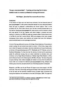
structure report compounds of DCPA when prepared in aqueous alcoholic solution, with the only three known examples limited to the salts with quinaldic acid (a monohydrate) (Smith et al., 2008a), 2-aminobenzoic acid (a dihydrate) (Smith et al., 2008b), hexamethylenetetramine (a monohydrate) (Smith et al., 2009) and with the drug quinacrine (a tetrahydrate) (Smith & Wermuth, 2009). The 1:1 stoichiometric reaction of DCPA with the substituted monocyclic heteroaromatic bases 2-aminopyrimidine, 3-(aminocarbonyl)pyridine (nicotinamide) and 4-(aminocarbonyl)pyridine (isonicotinamide) in methanol gave the anhydrous compounds 2-aminopyrimidinium 2-carboxy-4,5-dichlorobenzoate C4H6N3+ C8H3Cl2O4- (I), 3-(aminocarbonyl)pyridinium 2-carboxy-4,5-dichlorobenzoate C7H8NO2+ C8H3Cl2O4- (II) and the unusual adduct 4-(aminocarbonyl)pyridinium 2-carboxy-4,5-dichlorobenzoate 2-carboxymethyl-4,5-dichlorobenzoic acid (1/1/1) C6H7N2O+ C8H3Cl2O4- . C9H6Cl2O4 (III). This set of compounds shows examples of zero-, one- and two-dimensional hydrogen-bonded structures. All three compounds have at least one direct hetero-ring N+–H···Ocarboxyl hydrogen-bonding interaction (Figs. 1–3) (Tables 1–3), and as well all show low-dimensional hydrogen-bonded overall structures, two- in (II), one- in (III) and the first example of a cyclic zero-dimensional bis(cation–anion) species in (I) (Figs. 4–6). Associated with all of these DCPA structure types is the essentially planar monoanion species which is found in ca. 50% of the known 1:1 acid salts of DCPA with aromatic Lewis bases (Smith et al., 2008a). However structures (I)–(III) are sufficiently different as to be described separately. With compound (I), the primary cation-anion association is an asymmetric cyclic R22(8) pyrimidine heteroN···O, O'carboxyl association (Fig. 1). This is the high probability Type 4 hydrogen-bonding structural motif described by Allen et al. (1998). The cation-anion pairs so formed repeat across inversion centres via cyclic three-centre R21(4) amine -N+–H···O,O'carboxyl associations, enclosing R66(12) rings, giving discrete four-molecule 'heterotetramer' structural units (Fig. 4). Although other zero-dimensional structures are known among the DCPA proton-transfer compounds [others being discrete cation–anion 'heterodimers' (Etter & Adsmond, 1990), with brucine (Smith et al., 2007), hexamethylenetetramine and 1,10-phenanthroline (Smith et al., 2009), the formation of this bis-(cation–anion) structure type is driven more by the interactive features of 2-aminopyrimidine molecular synthon and finds a small incidence among its 1:1 salts with the aromatic acids, e.g. (3,4-dichlorophenoxy)acetic acid (Lynch et al., 1994) and phthalic acid (Smith et al., 1995). With (II), the nicotinamide cations form chain structures through homomeric amide N31–H···Ocarbonyl interactions which extend along the approximate planes in the unit cell (Fig. 5). These chains are linked along the b cell direction by associations involving proton donors of both the amide-N and the primary pyridinium groups to carboxyl oxygen acceptors of the anions (Table 3), giving a two-dimensional sheet structure. Compound (III) is an example of a (1:1:1) cation–anion adduct structure with the adduct molecule an unexpected methyl monoester of DCPA, arising from self synthesis in the methanol solvent under the conditions of the reaction. This phenomenon has no precedence among the proton-transfer compounds prepared under similar conditions in our laboratory. In (III), the primary hetero N+–H···Ocarboxyl hydrogen-bonded unit is extended into a zig-zag chain via an amide N–H···Ocarboxyl association which extends across the approximate planes in the unit cell (Fig. 6). The second amide-N, together with the amide carbonyl-O are involved in an asymmetric
publCIF
2
structure report cyclic R22(8) association with the peripherally linked DCPA methyl monoester adduct molecule (B). There is an absence in (I)–(III) of short intermolecular Cl···Cl interactions such as has been found in the DCPA compounds with 3- and 4-aminobenzoic acids (Smith et al., 2008a). The occurrence of this phenomenon particularly in dichloro- substituted aromatic compounds has previously been described (Sarma & Desiraju, 1986). However, in all three structures there are short Cl···Ocarboxyl associations [for (I), Cl4···O32ii, 3.0683 (14) Å: symmetry code (ii), -x + 1, -y + 2, -z + 1; for (II), Cl4···O11ii, 3.1582 (15) Å: symmetry code (iii), -x + 1, y + 1/2, -z + 5/2; for (III), Cl4···O22iii, 2.982 (5) Å: symmetry code (iii), x, -y + 1, z - 1/2. With the DCPA anions in this series the essential planarity is the result of the presence of the short intramolecular hydrogen bonds between the carboxyl groups [2.393 (8) Å in (III)–2.410 (2) Å in (II)]. Torsion angles associated with these groups (C2–C1–C11–O11 and C1–C2–C21–O22) are: for (I), -170.16 (16) and -179.70 (16) °; for (II), -178.68 (19), 172.58 (18) °; for (III), 173.0 (7), -175.6 (6) ° respectively. The planarity also means that there are short intramolecular aromatic ring C–H···Ocarboxyl interactions, typically C6–H6···O12, 2.676 (2) Å and C3–H3···O22, 2.643 (2) Å in (I). With the methyl ester adduct molecule in (III) the carboxylic acid group provides hydrogen-bonding links to the cation–anion chain structure rather than forming an intramolecular hydrogen bond and is therefore rotated out of the molecular plane [torsion angle C2B–C1B–C11B– O11B, -151.6 (6) °]. This present series provides a set of low-dimensional hydrogen- bonded structure types in the series of 1:1 proton-transfer compounds of 4,5-dichlorophthalic acid with aromatic Lewis bases. This low-dimensionality is largely associated with planarity in the internally hydrogen-bonded hydrogen phthalate anion species. Experimental Compounds (I)–(III) were synthesized by heating together for 10 min under reflux, 1 mmol quantities of 4,5dichlorophthalic acid and respectively 2-aminopyrimidine, nicotinic acid and isonicotinic acid in 50 mL of methanol. All compounds were obtained as small colourless plates or prisms [m. p. (I) 334 K; (II) 455–457 K; (III) 433–434 K], after partial room-temperature evaporation of solvent. (I) Crystal data C8H3Cl2O4·C4H6N3 Mr = 330.12 Triclinic, P1 a = 6.9738 (4) Å b = 9.4413 (4) Å c = 10.8900 (7) Å α = 97.420 (4)° β = 100.527 (5)°
publCIF
γ = 109.473 (5)° V = 650.50 (7) Å3 Z=2 Cu Kα radiation, λ = 1.54184 Å µ = 4.70 mm−1 T = 180 K 0.40 × 0.25 × 0.06 mm
3
structure report Data collection Oxford Diffraction Gemini-S Ultra CCD-detector diffractometer Absorption correction: Multi-scan SADABS (Sheldrick, 1996) Tmin = 0.263, Tmax = 0.750 4955 measured reflections
2542 independent reflections 2300 reflections with I > 2σ(I) Rint = 0.020
Refinement R[F2 > 2σ(F2)] = 0.033 2
wR(F ) = 0.095 S = 1.09 2542 reflections 206 parameters
0 restraints H atoms treated by a mixture of independent and constrained refinement Δρmax = 0.33 e Å−3 Δρmin = −0.27 e Å−3
Table 1 Hydrogen-bond geometry (Å, º) D—H···A O12—H12···O21 N1A—H1A···O22 N21A—H21A···O11i N21A—H21A···O12i N21A—H22A···O21
D—H 1.02 (4) 0.87 (2) 0.86 (2) 0.86 (2) 0.92 (3)
H···A 1.38 (3) 1.79 (2) 2.18 (3) 2.47 (3) 2.02 (3)
D···A 2.4037 (19) 2.6609 (19) 3.038 (2) 2.971 (2) 2.929 (2)
D—H···A 177 (3) 178.8 (19) 173 (3) 117 (2) 169.4 (19)
Symmetry code: (i) −x−1, −y+1, −z.
(II) Crystal data C8H3Cl2O4·C6H7N2O Mr = 357.14 Monoclinic, P21/c a = 11.4303 (3) Å b = 13.7933 (3) Å c = 9.2082 (2) Å β = 99.454 (2)°
V = 1432.06 (6) Å3 Z=4 Cu Kα radiation, λ = 1.54184 Å µ = 4.36 mm−1 T = 130 K 0.50 × 0.25 × 0.07 mm
Data collection Oxford Diffraction Gemini-S CCD-detector diffractometer Absorption correction: Multi-scan SADABS (Sheldrick, 1996) Tmin = 0.340, Tmax = 0.740 6939 measured reflections
publCIF
2798 independent reflections 2237 reflections with I > 2σ(I) Rint = 0.026
4
structure report Refinement R[F2 > 2σ(F2)] = 0.035 2
wR(F ) = 0.095 S = 0.96 2798 reflections 224 parameters
0 restraints H atoms treated by a mixture of independent and constrained refinement Δρmax = 0.34 e Å−3 Δρmin = −0.22 e Å−3
Table 2 Hydrogen-bond geometry (Å, º) D—H···A O12—H12···O21 N1A—H1A···O22 N31A—H31A···O31Ai N31A—H32A···O11ii
D—H 0.99 (4) 0.96 (2) 0.88 (3) 0.89 (3)
H···A 1.43 (4) 1.62 (2) 2.04 (3) 2.06 (3)
D···A 2.410 (2) 2.571 (2) 2.908 (2) 2.868 (2)
D—H···A 180 (6) 178 (3) 171 (2) 151 (2)
Symmetry codes: (i) x, −y+3/2, z+1/2; (ii) x, y+1, z.
(III) Crystal data C9H6Cl2O4·C8H3Cl2O4·C6H7N2O Mr = 606.18 Monoclinic, Cc a = 11.9645 (4) Å b = 26.1393 (6) Å c = 9.3213 (3) Å β = 122.509 (3)°
V = 2458.39 (15) Å3 Z=4 Cu Kα radiation, λ = 1.54178 Å µ = 4.90 mm−1 T = 130 K 0.56 × 0.14 × 0.07 mm
Data collection Oxford Diffraction Gemini-S Ultra CCD-detector diffractometer Absorption correction: Multi-scan SADABS (Sheldrick, 1996) Tmin = 0.454, Tmax = 0.710 6097 measured reflections
3034 independent reflections 2530 reflections with I > 2σ(I) Rint = 0.045
Refinement R[F2 > 2σ(F2)] = 0.046 wR(F2) = 0.128 S = 0.97 3034 reflections 363 parameters 1 restraint
publCIF
H atoms treated by a mixture of independent and constrained refinement Δρmax = 0.32 e Å−3 Δρmin = −0.44 e Å−3 Absolute structure: Flack (1983); 576 Friedel pairs Flack parameter: 0.03 (2)
5
structure report Table 3 Hydrogen-bond geometry (Å, º) D—H···A O12—H12···O21 N1A—H1A···O22i N41A—H41A···O12B N41A—H42A···O11 O11B—H11B···O41A
D—H 0.81 (8) 0.93 (8) 0.83 (7) 0.81 (8) 0.85 (9)
H···A 1.60 (9) 1.70 (7) 2.05 (8) 2.14 (8) 1.81 (8)
D···A 2.393 (8) 2.620 (8) 2.867 (10) 2.935 (7) 2.661 (8)
D—H···A 165 (10) 169 (5) 166 (5) 167 (9) 174 (8)
Symmetry code: (i) x+1/2, −y+3/2, z−1/2.
Hydrogen atoms potentially involved in hydrogen-bonding interactions in all compounds were located by difference methods and their positional and isotropic displacement parameters were refined. Other H atoms were included at calculated positions [C–H = 0.93 Å] and treated as riding with Uiso(H) = 1.2Ueq(C). The Flack (1983) absolute structure parameter obtained for (III) [0.03 (2): 617 Friedel pairs] has no chemical significance with this achiral compound. For all compounds, data collection: CrysAlis CCD (Oxford Diffraction, 2008). Cell refinement: CrysAlis RED (Oxford Diffraction, 2008) for (I), (III); CrysAlis RED (Oxford Diffraction, 2008 for (II). For all compounds, data reduction: CrysAlis RED; program(s) used to solve structure: SHELXS97 (Sheldrick, 2008); program(s) used to refine structure: SHELXL97 (Sheldrick, 2008); molecular graphics: PLATON (Spek, 2003); software used to prepare material for publication: PLATON. The authors acknowledge financial support from the School of Physical and Chemical Sciences, Queensland University of Technology, the School of Biomolecular and Physical Sciences, Griffith University and the School of Chemistry, University of Melbourne. References Allen, F. H., Raithby, P. R., Shields, G. P. & Taylor, R. (1998). Chem. Commun. pp. 1043–1044. Bozkurt, E., Kartal, I., Odabasoglu, M. & Büyükgüngör, O. (2006). Acta Cryst. E62, o4258–o4260. Etter, M. C. & Adsmond, D. A. (1990). J. Chem. Soc., Chem. Commun., pp. 589–591. Etter, M. C., MacDonald, J. C. & Bernstein, J. (1990). Acta Cryst. B46, 256–262. Flack, H. D. (1983). Acta Cryst. A39, 876–881. Lynch, D. E., Smith, G., Freney, D., Byriel, K. A. & Kennard, C. H. L. (1994). Aust. J. Chem. 47, 1097–1115. Mallinson, P. R., Smith, G. T., Wilson, C. C., Grech, E. & Wozniak, K. (2003). J. Amer. Chem. Soc. 125, 4259– 4270. Oxford Diffraction (2008). CrysAlis CCD. Version 1.171.32.5 and CrysAlis RED Version 1.171.32.5. Oxford Diffraction Ltd., Abingdon, England.
publCIF
6
structure report Sheldrick, G. M. (1996). SADABS. University of Göttingen, Germany. Sarma, J. A. R. P. & Desiraju, G. R. & (1986). Acc. Chem. Res. 19, 222–228. Sheldrick, G. M. (2008). SHELXL97 and SHELXS97. Acta Cryst. A64, 112–122. Smith, G., Gentner, J. M., Lynch, D. E., Byriel, K. A. & Kennard, C. H. L. (1995). Aust. J. Chem. 48, 1151– 1166. Smith, G. & Wermuth, U. D. (2009). Acta Cryst. C65, GA3177. Smith, G., Wermuth, U. D. & White, J. M. (2007). Acta Cryst. E63, o4276–o4277. Smith, G., Wermuth, U. D. & White, J. M. (2008a). Acta Cryst. C64, o123–o127. Smith, G., Wermuth, U. D. & White, J. M. (2008b). Acta Cryst. C64, o180–o183. Smith, G., Wermuth, U. D. & White, J. M. (2009). Unpublished data. Spek, A. L. (2003). J. Appl. Cryst. 36, 7–13. Figure 1 Figure 1. The molecular configuration and atom-numbering scheme for the 2-aminopyrimidinium cation and the 2-carboxy-4,5-dichlorobenzoate anion in (I) showing the cyclic R22(8) inter-species hydrogen-bonding associations as dashed lines. Non-H atoms are shown as 50% probability displacement ellipsoids. Figure 2 Figure 2. Molecular configuration and atom-numbering scheme for the 3-(aminocarbony)pyridinium cations and the 2-carboxy-4,5-dichlorobenzoate anion in (II). The dashed lines indicate the inter-species hydrogen bonds while non-H atoms are shown as 50% probability displacement ellipsoids. Figure 3 Figure 3. Molecular configuration and atom-numbering scheme for the 4-(aminocarbonyl)pyridinium cation, the 2-carboxy-4,5-dichlorobenzoate anion and the 2-(carboxymethyl)-4,5-dichlorobenzoic acid adduct molecule in (III). The dashed lines indicate the inter-species hydrogen bonds while non-H atoms are shown as 50% probability displacement ellipsoids. Figure 4 Figure 4. Hydrogen-bonding in the discrete cyclic centrosymmetric bis(cation–anion) 'heterotetramer' structural units in (I), shown as dashed lines. Non-interactive hydrogen atoms are omitted. For symmetry code (i), see Table 1. Figure 5 Figure 5. The hydrogen-bonding in the homomeric cation chains and the peripheral cation–anion extensions in the two-dimensional sheet structure of in a perspective view of the unit cell of (II). Non-interactive hydrogen atoms are omitted and hydrogen bonds are shown as dashed lines. For symmetry codes, see Table 2.
publCIF
7
structure report Figure 6 Figure 6. The one-dimensional hydrogen-bonded zig-zag chains formed by extension of the cation–anion pairs and the peripheraly attached methyl monoester adduct B-molecules, in the structure of (III), in a perspective view of the unit cell. Non-interactive hydrogen atoms are omitted. For symmetry code (ii): x - 1/2, y + 3/2, z - 1/2. For symmetry code (i), see Table 3.
publCIF
8