Clinical Orthopaedics and Related Research®
Clin Orthop Relat Res (2013) 471:1356–1364 DOI 10.1007/s11999-012-2649-0
A Publication of The Association of Bone and Joint Surgeons®
SURGICAL TECHNIQUE
Surgical Technique Talar Neck Osteotomy to Lengthen the Medial Column After a Malunited Talar Neck Fracture Thomas Suter MD, Alexej Barg MD, Markus Knupp MD, Heath Henninger PhD, Beat Hintermann MD
Received: 8 May 2012 / Accepted: 5 October 2012 / Published online: 17 October 2012 Ó The Association of Bone and Joint Surgeons1 2012
Abstract Background Treatment of malunited talar neck fractures is challenging, and few studies address anatomic reconstruction as an alternative to arthrodesis. We describe a new surgical approach attempting to improve function and avoid development of degenerative changes in the adjacent joints. Description of Technique Indications included malunited talar neck fractures. Through a dorsomedial approach, a correcting osteotomy with interposition of an autograft or
Each author certifies that he or she has no commercial associations (eg, consultancies, stock ownership, equity interest, patent/licensing arrangement etc) that might pose a conflict of interest in connection with the submitted article. All ICMJE Conflict of Interest Forms for authors and Clinical Orthopaedics and Related Research editors and board members are on file with the publication and can be viewed on request. Clinical Orthopaedics and Related Research neither advocates nor endorses the use of any treatment, drug, or device. Readers are encouraged to always seek additional information, including FDA approval status, of any drug or device before clinical use. Each author certifies that his or her institution approved the human protocol for this investigation, that all investigations were conducted in conformity with ethical principles of research, and that informed consent for participation in the study was obtained. This work was performed at the Clinic of Orthopaedic Surgery, Kantonsspital Liestal, Switzerland. T. Suter (&), A. Barg, M. Knupp, B. Hintermann Clinic of Orthopaedic Surgery, Kantonsspital Liestal, Rheinstrasse 26, 4410 Liestal, Switzerland e-mail:
[email protected] H. Henninger Harold K. Dunn Orthopaedic Research Laboratory, University Orthopaedic Center, University of Utah, Salt Lake City, UT, USA
123
allograft was performed and internally fixed using buttress plate and/or screws. Methods We retrospectively reviewed seven patients in whom the new technique was indicated for malunited talar neck fractures. The mean age of the patients was 42 years (range, 17–60 years). We analyzed the patients clinically and radiographically with a minimum followup of 2.5 years (mean, 4 years; range, 2.5–9.8 years). Results At followup, all patients experienced substantial pain relief. No development of avascular necrosis or radiographic arthritic changes were observed. Physical categories of the SF-36 score showed great improvements. The American Orthopaedic Foot and Ankle Society hindfoot score increased from 41 ± 19 preoperatively (range, 20–62) to 84 ± 11 (range, 68–97). The average talar-first metatarsal angle increased dramatically. All but one patient showed radiographic union of the talar osteotomy. Implant removal was performed in three patients. Conclusions Based on these observations, correctional osteotomy is a reasonable option for treating patients with malunited talar neck fractures by providing a pain-free foot with good function, recreating anatomy, and involving a low risk of postoperative complications. Further studies with longer followups are required to confirm these findings persist with time. Level of Evidence Level IV, therapeutic study. See the Instructions for Authors for a complete description of levels of evidence.
Introduction Talus fractures account for less than 1% of all fractures [26, 30]. The malunion rate of talus fractures varies from 9% to 47% [9, 26] (Table 1). Patients with malunited fractures
Volume 471, Number 4, April 2013
Talar Neck Osteotomy
1357
Table 1. Literature review addressing malunion rate of patient with talar fractures Study
Study type
Number of patients All
Talar neck fracture
Retrospective, single center
41
41
Fleuriau Chateau et al. [9]
Retrospective, single center
23
23
Frawley et al. [10]
Retrospective, single center
28
28
Dumont et al. [8]
Surgical procedures
Followup (years)*
Number of malunions
39 screw osteosyntheses
4 (1–6)
6
23 open reductions and internal fixations (with one or two 2.0- or 2.4-mm plates, with additional 2.0-, 2.7-, or 3.5-mm lag screws)
1.7 (0.5–4.3)
2 mild extension malunions
6 plaster immobilizations alone
(1–8)
3
7.5
6 varus malunions in talar neck fractures
2 K-wire osteosyntheses (12 with additional external fixation)
6 manipulations and plaster 10 open reductions and internal fixations (K-wire) 6 open reductions and internal fixations (screws) with or without supplementary K-wires
Ohl et al. [21]
Sanders et al. [28]
Retrospective, single center
20
Retrospective, single center
70
10
8 K-wires 7 3.5-mm or small Herbert screws
2 varus malunions in talar body fractures
5 combination of K-wires and screws 69
Open reduction and screw fixation
5.2 (2–10.5)
23 (3 treated with triple arthrodesis and 2 treated with subtalar fusion)
* Values are expressed as mean, with range in parentheses.
have uniformly poor results [26, 28], typically have painful overload of the lateral foot, and are greatly disabled in daily activities [5, 20]. Poor clinical outcomes may be attributable to shortening of the medial column [5, 18, 23]. The most common deformity after malunion of a talus fracture is varus malalignment of the talar neck [3, 5]. Varus malalignment of the talar neck results in substantial shortening of the medial column, with resulting locking of the hindfoot in varus and internal rotation [7]. Additionally, correlations exist between the degree of varus malalignment and the change in the position of the foot and the degree of subtalar motion [7]. Malalignment of 2 mm at the talar neck can result in considerable load redistribution between the posterior, middle, and anterior facets of the subtalar joints [29]. Numerous salvage procedures may treat malunited talar fractures, including arthrodeses of the tibiotalar, subtalar, and talonavicular joints [1, 5, 10]. Canale and Kelly [5] described five of 12 patients with varus malunion treated with a triple arthrodesis. Two patients had a poor result. Few studies address anatomic reconstruction to restore normal foot function, with preserved joint cartilage and without evidence of talar collapse or infection [13, 20, 25]. None compare their results with those of patients with arthrodesis. In a case report, Monroe and Manoli [20] described a talar neck osteotomy with insertion of a
tricortical iliac crest bone graft in varus deformity after nonoperative treatment of a Hawkins II talar neck fracture. Fifty-six months postoperatively, the patient had returned to heavy labor and his American Orthopaedic Foot and Ankle Society (AOFAS) score improved from 11 to 85. In two other reports [13, 25], a combined 11 patients received delayed surgical treatment for neglected or malreduced talar fractures using a corrective talar neck osteotomy with interposition of a tricortical iliac crest bone graft. One patient had avascular necrosis of the talus develop, which was treated by ankle fusion. The other 10 patients experienced satisfactory results after 4 years of followup but one patient required ankle fusion after 8 years. The AOFAS scores at final followup were substantially improved. Little is known about the complications of a talar neck osteotomy and its effect on painful malunited talar fractures. The aims of this report are: (1) to describe a surgical approach to address malunited talar fractures; (2) to provide intraoperative and postoperative details including the occurrence of any complications, including secondary surgeries; (3) to determine whether corrective osteotomy of the talar neck provides short-term pain relief; (4) to quantify short-term functional outcomes, including ROM and quality of life; and (5) to analyze the postoperative radiographic findings.
123
1358
Suter et al.
Clinical Orthopaedics and Related Research1
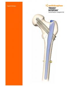
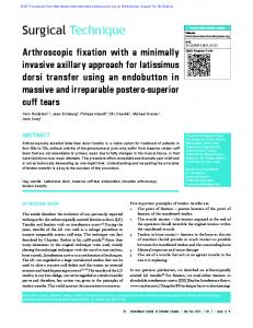
The surgical indications were malunited talar neck fractures with shortening and deformity of the medial column with hindfoot varus, forefoot varus, and adducted position. All patients had gait dysfunction, painful overload of the lateral foot during weightbearing, and difficulty wearing a brace or shoes. The procedure was contraindicated in the presence of skin lesions, ulcers on the foot, or the presence of avascular necrosis of the talus. With a tourniquet applied to the ipsilateral thigh, a dorsomedial approach was used. In five patients, implant removal was performed before the correctional osteotomy. A chisel was directed along the fracture plane after implant removal (Fig. 1A–B). In two patients with posteromedial tendon contractions (flexor digitorum longus tendon and posterior tibial tendon), additional posteromedial releases were performed before lengthening the medial columns. Additional lengthening of the Achilles tendon was required in two patients to achieve physiologic ROM. In one patient, an arthroscopy of the subtalar joint was performed to rule out posttraumatic osteoarthritis before the correctional osteotomy was performed (Table 2). To determine the amount of distraction and rotational correction required of the talar head with regard to the talar
body, we used a HintermannTM distractor (Integra LifeSciences Corporation, Plainsboro, NJ, USA), which was applied between two K-wires, and allowed manipulation of the distracting forces and rotational movement until an appropriate correction of the forefoot was achieved (Fig. 1C–D). Once the desired position was achieved, the size of the graft was determined as measured by a ruler. Human cancellous allograft blocks (Tutoplast1; Tutogen Medical GmbH, Neunkirchen am Brand, Germany) were used on a routine basis for interposition as the required size of graft was not considered critical for revascularization (Fig. 1E–F). Two patients requested the use of their own bone, so a tricortical autograft from the iliac crest was used. As a standard approach, two screws provided the required stability after the correcting osteotomy and graft insertion. However, if the bone graft did not provide enough distraction stability (eg, against compressive forces), the use of a buttress plate was considered. Analogously, if the head fragment was too short and thus the screw purchase alone was insufficient to provide stability against rotational forces, an additional plate was considered (Fig. 1G–H). In two cases of concomitant osteoarthritis of the adjacent joints, additional surgeries were performed: one subtalar arthrodesis and one arthrodesis of the subtalar and talonavicular joint (Table 2).
Fig. 1A–H After performing a dorsomedial approach, the future osteotomy is marked with two K-wires at the apex of the deformity. (A) Dorsoplantar and (B) lateral radiographs show the ankle intraoperatively. The special distractor is applied between two K-wires to perform (C) distractional and (D) rotational correction
of the alar head with regard to the talar body. (E) Dorsoplantar and (F) lateral intraoperative radiographs show the ankle after insertion of a fully cancellous allograft block (arrow). (G) Lateral and (H) dorsoplantar intraoperative radiographs show internal stabilization using a plate to stabilize the distraction osteotomy.
Surgical Technique
123
Volume 471, Number 4, April 2013
Talar Neck Osteotomy
1359
Table 2. Demographic data for seven patients with malunited talar neck fractures Patient
Sex
Age BMI (years)* (kg/m2)
Initial fracture treatment
Number of surgical procedures
Additional surgeriesà
Followup (years)
Revisions
1
Male
25
22
ORIF
1
Implant removal, posterior release, lengthening achilles tendon
3.4
None
2
Male
52
21
Nonoperative 3
None
2.7
Implant removal
3
Male
43
27
Nonoperative 0
Subtalar fusion
2.8
Implant removal, arthroscopic arthrolysis of the ankle
4
Male
49
26
Nonoperative 1
Implant removal
2.7
Subtalar and talonavicular fusion
5
Male
48
26
ORIF
1
Implant removal
9.8
None
6
Female 60
37
ORIF
1
Implant removal, posterior release, lengthening achilles tendon, subtalar and talonavicular fusion
3.9
None
7
Female 17
19
ORIF
2
Implant removal, arthroscopy of the subtalar joint
2.5
Implant removal
Mean ± SD
42 ± 15 25 ± 6
4.0 ± 2.6
* At the time of the talar neck osteotomy to lengthen the medial column; before the talar neck osteotomy; àin addition to the talar neck osteotomy; ORIF = open reduction and internal fixation.
The foot was placed in a stable walker (VACO1ped; OPED AG, Cham, Switzerland) or in a short leg cast (three patients) for 8 weeks with partial weightbearing up to 15 kg until bone union was achieved. A rehabilitation program was continued for at least 4 months after removal of the walker or cast, including intensive walking, stretching, and strengthening exercises.
Patients and Methods We retrospectively reviewed all seven consecutive patients with malunited talar neck fractures who underwent corrective osteotomy of the talar neck (lengthening of the medial column) between January 2002 and May 2009. The study group included two females and five males (Table 2). The mean ± SD age of the patients at the time of the surgical procedure was 42 ± 15 years (range, 17–60 years). All patients originally sustained closed talar neck fractures. The initial treatment in four patients was open reduction and internal fixation using K-wires or screw fixation. Three patients were treated nonoperatively with immobilization in casts for 6 to 8 weeks. The same clinical examination was performed preoperatively and postoperatively. The clinical examination involved assessment of ankle alignment and ROM with the
patient standing and ankle stability with the patient sitting. Tibiotalar ROM was determined with a goniometer placed along the lateral border of the leg and foot [2, 11]. All goniometer measurements in the weightbearing position were performed comparable with the method described by Lindsjo¨ et al. [17]. Accuracy of the goniometric measurements was assessed by direct comparison with ankle ROM measurements obtained from radiographs [2, 6, 11, 12]. ROM of the subtalar joint was determined with a goniometer placed posteriorly on the hindfoot [4]. Patients rated their pain on a VAS of 0 points (no pain) to 10 points (maximal pain) [14]. In addition, the AOFAS hindfoot score was calculated [15], and all patients completed SF-36 questionnaires on their quality of life [32] (Table 3). Affected hindfeet were evaluated preoperatively and postoperatively, based on weightbearing radiographs in two planes. The degrees of the degenerative changes in the tibiotalar and adjacent joints, and the talar-first metatarsal (TMT-I) angles, were evaluated. To assess the varus or valgus hindfoot deformity, a hindfoot alignment view as described by Saltzman and el Khoury was acquired [27]. The method for measuring the TMT-I angle has been described [33]. The AP TMT-I angle was considered negative if the first metatarsal was aligned in an adducted position with respect to the axis of the talus (Table 3). Two experienced orthopaedic residents (TS, AB) independently
123
1360
Clinical Orthopaedics and Related Research1
Suter et al.
Table 3. Preoperative data for seven patients with malunited talar neck fractures. Patient
VAS pain (points, maximum 10)
AOFAS score (points, maximum 100)
ROM (°) (tibiotalar joint) DF
PF
Total
EVE
1
7
62
20
30
50
10
15
25
39
73
2
2
8
22
10
15
25
5
10
15
39
73
8
3
6
53
18
31
49
10
20
30
40
73
15
4
8
20
16
27
43
10
15
25
37
73
16
5
7
48
15
30
45
10
10
20
40
71
6
6
9
22
15
29
44
5
10
15
30
69
24
7
7
59
11
22
33
10
15
25
44
71
12
Mean ± SD
7.4 ± 1.0
41 ± 19
15 ± 4
26 ± 6
41 ± 9
9±2
14 ± 4
22 ± 6
38 ± 4
72 ± 2
12 ± 7
ROM (°) (subtalar joint) INV
SF-36 score (points, maximum 100) Total
Physical health
TMT-I angle (°)
Mental health
AOFAS = American Orthopaedic Foot and Ankle Society; TMT-I = talar-first metatarsal; DF = dorsiflexion; PF = plantar flexion; EVE = eversion; INV = inversion.
analyzed the radiographs; the images were blinded and ordered randomly. For patients with suspicions of nonunion or avascular necrosis, CT or single photon emission CT was recommended [22]. Furthermore, CT was used to assess the integrity and contour of bone, in particular of subchondral bone, to plan the correcting osteotomy. The first clinical and radiographic followup was at 6 weeks to check the wound site and to assess bony union, position of the implants, and presence of avascular necrosis (using weightbearing radiographs in two planes). The next clinical and radiographic followups were at 4 months, 1 year, and annually thereafter. All patients were seen postoperatively in our outpatient clinic by two reviewers (TS, AB), who did not operate on any of the patients. No patients were lost to followup. The minimum followup was 2.5 years (mean, 4 years; range, 2.5–9.8 years). No patients were recalled specifically for this study; all data were retrieved from medical records and radiographs during followup in outpatient clinics. Major complications were considered to be pseudarthrosis or any subsequent operation to relieve pain or treat secondary deformities. Implant removal attributable to the will of the patient and not for mechanical reasons was not considered a major complication.
Results There were no intraoperative complications. No patients required postoperative blood transfusions, and no nerve lesions occurred. Wound healing occurred within 2 weeks of the surgery, free of adverse events, including any wound infections, dehiscence, wound edge necrosis, and breakdowns of the wound. No deep vein thromboses were seen.
123
We observed three major complications. In one patient, nonunion of the talar neck osteotomy occurred, and a subtalar and talonavicular fusion was performed 7 months after the lengthening osteotomy (Table 2). Thereafter, the patient was free of adverse events. An additional arthroscopic arthrolysis of the ankle was performed in one patient owing to postoperative scar formation at the time of removal of the implants. In a third patient, a buttress plate was used to provide required stability after the correcting osteotomy and graft insertion. Owing to an anterior impingement during dorsiflexion of the ankle, the plate was removed after bony healing of the osteotomy. In another patient, the implant was removed at the request of the patient. That subsequent operation did not relieve pain or treat secondary deformities and therefore was not considered a major complication. All patients experienced substantial pain relief (Table 4). Overall, the average pain score (VAS) decreased from 7.4 ± 1.0 (range, 6–9) to 1.7 ± 0.8 (range, 0–2). The average AOFAS hindfoot score increased from 40.9 ± 18.8 (range, 20–62) preoperatively to 83.9 ± 11.1 (range, 68–97) (Fig. 2). The physical examination of the affected joints at the latest followup showed no joint swelling, instability, or axial deformity of the affected joints. All patients were able to wear commercial shoes. The average ROM of the tibiotalar joint increased from 41.3 ± 9.1 (range, 25–50) preoperatively to 44.3 ± 7.7 (range, 32–53) postoperatively. The average ROM of the subtalar joint decreased from 22 ± 6 (range, 15–30) preoperatively to 17 ± 17 (range, 0–35) postoperatively (Table 4). At latest followup, all patients were satisfied with the results and stated they would undergo the surgery again. The physical categories of the SF-36 score showed substantial improvements postoperatively, and the preoperative and postoperative mental categories of SF-36 were
* Maximum 94 (owing to subtalar fusion); AOFAS = American Orthopaedic Foot and Ankle Society; TMT-I = talar-first metatarsal; DF = dorsiflexion; PF = plantar flexion; EVE = eversion; INV = inversion.
13 ± 5
18 6 No
1±5 74 ± 2
73 72 35 20 15 36 25 11
15 ± 3 29 ± 5 44 ± 8 7 ± 7 10 ± 10 17 ± 17 70 ± 8 84 ± 11
96 2 2.5 7
Mean ± SD 4.0 ± 2.6 2 ± 1
6
12 18
4 6 73
No
86 62
63 0
25 0 15 0
0 0
10 0 45 45
33
30 30
16 68* 2 2.7
9.8 3.9
4
0 2
97 72*
15 15
49
79 73
No No
12
Talar Neck Osteotomy
5 6
10
14 1
2 No
No 74
72 67
66 25 10
0 53 2
2
86* 2.8 3
81 2.7 2
32
0 35 18
15 20 12
0
5 3 No 74 74 15 50 2
87 3.4 1
Total
20 30 20
35
PostDifference from operative pre-operative Mental health Physical health EVE PF DF
INV
ROM (°) (subtalar joint) ROM (°) (tibiotalar joint) FollowVAS pain (points, AOFAS score up (years) maximum 10) (points, maximum 100) Patient
Table 4. Clinical and radiographic postoperative results for seven patients with malunited talar neck fractures
Total
SF-36 score Progress of (points, maximum degenerative 100) changes
TMT-I angle (°)à
Volume 471, Number 4, April 2013
1361
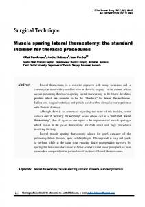
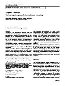
Fig. 2 Preoperative (gray columns) and postoperative (black columns) AOFAS hindfoot scores for all patients are shown. The data are presented as mean ± SD. The AOFAS hindfoot score increased from 40.9 ± 18.8 (range, 20–62) preoperatively to 83.9 ± 11.1 (range, 68–97).
Fig. 3 Preoperative (gray columns) and postoperative (black columns) quality of life for all patients, assessed by the SF-36, are shown. The data are presented as mean ± SD. The physical categories of the SF-36 score showed substantial improvements postoperatively, while the preoperative and postoperative mental categories of SF-36 were comparable.
comparable (Fig. 3). The average summarized components of the physical and mental outcomes scores improved from 38.4 ± 4.2 to 69.8 ± 8.1 and from 71.8 ± 1.6 to 74.0 ± 2.3, respectively (Table 4). All but one patient showed radiographic evidence of union of the talar osteotomy within 2 to 3 months postoperatively (Fig. 4). There was no loss of correction in any of the patients. Radiographically, no avascular necrosis development was observed after correction. Radiographic evidence of arthritic changes did not progress in any patient. A solid fusion on the site of the arthrodesis was
123
1362
Suter et al.
Clinical Orthopaedics and Related Research1
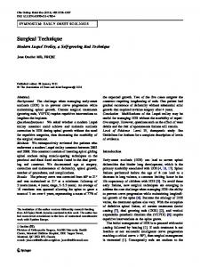
Fig. 4A–E (A) An AP radiograph shows the left foot and preoperative (B) AP and (C) lateral radiographs show the right foot of a 17-year-old girl with a malunited talar neck fracture and consecutive shortening of the medial column with supination and adduction deformities of the forefoot. Postoperative (D) AP and (E) lateral radiographs show the foot after partial removal of the implants with a solid union of the lengthening osteotomy of the talus. Radiographically, there was no loss of correction or development of avascular necrosis observed. The plantigrade position of the forefoot can be seen, and the corrected cyma line, compared with the preoperative situation.
detected in two patients, one patient with an additional subtalar arthrodesis and the other after talonavicular and subtalar arthrodesis. The average TMT-I angle increased from 11.9 ± 7.3 (range, 2 to 24) preoperatively to 0.9 ± 4.7 (range, 6 to 6) postoperatively (Table 4).
Discussion Treatment of malunited talar neck fractures is challenging, and few studies address anatomic reconstruction as an alternative to arthrodeses. We describe a new surgical technique to address talar malunion and our experience with this technique in terms of intraoperative and postoperative complications, including surgical revision for any reason; and the ability of the procedure to provide pain relief, improvement in foot and ankle ROM, and improvement in patient-reported quality of life. An important concern regarding talar neck lengthening osteotomies is the high rate of postoperative complications. We observed no intraoperative complications and a low rate of postoperative complications in our patients. All patients experienced substantial functional improvement regarding their ROM and quality of life. Although the results were encouraging, this study had limitations. First, only a small number of patients were
123
included, as fractures of the talar neck are rare. Although the use of lengthening osteotomies as an alternative to correcting arthrodesis of the tibiotalar, subtalar, and/or talonavicular joints is expanding, previous reports also have been limited to small patient numbers and short-term followups. Second, additional surgeries were performed and different after-treatments were used owing to differences in the initial state of each patient’s ankle. Because of the broad spectrum of injury, no single operative technique or after-treatment was applicable to all patients. A finite population with similar operative conditions may prove difficult to isolate. Third, the functional outcome in the cohort was partially assessed using the AOFAS hindfoot score. A policy statement from the AOFAS research committee recommending against use of this instrument was published in 2011 [24]. We have included results using the score from 2002 to 2006 which predated this notice. Since 2006 in our clinic, we modified the score to better validate it and applied it to our study after 2006 [16]. Finally, followup in this study was limited to an average of 4 years. Patients should be evaluated at 10 years for valid conclusions to be drawn regarding long-term viability of talar neck lengthening osteotomies in patients with malunited talar neck fractures. Major concerns regarding a talar neck osteotomy (with the purpose of lengthening the medial column) include the
Volume 471, Number 4, April 2013
potential risk of nonunion. In our series, there was one patient (Patient 4) who had a nonunion after talar neck lengthening osteotomy. This patient initially had a pseudarthrosis after nonoperative treatment of a talar neck fracture (Hawkins Type II); therefore an open reduction and internal fixation was performed 10 months before lengthening of the medial column. After subtalar and talonavicular fusion was performed, the postoperative followup was free of adverse events. Another possible complication resulting from osteotomy of a malunited talar neck fracture is the potential risk of avascular necrosis [8, 21, 31]. Huang and Cheng [13] reported that one patient had avascular necrosis of the talus develop after a correcting osteotomy in a malunited talar neck fracture and was treated with an ankle fusion. An incision medial to the tibialis anterior was used, which started at the navicular tuberosity. This incision was extended proximally to facilitate fixation of a concomitant malleolar fracture or to perform a malleolar osteotomy and reduction of a talar body fracture. In the current series, no patient had avascular necrosis develop, which was comparable to other studies [19, 20, 25]. A dorsomedial incision was performed to expose the malunited talar neck, as it is used routinely for total ankle replacement. Therefore, not only were the deltoid branches protected, but the periosteal branches of the dorsalis pedis and peroneal arteries were as well. Other advantages of this approach were observation of the whole deformity and the ability to control rotation of the talar head after correction of the talar neck malunion. Until now only autologous bone grafting from the iliac crest was used to fill the osteotomy to lengthen the medial column [13, 19, 20, 25]. In the current study an allograft was used in five patients. We observed bony union in all but one patient; an autograft was used in this patient (Patient 4). It is known that pain is particularly relevant in the daily life of patients with malunited talar neck fractures [5, 20]. In our study, all patients experienced pain relief postoperatively, and all patients had a VAS score less than 2 at the latest followup. However, in patients with subtalar and/or talonavicular arthrodesis the pain relief may occur partially because of the arthrodesis and not owing to correction surgery alone. It is difficult to compare the functional results of this investigation with results of other studies because of some important differences in the cohorts. Huang and Cheng [13] reported on two patients with neglected or malreduced talar fractures, in which a corrective osteotomy of the talar neck was performed (in one patient and combined with subtalar fusion). Other surgical procedures included open reduction (three patients), open reduction with bone grafting (three patients), and an open reduction combined with ankle fusion (one patient). The AOFAS ankle-hindfoot score in this heterogeneous study group averaged 77.4. Rammelt
Talar Neck Osteotomy
1363
et al. [25] found excellent results in a series of 10 patients who were followed for a mean of 4 years. That study mentioned only five patients had malunions; the remainder had a nonunion or partial avascular necrosis after talar neck or talar body fractures. They reported the AOFAS anklehindfoot score increased substantially from a mean of 38 to a mean of 86 at followup. We observed reliable and satisfying clinical results after talar neck lengthening osteotomy in patients with malunited talar neck fractures. A correcting osteotomy is a reliable method to restore function, decrease pain, and realign the hindfoot to avoid development of degenerative changes in the adjacent joints. This type of surgery, however, should be performed by foot and ankle surgeons with adequate experience to minimize the risk of intraoperative and postoperative complications, including the risk of avascular necrosis. Long-term followup of additional patients is needed to confirm our findings. Acknowledgments We thank Lilianna Bolliger MSc, for aid in the collection of the data and with preparation of the radiographic and intraoperative pictures.
References 1. Asencio G, Rebai M, Bertin R, Megy B, Daude O. [Pseudarthrosis and non-union of disjunctive talar fractures] [in French]. Rev Chir Orthop Reparatrice Appar Mot. 2000;86:173–180. 2. Barg A, Elsner A, Anderson AE, Hintermann B. The effect of three-component total ankle replacement malalignment on clinical outcome: pain relief and functional outcome in 317 consecutive patients. J Bone Joint Surg Am. 2011;93:1969–1978. 3. Baumhauer JF, Alvarez RG. Controversies in treating talus fractures. Orthop Clin North Am. 1995;26:335–351. 4. Buckley RE, Hunt DV. Reliability of clinical measurement of subtalar joint movement. Foot Ankle Int. 1997;18:229–232. 5. Canale ST, Kelly FB Jr. Fractures of the neck of the talus: longterm evaluation of seventy-one cases. J Bone Joint Surg Am. 1978;60:143–156. 6. Coetzee JC, Castro MD. Accurate measurement of ankle range of motion after total ankle arthroplasty. Clin Orthop Relat Res. 2004;424:27–31. 7. Daniels TR, Smith JW, Ross TI. Varus malalignment of the talar neck: its effect on the position of the foot and on subtalar motion. J Bone Joint Surg Am. 1996;78:1559–1567. 8. Dumont C, Fuchs M, Burchhardt H, Tezval M, Wachowski MM, Sturmer KM. [What are the clinical results of operated fractures of the talus?] [in German]. Z Orthop Unfall. 2007;145:212–220. 9. Fleuriau Chateau PB, Brokaw DS, Jelen BA, Scheid DK, Weber TG. Plate fixation of talar neck fractures: preliminary review of a new technique in twenty-three patients. J Orthop Trauma. 2002;16:213–219. 10. Frawley PA, Hart JA, Young DA. Treatment outcome of major fractures of the talus. Foot Ankle Int. 1995;16:339–345. 11. Hintermann B, Barg A, Knupp M, Valderrabano V. Conversion of painful ankle arthrodesis to total ankle arthroplasty. J Bone Joint Surg Am. 2009;91:850–858. 12. Hintermann B, Valderrabano V, Dereymaeker G, Dick W. The HINTEGRA ankle: rationale and short-term results of 122 consecutive ankles. Clin Orthop Relat Res. 2004;424:57–68.
123
1364
Suter et al.
13. Huang PJ, Cheng YM. Delayed surgical treatment for neglected or mal-reduced talar fractures. Int Orthop. 2005;29:326– 329. 14. Huskisson EC. Measurement of pain. Lancet. 1974;2:1127–1131. 15. Kitaoka HB, Alexander IJ, Adelaar RS, Nunley JA, Myerson MS, Sanders M. Clinical rating systems for the ankle-hindfoot, midfoot, hallux, and lesser toes. Foot Ankle Int. 1994;15:349– 353. 16. Knupp M, Stufkens SA, Bolliger L, Barg A, Hintermann B. Classification and treatment of supramalleolar deformities. Foot Ankle Int. 2011;32:1023–1031. 17. Lindsjo U, Danckwardt-Lilliestrom G, Sahlstedt B. Measurement of the motion range in the loaded ankle. Clin Orthop Relat Res. 1985;199:68–71. 18. Lorentzen JE, Christensen SB, Krogsoe O, Sneppen O. Fractures of the neck of the talus. Acta Orthop Scand. 1977;48:115–120. 19. Matsumura T, Sekiya H, Hoshino Y. Correction osteotomy for malunion of the talar head: a case report. J Orthop Surg (Hong Kong). 2008;16:96–98. 20. Monroe MT, Manoli A 2nd. Osteotomy for malunion of a talar neck fracture: a case report. Foot Ankle Int. 1999;20:192–195. 21. Ohl X, Harisboure A, Hemery X, Dehoux E. Long-term followup after surgical treatment of talar fractures: twenty cases with an average follow-up of 7.5 years. Int Orthop. 2011;35:93–99. 22. Pagenstert GI, Barg A, Leumann AG, Rasch H, Muller-Brand J, Hintermann B, Valderrabano V. SPECT-CT imaging in degenerative joint disease of the foot and ankle. J Bone Joint Surg Br. 2009;91:1191–1196.
123
Clinical Orthopaedics and Related Research1 23. Peterson L, Goldie IF, Irstam L. Fracture of the neck of the talus: a clinical study. Acta Orthop Scand. 1977;48:696–706. 24. Pinsker E, Daniels TR. AOFAS position statement regarding the future of the AOFAS Clinical Rating Systems. Foot Ankle Int. 2011;32:841–842. 25. Rammelt S, Winkler J, Heineck J, Zwipp H. Anatomical reconstruction of malunited talus fractures: a prospective study of 10 patients followed for 4 years. Acta Orthop. 2005;76:588–596. 26. Rammelt S, Zwipp H. Talar neck and body fractures. Injury. 2009;40:120–135. 27. Saltzman CL, el-Khoury GY. The hindfoot alignment view. Foot Ankle Int. 1995;16:572–576. 28. Sanders DW, Busam M, Hattwick E, Edwards JR, McAndrew MP, Johnson KD. Functional outcomes following displaced talar neck fractures. J Orthop Trauma. 2004;18:265–270. 29. Sangeorzan BJ, Wagner UA, Harrington RM, Tencer AF. Contact characteristics of the subtalar joint: the effect of talar neck misalignment. J Orthop Res. 1992;10:544–551. 30. Santavirta S, Seitsalo S, Kiviluoto O, Myllynen P. Fractures of the talus. J Trauma. 1984;24:986–989. 31. Vallier HA, Nork SE, Barei DP, Benirschke SK, Sangeorzan BJ. Talar neck fractures: results and outcomes. J Bone Joint Surg Am. 2004;86:1616–1624. 32. Ware JE Jr, Sherbourne CD. The MOS 36-item short-form health survey (SF-36): I. Conceptual framework and item selection. Med Care. 1992;30:473–483. 33. Younger AS, Sawatzky B, Dryden P. Radiographic assessment of adult flatfoot. Foot Ankle Int. 2005;26:820–825.