Am J Physiol Regulatory Integrative Comp Physiol 280: R123–R131, 2001.
Survival of water stress in annual fish embryos: dehydration avoidance and egg envelope amyloid fibers JASON E. PODRABSKY,1 JOHN F. CARPENTER,2 AND STEVEN C. HAND1 1 Section of Integrative Physiology and Neurobiology, Department of Environmental, Population, and Organismic Biology, University of Colorado, Boulder 80309–0334; and 2Department of Pharmaceutical Sciences, University of Colorado Health Sciences Center, Denver, Colorado 80262 Received 8 February 2000; accepted in final form 10 August 2000
Podrabsky, Jason E., John F. Carpenter, and Steven C. Hand. Survival of water stress by annual fish embryos: dehydration avoidance and egg envelope amyloid fibers. Am J Physiol Regulatory Integrative Comp Physiol 280: R123–R131, 2001.—Diapausing embryos of Austrofundulus limnaeus survive desiccating conditions by reducing evaporative water loss. Over 40% of diapause II embryos survive 113 days of exposure to 75.5% relative humidity. An early loss of water from the perivitelline space occurs during days 1–2, but thereafter, rates of water loss are reduced to near zero. No dehydration of the embryonic tissue is indicated based on microscopic observations and the retention of bulk (freezable) water in embryos as judged by differential scanning calorimetry. Such high resistance to desiccation is unprecedented among aquatic vertebrates. Infrared spectroscopy indicates frequent intermolecular contacts via -sheet (14%) in hydrated egg envelopes (chorions). These -sheet contacts increase to 36% on dehydration of the egg envelope. Interestingly, the egg envelope is composed of protein fibrils with characteristics of amyloid fibrils usually associated with human disease. These features include a high proportion of intermolecular -sheet, positive staining and green birefringence with Congo red, and detection of long, unbranched fibrils with a diameter of 4–6 nm. The high resistance of diapause II embryos to water stress is not correlated with ontogenetic changes in the egg envelope. Austrofundulus limnaeus; diapause; chorion
Austrofundulus limnaeus persist in ephemeral pond habitats in coastal desert and inland savanna regions of Venezuela (32). Survival of desiccating conditions is accomplished not by the adult or juvenile forms but by diapausing embryos that are presumably drought tolerant (37). Survival of environmental water stress in embryos of annual killifish has received little attention, despite the obvious requirement and biological significance to the survival of these species. In this paper, we investigated the survival and hydration state of A. limnaeus embryos exposed to varying degrees of dehydration stress, the role of the egg envelope (chorion) to survival under xeric conditions, and the structure of the egg envelope proteins.
POPULATIONS OF THE ANNUAL KILLIFISH
Address for reprint requests and other correspondence: J. E. Podrabsky, Hopkins Marine Station of Stanford Univ., Oceanview Blvd., Pacific Grove, CA 93950 (E-mail:
[email protected]). http://www.ajpregu.org
Fertilized embryos of annual killifish may spend the majority of their lives (from several months to years) in a state of diapause (29, 37). In embryos of A. limnaeus, diapause is a state of developmental and metabolic arrest (26) that is part of the natural developmental program (37). Continuous development in annual killifish may be interrupted at three distinct developmental stages, termed diapause I, II, and III (37). Entry into diapause precedes environmental insult, and as a result, embryos will enter diapause even under conditions that appear optimal (i.e., normoxic and fully hydrated) for development. Although there are no data describing the distribution of A. limnaeus embryos in pond sediments, these embryos are likely buried within a few centimeters of the soil surface, too shallow to access moisture resources often found deep in the soil. Diapausing embryos of A. limnaeus are likely exposed to a highly variable and unpredictable environment including intense dehydration pressures for extended periods of time while encased in the dry mud. Ponds inhabited by A. limnaeus may experience several cycles of innundation and drying during a single annual cycle or remain dry for years at a time (J. Thomerson, personal communication). The life history of A. limnaeus and the nature of their ephemeral pond habitat suggest that embryos must either enter a state of anhydrobiosis (5, 6) or else dramatically reduce evaporative water loss to survive desiccating conditions. One mechanism for survival of water stress is to allow the removal of virtually all cellular water and enter a state of anhydrobiosis (5, 6). Anhydrobiotes include invertebrates such as nematodes, tardigrades, and encysted embryos of the brine shrimp Artemia franciscana. Survival in the dried state is dependent on the stabilization of macromolecules by trehalose, which can in effect replace water associated with cellular structures (15). A slow transition (on the order of days) from wet to dry is often required for survival of anhydrobiosis and is thought to allow organisms time to make the biochemical adjustment (e.g., accumulation of trehalose) necessary for survival (36). The costs of publication of this article were defrayed in part by the payment of page charges. The article must therefore be hereby marked ‘‘advertisement’’ in accordance with 18 U.S.C. Section 1734 solely to indicate this fact.
0363-6119/01 $5.00 Copyright © 2001 the American Physiological Society
R123
R124
DEHYDRATION AVOIDANCE IN EMBRYOS OF AN ANNUAL KILLIFISH
Some aquatic vertebrates and soil-dwelling arthropods have developed mechanisms for preventing water loss when faced with dehydration stress. Examples include desert-dwelling amphibians such as spadefoot toads (24, 35), the African lungfish Protopterus aethiopicus (12), and soil-dwelling Collembola (1). Avoidance of dehydration is usually achieved by burrowing deep into the soil where water resources are available and water loss is less severe. Additionally, many species produce cocoons or secrete cutaneous lipids that substantially reduce water loss (33). Mechanisms for the prevention of water loss are typically only effective at mild or moderate dehydration pressures [⬎98% relative humidity (RH), water potentials ⬎⫺2.8 MPa]. In this study, we exposed embryos of A. limnaeus to a slow dehydration regime to evaluate how these embryos survive water stress. This study addresses four issues concerning survival of annual killifish embryos under xeric conditions: 1) the stages of development and diapause that are tolerant to water deprivation, 2) the mechanism, water loss or retention, that allows embryos to survive, 3) the role of the egg envelope in survival of water stress, and 4) the structure of the proteins comprising the egg envelope. MATERIALS AND METHODS
Developmental stages investigated. Three developmental stages of fertilized embryos are compared in this study: dispersion/reaggregation (D/R) stage, diapause II, and diapause III embryos. D/R stage embryos are the earliest stage investigated. This stage occurs between 6 and 8 days postfertilization (DPF) before the formation of the embryonic axis. Diapause II is the next stage investigated and occurs at ⬃24–25 DPF (25). Diapause II is accompanied by a substantial metabolic depression that may last for several months or more (25). Diapause II embryos have 38–40 pairs of somites, the foundations of the central nervous system, and a functional tubular heart (25, 36). Diapause III embryos are the latest stages of development investigated. Metabolic depression was also observed in these embryos. Diapause III embryos are fully developed and ready to hatch given the appropriate stimuli (36). Embryos of A. limnaeus enter diapause III ⬃23–25 days postdiapause II. Evaporative water loss and survival. Embryos were collected and maintained before experiments, as described previously (25, 26). Embryos were exposed to atmospheres of controlled RH for various durations of time in glass desiccators (volume 2–5 liters) at 25°C. Groups of 30 embryos were removed from their aqueous rearing medium (embryo medium: 10 mM NaCl, 2.15 mM MgCl2, 0.8 mM CaCl2, 0.14 mM KCl, 1.3 M MgSO4, and 10 mg/l gentamycin sulfate) and placed in Petri dishes (diameter 50 mm) that held a sterilized filter pad (Fisher Scientific) soaked in 2 ml of embryo medium (25). Two milliliters of embryo medium were chosen because it allowed for a slow initial dehydration. Petri dishes were then introduced into glass desiccators to expose embryos to one of three RHs: 85, 75.5, or 50%. These RHs in the air phase were controlled with saturated solutions of KH2PO4, NaCl, and Ca(NO3)2, respectively (34). The solutions were continuously stirred with a Teflon-coated magnetic stir bar. At 50% RH, the filter pads lost most of their water after 2 days, whereas it took 4 days at 75.5% RH and ⬃8 days at 85% RH (data not shown). At various time intervals, Petri dishes containing 30 embryos were removed from the desiccators,
and mortality of embryos was recorded. Embryos were considered dead when they failed to retain water and were physically shriveled. Embryos that experience a severe loss of tissue water never recover. Dead embryos were not used in determinations of water content unless mortality rates were high, in which case the embryos were processed for comparative purposes. Water content (g H2O/g dry mass) was determined for 10 embryos randomly selected from each pad by subtracting dry mass from wet mass. Dry mass was obtained by drying the embryos at 60°C to constant mass. Differential scanning calorimetry. Differential scanning calorimetry (DSC) was performed using a Perkin Elmer model DSC 7 calorimeter (Norwalk, CT). Five embryos that had been incubated for 8 days at 75.5% RH were sealed in an aluminum sample pan for DSC analysis. Temperatures were scanned from ⫺40°C to 25°C at a scan rate of 10°C/min. Isolation and solubilization of egg envelopes. Egg envelopes were isolated manually with fine-tipped forceps. Unfertilized egg envelopes were isolated in a saline solution containing 10 mM EDTA, 131 mM NaCl, 2.5 mM KCl, and 10 mM Tris 䡠 HCl (pH 7.3) to help prevent spontaneous hardening (31). Isolation of egg envelopes from fertilized stages was performed in embryo medium. All isolated egg envelopes were washed three times in 20 ml of reagent-grade water before use. The dry mass of isolated egg envelopes and the solubility of egg envelope proteins were determined on the same samples. Isolated envelopes were dried to constant mass at 60°C before determination of protein solubility. Solubilization of egg envelopes was achieved by homogenizing 10–30 envelopes in 100–400 l of 8 M guanidine HCl containing 50 mM Tris 䡠 HCl, pH 8 (31). Homogenates were boiled for 30 min in the presence or absence of 5% -mercaptoethanol to aid solubilization. Solubilized proteins from egg envelopes of unfertilized eggs were then dialyzed against 10 mM sodium phosphate buffer (pH 7) overnight at 4°C. Dialysis against the dilute phosphate buffer caused some of the protein to precipitate from solution. Precipitated protein was isolated by centrifugation (10,000 g for 10 min) and resuspended in 10 mM sodium phosphate buffer, pH 7. The protein secondary structure in the precipitated protein was investigated using infrared (IR) spectroscopy. The secondary structure of solubilized proteins in the supernatant fraction was analyzed using far-ultraviolet (UV)-circular dichroism (CD) spectroscopy and SDS-PAGE electrophoresis. IR spectroscopy. IR spectra were acquired using a Bomem MB-104 spectrometer (Quebec, Canada). Solutions were prepared immediately before use by homogenization of 10 egg envelopes in 50 l of 10 mM sodium phosphate (pH 7) using a ground glass tissue homogenizer. Homogenates were placed in a liquid sampling cell with CaF2 windows and a path length of 6 m. Background spectra were collected under the same conditions using only sample buffer (10 mM sodium phosphate). For collection of IR spectra from dried egg envelopes, envelopes were isolated as described above and dried over P2O5 for 12–24 h. Dried egg envelopes were crushed and mixed with IR-grade potassium bromide (KBr). A disc of the material was prepared by subjecting the powder to 5.4 metric tons of pressure. An interferogram (256 scans) was generated for each sample in single-beam mode with a 4 cm⫺1 resolution. For aqueous samples, the contributions of liquid and gaseous water were subtracted using previously established criteria (8). Protein spectra in the amide 1 region (1,600–1,700/cm) were transformed to the second derivative and smoothed by a seven-point Savitsky-Golay smooth function to remove noise. Spectra were then baseline adjusted
DEHYDRATION AVOIDANCE IN EMBRYOS OF AN ANNUAL KILLIFISH
R125
coated grids (400 mesh). After 1 min, the grids were negatively stained with 2% uranyl acetate for 1 min and allowed to air dry. Samples were observed at 80 kV on a JEOL transmission electron microscope (model 100C, Peabody, MA) at magnifications of ⫻50,000–130,000. RESULTS
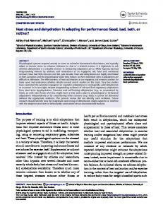
Fig. 1. Water content of Austrofundulus limnaeus embryos exposed to 50, 75.5, and 85% relative humidity (RH) for 113 days. Symbols are means ⫾ SE (n ⫽ 3).
and area normalized (18) using Grams v.4.04 software (Bomem). Far-UV-CD spectroscopy. Far-UV-CD spectra were obtained at 25°C with an Aviv 62DS CD spectrometer and thermoelectric temperature-control unit (Lakewood, NJ). Samples were placed in a quartz cell with a 1-mm path length. CD spectra were obtained for two samples of solubilized egg envelopes isolated from unfertilized eggs. Protein concentration in the samples was 0.35–0.50 mg/ml in 10 mM sodium phosphate (pH 7). Background spectra were recorded under identical conditions and subtracted from the protein spectrum. Congo red staining. Congo red staining was performed on egg envelope aggregates formed in vitro (see MATERIALS AND METHODS) and on homogenized/sonicated egg envelopes isolated from unfertilized eggs and fertilized embryos (11 h postfertilization). Samples were applied to a glass microscope slide, allowed to air dry, and stained with Congo red (27). The slides were incubated at room temperature for 20 min in a saturated solution of NaCl in 80% ethanol and 0.1% NaOH. This step was followed by a 50-min incubation in a saturated solution of Congo red and NaCl prepared in 80% ethanol and 0.1% NaOH. The slides were rinsed with three changes of 100% ethanol, air dried, and the stained tissue was mounted under a glass coverslip with acrylic mounting medium. Stained slides were observed using a light microscope with crossed polarization filters at 630⫻ magnification. Red staining under bright-field optics and green birefringence under crossed polarizers were considered diagnostic for amyloid protein structure (2, 27). Electron microscopy. Protein aggregates formed in vitro and homogenates of egg envelopes (as prepared above for Congo red staining) were applied to formvar and carbon-
Evaporative water loss and survival. Embryos of A. limnaeus survive desiccating conditions by substantially reducing water loss and retaining a large portion of their total water (Fig. 1). Diapause II embryos are the most resistant developmental stage to water loss and retain ⬃50% of their initial water even after 32 days of exposure to dehydrating conditions in 50% RH. Embryos in the dispersion and reaggregation (DR) stage of development and diapause III embryos are almost completely dehydrated after only 2 days of exposure to 50% RH (Fig. 1). At 75.5% RH (Fig. 1), diapause II embryos display a similar pattern of water retention to that seen at 50% RH. Diapause III embryos are essentially dehydrated after 8 days, and DR-stage embryos have an intermediate ability to resist water loss. The latter embryos retain ⬃50% of their water during the first 8 days of exposure and are not completely dehydrated until day 16. Finally, at 85% RH, both DR-stage and diapause II embryos retain ⬃60% of their water, whereas diapause III embryos retain 65% of their water during 37 days of exposure. It is appropriate to note that the water retained by diapause II embryos exposed for 8 days to 75.5% RH is freezable and, therefore, likely to be bulk water. DSC indicates a large heat-flow signal near 0°C during warming of samples previously cooled to ⫺40°C, which indicates the melting of water that froze during cooling (Fig. 2). The rate of water loss is initially high in diapause II embryos exposed to all levels of RH (Fig. 3). Maximal rates ranged from 0.018 to 0.035 g H2O 䡠 g⫺1 dry mass 䡠 h⫺1. After the initial 4 days of exposure, water loss is reduced to extremely low but nonzero values (t-test, using 0 as the parametric value, df ⫽ 2, P ⬍ 0.05). After 32 days of exposure, water loss is indistinguishable from zero (t-test as above). The less dehydration-resistant embryos (DR stage and diapause III)
Fig. 2. Differential scanning calorimetry indicates a large endothermic event near 0°C indicative of melting water in diapause II embryos of Austrofundulus limnaeus subjected to 8 days of dehydration stress at 75.5% RH.
R126
DEHYDRATION AVOIDANCE IN EMBRYOS OF AN ANNUAL KILLIFISH
Fig. 3. Rate of water loss in diapause II embryos exposed to environmental dehydration stress. Rates are indistinguishable from 0 after 32 days of exposure (t-test, using 0 as the parametric value, df ⫽ 2, P ⬍ 0.05). Symbols are means ⫾ SE (n ⫽ 3).
continue to lose water at a significant rate until dehydrated (data not shown). The initial and rapid loss of water in diapause II embryos is thought to come primarily from the perivitelline fluid. This suggestion is substantiated by the observation that the volume of the embryonic tissues and yolk do not change appreciably during the initial phase of water loss (Table 1), whereas the perivitelline space, as observed microscopically, is greatly reduced (Fig. 4). Calculations indicate that loss of water from the perivitelline space can Table 1. Diameter of diapause II embryos in aqueous medium vs. dehydrating conditions (75.5% RH) Incubation Condition
Diameter
Aqueous medium, whole embryo (i.e., egg envelope, perivitelline space, embryo tissue, and yolk) Aqueous medium (embryo and yolk only) 75.5% RH, whole embryo
1.69 ⫾ 0.032* 1.52 ⫾ 0.025† 1.56 ⫾ 0.031†
Values are means ⫾ SE (n ⫽ 10 embryos). Diameter in mm. Diameter was determined by measuring the diameter of each embryo twice and then averaging the values. The second measurement was always made perpendicular to the first. Means with the same symbol are not statistically different (Fisher’s protected least-significant difference; P ⬍ 0.05). Embryos exposed to 75.5% relative humidity (RH) for 5 days.
Fig. 4. Photomicrographs of diapause II embryos of Austrofundulus limnaeus incubated in aqueous embryo medium (A) and after 5 days of dehydration in 75.5% RH (B). Note the lack of the perivitelline space (PVS) in the embryo exposed to desiccating conditions (B). E, embryo; O, oil droplet; Y, large yolk mass. The outer edge of the egg envelope (EV) and the PVS in A are indicated.
account fully for the initial water loss on exposure to desiccating conditions (see DISCUSSION). For all stages of development, survival of water stress is highest in 85% RH, intermediate in 75.5% RH, and lowest in 50% RH (Fig. 5). Diapause II embryos have the highest survivorship, and 40% of these embryos survive for over 100 days at 75.5% RH. Diapause III embryos show the least resistance and all die after 8 days at 75.5% RH, whereas DR-stage embryos are all dead after 16 days of exposure. Embryos placed on filter pads preequilibrated to the various RH conditions (but not moistened as in the above experiments) always died within 24–48 h of exposure (data not shown). Thus embryo contact with a wetted surface early in the dehydration experiments is required to obtain the survivorship data reported here. Protein structure of the egg envelope. The dry mass of the egg envelope does not change as a result of fertilization and hardening nor during subsequent development (ANOVA, P ⬎ 0.05; Table 2). Approximately 80% of the dry weight of an unfertilized egg envelope is soluble protein in 8 M guanidine HCl (Table 2). As judged by SDS-PAGE, two major proteins (34 and 24 kDa) are extracted in this manner (Fig. 6). Only ⬃10%
DEHYDRATION AVOIDANCE IN EMBRYOS OF AN ANNUAL KILLIFISH
Fig. 5. Survival of embryos of Austrofundulus limnaeus during exposure to dehydrating conditions for 113 days. Symbols are means ⫾ SE (n ⫽ 3).
of the egg envelope dry weight is soluble protein after hardening, even in the presence of both 8M guanidine HCl and 5% -mercaptoethanol (Table 2). Furthermore, the solubility does not differ among prediapause II, diapause II, and diapause III embryos. To investigate the secondary structure of the proteins in homogenates of egg envelopes we chose IR spectroscopy, which can be used to investigate proteins in any state, including those with large insoluble particles (10). For egg envelopes isolated from unfertilized
R127
Fig. 6. SDS-PAGE gel of solubilized egg envelope proteins. Two major protein bands are observed at 34 and 24 kDa. Stds, standards; me, -mercaptoethanol.
eggs and hardened embryos (11 h postfertilization), the second-derivative IR spectra in the conformationally sensitive amide I region were very similar (Fig. 7). The deconvoluted spectra indicate that the egg envelope proteins of A. limnaeus contain mostly -sheet (47%) and turns (30%; Fig. 8, Table 3) with a small fraction of ␣-helix (10%). Surprisingly, the spectra also exhibit bands at 1,613 and 1,690/cm, which indicate that the egg envelope contains 14% intermolecular -sheet (10). This structure is often found in nonnative aggregated states of proteins that are formed by perturbing condi-
Table 2. Dry mass and solubility of the egg envelope do not change as fertilized embryos develop
Stage
DPF
Dry Mass, g/egg envelope
Unfertilized Prediapause II Diapause II Diapause III
0 20 42 33–116
41.6 ⫾ 1.7* 44.2 ⫾ 2.3* 45.7 ⫾ 0.94* 46.6 ⫾ 2.6*
%Solubilitya
%Solubilityb
76 ⫾ 14† 9.4 ⫾ 1.3* 8.8 ⫾ 2.8* 8.0 ⫾ 2.5*
80 ⫾ 8.9† 11 ⫾ 1.1* 9.9 ⫾ 2.3* 9.3 ⫾ 2.8*
Values are means ⫾ SE; n ⫽ 3. Age of embryos is given in days postfertilization (DPF) at the time egg envelopes were isolated. a Percentage of egg envelope dry mass that is soluble in 8 M guanidine HCl. b Percentage of egg envelope dry mass that is soluble in the presence of 8 M guanidine HCl containing 5% -mercaptoethanol. Means with the same symbol are not statistically different (Tukey’s honestly significant difference, P ⬍ 0.05).
Fig. 7. Second-derivative infrared (IR) spectra in the amide-I region of isolated egg envelopes from unfertilized eggs, fertilized embryos (11 h postfertilization), and in vitro aggregates of egg envelope proteins. dI2(dx2)⫺1, Second-derivative of infrared absorbance.
R128
DEHYDRATION AVOIDANCE IN EMBRYOS OF AN ANNUAL KILLIFISH
Fig. 8. Deconvoluted second-derivative IR spectra of hydrated (top) and dehydrated (bottom) egg envelopes isolated from fertilized embryos 11 h postfertilization (posthardening).
tions in vitro [e.g., thermally induced precipitation, (10)] or pathological processes in vivo causing amyloid fibril formation (19, 22). To determine the macroscopic structure associated with the intermolecular -sheet, we investigated egg envelope homogenates with transmission electron microscopy (TEM). The micrographs (Fig. 9) revealed bundles of unbranched fibrillar structure with a diameter of ⬃4–6 nm and indeterminate length. This structure is a hallmark of amyloid fibrils (22). Another diagnostic test for these fibrils is green birefringence of Congo red-stained fibrils under cross-polarized light (22, 30). Remarkably, the fibrils from egg envelopes display this staining behavior (Fig. 10) and appear similar to typical pathological fibrils formed in disease processes such as Alzheimer’s disease (22) and systemic amyloidosis (30). Table 3. Protein 2° structure of hydrated and dried egg envelopes Hydrated 2° Structure
Intermolecular -sheet Turn Turn ␣-Helix Intramolecular -sheet Intermolecular -sheet
Dried
Wavenumber, per cm
% Structure
Wavenumber, per cm
% Structure
1,690 1,680 1,667 1,653
6 15 14 10
1,694 1,681 1,673 1,658
29 6 6 21
1,635
47
1,631
31
1,613
8
1,613
7
Wavenumber (per cm) assignments for secondary structure are based on data from Ref. 7. 2°, Secondary.
Fig. 9. Transmission electron micrographs (⫻100,000) illustrating the fibrillar structure of egg envelope protein aggregates formed in vitro (top) and in vivo (bottom). The scale bar is equal to 100 nm. Individual fibers are ⬃4–6 nm in diameter with indeterminate length.
To test whether amyloid fibrils from egg envelope proteins could form in vitro, we solubilized isolated egg envelopes in 8 M guanidine HCl and removed the denaturant by dialysis. This process often fosters aggregation and precipitation of proteins during refolding procedures (11). Over 80% of the egg envelope protein precipitated during dialysis to remove the denaturant. The IR spectra of the precipitates were similar to those of the original egg envelope homogenates and again confirmed the presence of a substantial fraction of intermolecular -sheet structure (Fig. 7). Furthermore, in these in vitro precipitated preparations, both Congo red staining and TEM showed the presence of amyloid fibrils that were virtually indistinquishable from those in egg envelope homogenates (Figs. 9 and 10). The secondary structure of the soluble protein (comprised of 2 major proteins; Fig. 6) remaining after dialysis was studied with far-UV-CD spectroscopy. This method was chosen because it can be used with relatively low protein concentrations compared with the requirements of IR spectroscopy. The far-UV-CD spectra (Fig. 11) have a prominent minimum ellipticity at 205 nm, which indicates that the proteins are mostly random coil (23).
DEHYDRATION AVOIDANCE IN EMBRYOS OF AN ANNUAL KILLIFISH
R129
tered relative to that for the hydrated proteins (Figs. 8 and 12). There is a large increase in bands associated with intermolecular -sheet at 1,613 and 1,694/cm, concomitant with reduction in absorbances for turn and intramolecular -sheet (Table 3). Rehydration of the envelopes fully reversed these structural alterations (Fig. 12). DISCUSSION
Fig. 10. Light microscopy of egg envelope homogenates and in vitro aggregates, both stained with Congo red. Photographs on the left were obtained with bright-field optics, whereas the images on the right were obtained with cross polarization. Protein aggregates formed in vitro from precursors previously solubilized in 8 M guanidine HCl (top). Egg envelopes isolated from unfertilized eggs (middle). Egg envelopes isolated from fertilized (hardened) embryos at 11 h postfertilization (bottom). Magnification is ⫻630.
Considering the potential role of the egg envelope in desiccation resistance of the embryos, we next investigated the effects of drying over phosphorous pentoxide on the secondary structure of the envelope proteins. The IR spectrum of the dried sample was grossly al-
Fig. 11. Circular dichroism spectra of soluble proteins from the egg envelope indicate random coil structure. Protein concentration of samples was 0.35–0.5 mg/ml. The soluble proteins are a mixture of 2 major and a few minor proteins as illustrated in Fig. 6.
Survival of environmental dehydration. Survival of A. limnaeus embryos under severely desiccating conditions is achieved by a large reduction in evaporative water loss (Fig. 1). A slow transition from wet to dry is required for survival and may indicate that the embryos make biochemical or physiological adjustments that foster water retention and survival. All developmental stages of embryos of A. limnaeus investigated in this study appear to possess a substantial ability to reduce water loss when faced with dehydrating conditions of 85% RH. This suggests that short-term or moderate drying of the soil may be tolerated by diapausing as well as developing embryos. However, diapause II embryos are the most resistant stage to water stress, and 40% survive for over 100 days at 75.5% RH. These data indicate that diapause II embryos are likely the life history stage responsible for long-term survival of environmental dehydration and the persistence of populations of A. limnaeus in ephemeral habitats. Retention of cellular water during environmental dehydration. Considering the severely dehydrating conditions to which the embryos of A. limnaeus are likely exposed in nature, retention of water was an unanticipated mechanism for survival. Surface soils (⬍0.5 m) in desert regions can become severely dried. Soils from an ephemeral stream bed in the Chihuahuan desert have been reported to be extremely dry, with water potentials reaching ⫺13 to ⫺16 MPa after long periods (mo) without rain (28). Many amphibians and fish that inhabit xeric or ephemeral environments have mechanisms that reduce water loss (see introduction), but these animals can only resist water loss under relatively mild dehydration stresses. In contrast, diapause II embryos of A. limnaeus can retain water under extremely dehydrating conditions for ⬎113 days at
Fig. 12. Second-derivative IR spectra of hydrated and dehydrated egg envelopes isolated from diapause II embryos. Changes in secondary structure caused by dehydration are reversible on rehydration.
R130
DEHYDRATION AVOIDANCE IN EMBRYOS OF AN ANNUAL KILLIFISH
25°C. Such high resistance to desiccation is unprecedented among aquatic vertebrates. Embryos lose ⬃50% of their water during the initial period of dehydration (Fig. 1), but importantly, this water does not appear to be associated with embryonic tissues or yolk. Using an average egg diameter of 1.73 ⫾ 0.026 mm and an average yolk diameter of 1.46 ⫾ 0.022 mm (25), we estimated the volume of the perivitelline space to be 1.082 l. This value corresponds to ⬃0.76 mg of water when one considers that 70% of the perivitelline fluid is water and the balance is solutes, such as proteins and salts for example (14). At 75.5% RH, diapause II embryos have lost on average ⬃0.76 mg of water/embryo after 16 days of exposure to dehydrating conditions. This value represents 100% of the estimated water in the perivitelline space. Therefore, it is probable that all of the water lost during the initial phases of dehydration is from the perivitelline space and not from the embryo or yolk. This interpretation would explain why the water content of diapause II embryos always approaches the same value over a large range of dehydration pressures. The large amount of freezable water present in embryos exposed to 8 days of dehydration at 75.5% RH (Fig. 2) also suggests a hydrated embryonic compartment in diapause II embryos. At 113 days of exposure to 75.5% RH, only 75% of the water loss is explained by the perivitelline space. Thus the smaller amounts of water lost after 16 days must come from the embryonic/yolk compartment. Mechanisms for water retention. The unchanged structure of the egg envelope among DR-stage, diapause II, and diapause III embryos suggests that mechanisms other than changes in the egg envelope proteins must be involved in the reduced water loss observed in diapause II embryos. Damage to the egg envelope during water stress, either mechanically or by fungal infection, causes rapid water loss and death of embryos (data not shown). Thus the egg envelope is required for survival under desiccating environmental conditions, but it does not appear to be solely responsible for resistance to water loss. Dehydration greatly increased the intermolecular -sheet content of the egg envelope proteins, a process that was reversible on rehydration (Fig. 12). These -sheet structures are indicative of increased intermolecular contacts and interactions (2, 9, 11, 22). Dehydration-induced increases in intermolecular interactions could serve to decrease the water permeability of the egg envelope. Further investigation of the permeability of isolated egg envelopes in hydrated and dehydrated conditions may clarify the role of the structural changes in the egg envelope during dehydration. Loss of perivitelline-space water and retention of embryonic water suggests that the plasma membranes of the enveloping cell layer may be important in the retention of water during environmental dehydration in embryos of A. limnaeus. The permeability of the plasma membranes of early fish embryos to water is among the lowest reported for aquatic animals (13). However, the low permeability to water and ions in
typical fish embryos is obviously not sufficient to retard evaporative water loss to the degree we observe in A. limnaeus; otherwise, we would expect survival of desiccating conditions to be more common among fish embryos. Secretion of substances into the perivitelline space (either at fertilization or during subsequent development) could play a role in the dehydration resistance of diapause II embryos of A. limnaeus. The perivitelline space is known to have colloidal properties (21) that are thought to be responsible for the accumulation of a substantial internal hydrostatic pressure in eggs of Fundulus on fertilization (17). The perivitelline space of fish embryos contains significant amounts of protein (20% by weight) (14), lipid (5% by weight) (14), polysaccharides and mucopolysaccharides (21), and polysialoglycoproteins (20). It is possible that certain constituents of the perivitelline space of diapause II embryos prevent water loss by creating a hydrophobic barrier on drying. It is also possible that the constituents of the perivitelline space may vitrify (form a glass) (4) when severely desiccated, thus substantially slowing water loss from the embryonic and yolk compartments. The later possibility is supported by observations that the outer layers of the embryo (egg envelope and perivitelline space) become very brittle and glasslike after the initial dehydration period is complete (Fig. 4), whereas the internal compartments (yolk and embryonic cells) appear to remain relatively hydrated as demonstrated by DSC (Fig. 2). Amyloid protein structure of the egg envelope. An unexpected result of the current study was the observation that the egg envelope of A. limnaeus is composed of proteins that share many characteristics of amyloid fibrils that are associated with numerous human diseases (3, 22). This conclusion is substantiated by three major pieces of evidence: 1) positive staining and green birefringence observed with Congo red (Fig. 10), 2) the fibrillar structure of the aggregates demonstrated with TEM (Fig. 11), and 3) the characteristic intermolecular -sheet secondary structure (Figs. 5, 8, 9). To our knowledge this is the first report of a nonpathological amyloid fibril. Sequence analyses and in vitro studies with synthetic peptide fragments suggest that the OsmB protein from Escherichia coli has the potential to form fibrils (16), although in vivo amyloidlike fibrils have not been observed. In A. limnaeus and other fish embryos, this fibrillar structure appears to protect against mechanical disruption as well as to serve as a barrier to polyspermy, microbes, low molecular weight solutes, and, in the case of A. limnaeus, perhaps water. We have found that the soluble protein from the egg envelope is predominantly composed of random coil. Furthermore, on the basis of our in vitro results, the egg envelope proteins show a propensity to form amyloid fibrils instead of amorphous precipitates. Thus all that may be needed to foster fibril formation in the maternal ovaries is a sufficient concentration of precursor protein molecules. It is well established for many protein-aggregation, precipitation, and fibrilforming processes that the rate and extent correlates directly with protein concentration (11).
DEHYDRATION AVOIDANCE IN EMBRYOS OF AN ANNUAL KILLIFISH
Diapause II embryos of A. limnaeus can survive under extremely dehydrating conditions by reducing evaporative water loss and retaining cellular water. The protein secondary structure, dry mass, and solubility of the egg envelope do not change during development and therefore do not correlate with increased resistance to environmental dehydration in diapause II embryos. It appears that additional mechanisms such as vitrification of the perivitelline space must be important for reduction of evaporative water loss during exposure to water stress in diapause II embryos of A. limnaeus. NOTE ADDED IN PROOF
Recently, amyloid fibrils have been reported to play a protective role in the chorion of the silkmoth (Bombyx mori) embryo (Iconomidou VA, Vriend G, and Hamodrakas SJ. Amyloids protect the silkmoth oocyte and embryo. FEBS Lett 479: 141–145). We thank Yong-sung Kim for assistance with CD measurements and Dr. Suchart Chonprasert for help with DSC. The efforts of Natalia Gomez and Tom Giddings in preparing the TEM samples are greatly appreciated. This work was supported by National Science Foundation Grants IBN-9723746 to S. C. Hand and BES-9816975 to J. F. Carpenter. Present address for S. C. Hand: Department of Biological Sciences, Louisiana State University, Baton Rouge, LA 70803. REFERENCES 1. Bayley M. and Holmstrup M. Water vapor absorption in arthropods by accumulation of myoinositol and glucose. Science 285: 1909–1911, 1999. 2. Chiti F, Webster P, Taddei N, Clark A, Stefani M, Ramponi G, and Dobson CM. Designing conditions for in vitro formation of amyloid protofilaments and fibrils. Proc Natl Acad Sci USA 96: 3590–3594, 1999. 3. Conway KA, Harper JD, and Lansbury PT Jr. Fibrils formed in vitro from ␣-synuclein and two mutant forms linked to Parkinson’s Disease are typical amyloid. Biochemistry 39: 2552– 2563, 2000. 4. Crowe JH, Carpenter JF, and Crowe LM. The role of vitrification in anhydrobiosis. Annu Rev Physiol 60: 73–103, 1998. 5. Crowe JH and Clegg JS (Eds.). Anhydrobiosis. Stroudsburg: Dowden, Hutchinson and Ross, 1973, 1–477. 6. Crowe JH and Clegg JS (Eds.). Dry Biological Systems. New York: Academic, 1978, 1–357. 7. Dong A and Caughey WS. Infrared methods for study of hemoglobin reactions and structures. Methods Enzymol 232: 139–175, 1994. 8. Dong A, Huang P, and Caughey WS. Protein secondary structures in water from second-derivative amide I infrared spectra. Biochemistry 29: 3303–3308, 1990. 9. Dong A, Matsuura J, Manning MC, and Carpenter JF. Intermolecular -sheet results from trifluorethanol-induced nonnative ␣-helical structure in -sheet predominant proteins: infrared and circular dichroism spectroscopic study. Arch Biochem Biophys 355: 275–281, 1998. 10. Dong A, Prestrelski SJ, Allison SD, and Carpenter JF. Infrared spectroscopic studies of lyophilization- and temperature-induced protein aggregation. J Pharm Sci 84: 415–424, 1995. 11. Fink AL. Protein aggregation: folding aggregates, inclusion bodies and amyloid. Folding & Design 3: R9–R23, 1998. 12. Fishman AP, Galante RJ, Winokur A, and Pack AI. Estivation in the African Lungfish. Proc Am Philos Soc 136: 61–72, 1992. 13. Guggino WB. Water balance in embryos of Fundulus heteroclitus and F. bermudae in seawater. Am J Physiol Regulatory Integrative Comp Physiol 238: R36–R41, 1980. 14. Hamor T and Garside ET. Quantitative composition of the fertilized ovum and constituent parts in the Atlantic salmon Salmo salar L. Can J Zool 55: 1650–1655, 1977.
R131
15. Hoekstra FA, Wolkers WF, Buitink J, Golovina EA, Crowe JH, and Crowe LM. Membrane stabilization in the dry state. Comp Biochem Physiol 117A: 335–341, 1997. 16. Jarrett JT and Lansbury PT Jr. Amyloid fibril formation requires a chemically discriminating nucleation event: studies of an amyloidogenic sequence from the bacterial protein OsmB. Biochemistry 31: 12345–12352, 1992. 17. Kao C-Y and Chambers R. Internal hydrostatic pressure of the Fundulus egg I. The activated egg. J Exp Biol 31: 139–149, 1954. 18. Kendrick BS, Dong A, Allison SD, Manning MC, and Carpenter JF. Quantitation of the area of overlap between secondderivative amide I infrared spectra to determine the structural similarity of a protein in different states. J Pharm Sci 85: 155–158, 1996. 19. Kim Y-S, Wall JS, Meyer J, Murphy C, Randolph TW, Manning MC, Solomon A, and Carpenter JF. Thermodynamic modulation of light chain amyloid fibril formation. J Biol Chem 275: 1570–1574, 2000. 20. Kitajima K, Inoue Y, and Inoue S. Polysialoglycoproteins of Salmonidae fish eggs. Complete structure of 200-kDa polysialoglycoprotein from the unfertilized eggs of rainbow trout (Salmo gairdneri). J Biol Chem 261: 5262–5269, 1986. 21. Laale HW. The perivitelline space and egg envelopes of bony fishes: a review. Copeia 1980: 210–226, 1980. 22. Lansbury PT Jr. Evolution of amyloid: what normal protein folding may tell us about fibrillogenesis and disease. Proc Natl Acad Sci USA 96: 3342–3344, 1999. 23. Manning MC. Underlying assumptions in the estimation of secondary structure content in proteins by circular dichroism spectroscopy–a critical review. J Pharm Biomed Anal 7: 1103– 1119, 1989. 24. Mayhew WW. Adaptations of the amphibian, Scaphiopus couchi, to desert conditions. Am Mid Nat 74: 95–109, 1965. 25. Podrabsky JE. Husbandry of the annual killifish Austrofundulus limnaeus with special emphasis on the collection and rearing of embryos. Env Biol Fish 54: 421–431, 1999. 26. Podrabsky JE and Hand SC. The bioenergetics of embryonic diapause in an annual killifish, Austrofundulus limnaeus. J Exp Biol 202: 2567–2580, 1999. 27. Puchtler H, Sweat F, and Levine M. On the binding of congo red by amyloid. J Histochem Cytochem 10: 355–364, 1962. 28. Scanlon BR. Water and heat fluxes in desert soils 1. Field studies. Water Resour Res 30: 709–719, 1994. 29. Simpson BRC. The phenology of annual killifishes. Symp Zool Soc Lond 44: 243–261, 1979. 30. Solomon A, Weiss DT, Murphy CL, Hrncic R, Wall JS, and Schell M. Light chain-associated amyloid deposits comprised of a novel k constant domain. Proc Natl Acad Sci USA 95: 9547– 9551, 1998. 31. Sugiyama H, Murata K, Iuchi I, and Yamagami K. Evaluation of solubilizing methods of the egg envelope of the fish, Oryzias latipes, and partial determination of amino acid sequence of its subunit protein, ZI-3. Comp Biochem Physiol 114B: 27–33, 1996. 32. Taphorn DC and Thomerson JE. A revision of the South American cyprinodont fishes of the genera Rachovia and Austrofundulus, with the description of a new genus. Acta Biol Venez 9: 377–452, 1978. 33. Toledo RC and Jared C. Cutaneous adaptations to water balance in amphibians. Comp Biochem Physiol 105A: 593–608, 1993. 34. Winston PW and Bates DH. Saturated solutions for the control of humidity in biological research. Ecology 41: 232–237, 1960. 35. Withers PC and Guppy M. Do Australian desert frogs coaccumulate counteracting solutes with urea during aestivation? J Exp Biol 199: 1809–1816, 1996. 36. Womersley C and Ching C. Natural dehydration regimes as a prerequisite for the successful induction of anhydrobiosis in the nematode Rotylenchulus reniformis. J Exp Biol 143: 359–372, 1989. 37. Wourms JP. The developmental biology of annual fishes III. Pre-embryonic and embryonic diapause of variable duration in the eggs of annual fishes. J Exp Zool 182: 389–414, 1972.