tissues makes it a useful molecular marker in cancer ... November 09, 2011, IP: 92.42.55.140] || Click here to download free Android application for this · journal ...
[Downloaded free from http://www.cancerjournal.net on Wednesday, November 09, 2011, IP: 92.42.55.140] || Click here to download free Android application for this journal
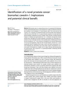
Original Article
Survivin-deltaEx3: A novel biomarker for diagnosis of papillary thyroid carcinoma ABSTRACT Context: The most important problem in the case of thyroid nodules is the lack of suitable criteria for detecting malignant thyroid tumors from other nodules in the early stage. Variable expressions level of survivin, an inhibitory protein in apoptotic pathway, and its splice variants in malignant carcinoma versus well-differentiated normal tissues candidate them as reliable biomarkers in cancers. Aim: To semi-quantitative detection of survivin and its splice variant, survivin-deltaEx3, in thyroid nodules. Setting and Design: We evaluated the expression level of mentioned biomarkers in thyroid nodules including carcinoma. Materials and Methods: Samples were collected from 61 thyroid nodules including malignant, adenoma, non-tumoral (goiter and thyroidities) as well as non-neoplastic normal tissues. Transcriptional levels were measured using semi-quantitative reverse transcriptasepolymerase chain reaction (RT-PCR) and the results were normalized to 2microglubin (2m) gene. Statistical Analysis Used: Independent sample t-test was used to assess the significant variation of expression between different groups. Result: Our data for a first time revealed that survivin-deltaEx3 is significantly up-regulated from normal to malignant thyroid carcinoma tissues (approximately ten fold). Conclusion: High expression level of survivin and survivin-deltaEx3 in malignant papillary thyroid carcinoma suggested survivin gene expression and its splice variant, survivin-deltaEx3, can be potential new markers in diagnosis of human papillary thyroid carcinoma. KEY WORDS: Malignant carcinoma, reverse transcriptase-polymerase chain reaction, semi quantitative polymerase chain reaction, splice variant, survivin
INTRODUCTION Thyroid cancer, the most common type of endocrine malignancy, accounts for the majority of endocrine cancer related to death each year.[1] Well-differentiated thyroid cancers arise from follicular cells and encompasses papillary, follicular and hurthle carcinomas. Other histological types of thyroid cancer are medullary and anaplastic.[2,3] Papillary thyroid cancer is the most common type of thyroid tumors, making up about 70-80% of all thyroid cancers, and can occur at any age.[4] Involvement of the lymph node is relatively common in papillary carcinoma and lymphatic spread is the major target of the metastasis.[5] Because of the heterogeneous nature of tumoral and non-tumoral thyroid nodules as well as the lack of suitable clinico-pathological parameters, diagnostic criteria for thyroid cancer are highly variable.[6] Therefore, finding any appropriate molecular marker involved in thyroid cancer will be useful in exact diagnosis of malignant tumors, especially managing appropriate treatments.[7,8] As known, derangement in apoptosis would be involved in tumorigenesis.[9] Apoptosis has been identified as an intricate and critical mechanism for active cell elimination and tissue
homeostasis. Defects in apoptosis pathway may lead to uncontrolled cell proliferation and tumorigenesis. Therefore, inhibitors of apoptosis can be involved in tumorigenesis and cancer progression.[10] Survivin is a member of the inhibitor of apoptosis protein family (IAPs) that has attracted attentions from several viewpoints of research. It plays a key role in the regulation of apoptosis and cell division. Five different variants are produced from survivin pre-mRNA during alternative splicing process. In addition to wild type of survivin, two survivin isoforms (i.e., survivin-2B and survivin-deltaEx3) are generated by insertion of an alternative exon 2 and removal of exon 3, respectively.[11-13] The other isoforms have been identified as survivin-3B and survivin-2.[14] Survivin is abundantly expressed both during normal fetal development and in a broad spectrum of human cancers but is barely detectable in normal well differentiated tissues.[11] The differential expression of survivin in some types of cancers versus normal differentiated tissues makes it a useful molecular marker in cancer diagnosis and a promising therapeutic target.[15-17]
Somayeh Vandghanooni1,2, Morteza Eskandani2, Vahid Montazeri3, Monireh Halimi3, Esmaeil Babaei1,4, Mohamad Ali Hosseinpour Feizi1 1 Department of Biology-Genetics, University of Tabriz, 2 Research Center for Pharmaceutical Nanotechnology, Student Research Committee, Tabriz University of Medical Sciences, 3Pathology Department, Imam Khomeini Hospital, Tabriz University of Medical Sciences, Tabriz, 4Department of Genetics, Tarbiat Modares University, Tehran, Iran
For correspondence: Dr. Babaei, Dr. Hosseinpour Feizi, Department of Biology-Genetics, University of Tabriz, P.O. Box 5166616471, Tabriz, Iran. E-mail: e_babaei@ madarres.ac.ir
Access this article online Website: www.cancerjournal.net DOI: 10.4103/0973-1482.87038 PMID: *** Quick Response Code:
In the present study, the expression level of survivin and its splice variant, survivin-deltaEx3, were
Journal of Cancer Research and Therapeutics - July-September 2011 - Volume 7 - Issue 3
325
[Downloaded free from http://www.cancerjournal.net on Wednesday, November 09, 2011, IP: 92.42.55.140] || Click here to download free Android application for this journal Vandghanooni, et al.: Novel biomarker for thyroid carcinoma
evaluated in 61 specimens classified in 4 groups; papillary thyroid carcinoma, benign tumors, non-tumoral nodules including goiter and thyroidities and non-neoplastic samples obtained from margin of carcinoma specimens. However the aim of this study is investigation of the expression levels of survivin and Survivin-deltaEx3 in thyroid nodules and evaluation of their value as diagnostic markers in papillary thyroid carcinomas.
classified as malignant tumors, benign tumors, non-tumoral nodules including goiter and thyroiditis and finally adjacent non-neoplastic tissues for obtaining normal tissues from each patients [Figure 1]. Clinico-pathological diagnosis of malignant papillary carcinomas was based on the some features of the cells such as neoplasticity with glass appearance nuclear and papillae formation.
MATERIALS AND METHODS
For RT-PCR reaction, total RNA was extracted with the RNeasy Micro Kit (Qiagen, Hilden, Germany) exactly according to the manufacturer’s instructions. However, the integrity of the extracted RNA was evaluated by agarose gel electrophoresis and purity of RNA was examined by optical density measurement (A260/A280 ratio).
The research was conducted in accordance with the Helsinki Declaration and research was approved by the ethics committee of hospital and the University of Tabriz. Moreover, informed consent was obtained from all patients undergoing surgery. All the specimens were obtained in surgical operation from Imam Khomeini hospital, Tabriz, Iran. Biopsies were snap frozen in liquid nitrogen and stored at 80ºC until required for Ribonucleic acid (RNA) extraction. The histopathological analyses were performed according to the World Health Organization[18] and tissue samples were
The same amount of each extracted RNA sample (5 μg) was used for cDNA synthesis using Oligo-dT primers and revert AidTMMMulv Reverse transcriptase (fermentase co. Lituania). Thereafter, two rounds of PCR reaction were performed to amplify survivincDNA (accession number: NM-001168). First round of PCR was performed in a final volume of 50μl on
Figure 1: Histological staining of 4 categorized groups of thyroid tissues, (a) non-cancerous samples collected from margin of malignant tumors, it is appear that in this case the histological features of samples because of its follicles (a 1) and the other features is normal, (b) the goiter (nontumoral thyroid tissue), Colloid rich follicles (b 1) lined by flatten, inactive epithelium and areas of follicular epithelial hypertrophy and hyperplasia (b 2) is seen in this transversal section, (c) adenoma tissues (benign), cellular areas (c 1) with few or no follicles formed, the size of the follicles approaches that of non-neoplastic glands, the tumor cells are polygonal with normochromatic and round to oval nucleoli (c 2) is the highest features of this slide, d: papillary thyroid tissues, Papillae (d 1) formed by a central fibrovascular stalk and covered by neoplastic epithelial cells (d 2) and Pale, clear, empty or ground glass appearance: empty of nucleus with irregular thickened inner aspect of nuclear membrane (d 3) 326
Journal of Cancer Research and Therapeutics - July-September 2011 - Volume 7 - Issue 3
[Downloaded free from http://www.cancerjournal.net on Wednesday, November 09, 2011, IP: 92.42.55.140] || Click here to download free Android application for this journal Vandghanooni, et al.: Novel biomarker for thyroid carcinoma
Figure 2: Determining the optimum number of polymerase chain reaction cycles for survivin variants and 2m amplification. A mixed Ribonucleic acid (RNA) from six tumor, three non-tumor and three apparently normal thyroid tissues (1 μg from each sample) has been prepared, reverse transcribed and subjected to a first round of polymerase chain reaction amplification for 35 cycles and a nested polymerase chain reaction amplification for 15-35 cycles for survivin variants and 20-40 cycles for 2m
Figure 3: Reverse transcriptase-polymerase chain reaction analysis of survivin and its variant in malignant, benign, non-tumoral and noncancerous samples obtained from margin of carcinoma P.C: Positive control (breast cancer tissues that previously had showed high expression level of survivin and survivin-deltaEx3, used as a positive control), N.C: Negative control (distilled water used instead of cDNA in polymerase chain reaction), M: Molecular marker, MT: Malignant tumor, BT: Benign tumor, NT: Non tumoral samples and NN: Non-neoplastic samples obtained from Margin of Carcinoma
Techne thermal cycler using HFP 5-TGGCAGCCCTTTCTCAAG-3 (as forward primer) and HRP 5- GAAGAAACACTG GGCCAA G-3 (as reverse primer). The reaction was performed as initial denaturation at 94C for 120 s, followed by 35 cycles denaturation at 94C for 30 secs, annealing for at 57C for 30secs, extension at 72C for 60secs and a final extension at 72C for 5 mins. Second round of PCR was performed similar to the one described for the first round except for using 28 cycles of reactions as well as internal: HFPN 5-ACCACCGCATCTCTACATTC-3 (as forward primer) and HRPN 5-GTTCCTCTA TGGGGTCGTC-3 (as reverse primer). These primers amplified 556bp and 438bp segments for survivin and surviving-deltaEx3, respectively. 2m (accession number: NM_004048) was amplified with identical conditions of first round of PCR of survivin except for using specific primers including: HBF 5-CTACTCTCTCTTTCTGGCCTG-3 (as forward primer) and HBR 5-GACAAGTCTGAATGCTCCAC-3 (as reverse primer), as well as 30 cycles of reaction. These primers amplified a 191bp segment from 2m cDNA. In this study, to quantifying PCR reaction, products were separated on a 1.5% agarose gel and visualized by ethidium
bromide staining. The amount of DNA was quantized by measuring the intensity of light emitted from corresponding bands under UV light using Labimage software (version 2.6; Kapelan GmbH Co., Germany). To ensure that equal amounts of RNA were used for each reaction and those potential differences in signal intensity were not due to differences in the amounts of starting RNA, 2m was used as an internal control for each reaction, RT-PCR was performed in separate tubes under similar conditions (except for the cycle number) for survivin, survivin-deltaEx3 and 2m with results expressed as Survivin/2m and survivin-deltaEx3/ 2m expression ratios, Also, amplified products from all sample sets were loaded onto a single agarose gel and electrophoresed. Finally, pictures were then captured under identical brightness/contrast conditions, thus maximizing the accuracy of quantification. To further confirm the accuracy of the Survivin PCR products, the amplification products were purified from the gel with a kit (Qiagen, Hilden, Germany) and sequenced on both forward and reverse direction and analyzed on a DNA sequencer (Model 3100 sequencer; ABI) according to the manufacture’s specifications, with the intermediately Neday e Fan company in Iran and matched with the NCBI survivin mRNA sequence
Journal of Cancer Research and Therapeutics - July-September 2011 - Volume 7 - Issue 3
327
[Downloaded free from http://www.cancerjournal.net on Wednesday, November 09, 2011, IP: 92.42.55.140] || Click here to download free Android application for this journal Vandghanooni, et al.: Novel biomarker for thyroid carcinoma
(NM-001168). Finally, all experiments were replicated three times and the semi quantitative numerical results were analyzed by performing independent sample t-test using SPSS 15, with P