Keren, D. F., P. J. Scott, and D. Bauer. 1980. Variables affecting the local ... McOrist, S., S. Jasni, R. A. Mackie, N. MacIntyre, N. Neef, and. G. H. K. Lawson. 1993.
JOURNAL OF CLINICAL MICROBIOLOGY, Aug. 1994, p. 1980-1985
Vol. 32, No. 8
0095-1137/94/$04.00+0 Copyright C 1994, American Society for Microbiology
Enzyme-Linked Immunosorbent Assay for Measuring Ileal Symbiont Intracellularis-Specific Immunoglobulin G Response in Sera of Pigs P. K. HOLYOAKE,* R. S. CUTLER,t I. W. CAPLE,t AND R. P. MONCKTON Regional Veterinary Laboratory, Department of Agriculture, Bendigo, Victoria 3550, Australia Received 15 February 1994/Returned for modification 6 April 1994/Accepted 29 April 1994
Proliferative enteritis (PE) is a common intestinal disease on pig farms. The disease is caused by ileal symbiont (IS) intracellularis (Campylobacter-like organism) bacteria. An enzyme-linked immunosorbent assay (ELISA) was developed to measure IS intracellularis-specific immunoglobulin G (IgG) response in the sera of pigs. The antigen used in the ELISA was filtered, percoll gradient-purified IS intracellularis extracted from the intestines of pigs affected with proliferative hemorrhagic enteropathy. The antibody responses of pigs challenged with intestinal homogenates from pigs affected with proliferative hemorrhagic enteropathy containing IS intracellularis or percoll-gradient purified IS intracellularis were low and variable. The low IgG titers measured in challenged pigs support previous findings that IgG plays a minor role in the immune response of pigs to IS intracellularis. On a farm in which infection was endemic, pigs seroconverted at between 7 and 24 weeks of age. High IgG titers, indicative of maternally acquired antibody, were present in 3-week-old pigs. The IgG titers in piglets were lowest at 6 weeks of age, which approximates the age of onset of clinical disease. These results suggest that IgG plays a role in determining the susceptibilities of pigs to natural infection. Measurements of seroconversion by the ELISA might aid in epidemiological investigations of PE in naturally infected herds. However, the variable antibody responses in experimentally challenged pigs would seem to limit its usefulness as an antemortem diagnostic test for PE.
Porcine proliferative enteritis (PE) is a fickle disease with variable clinical expression. Affected pigs show a variety of clinical signs, ranging from reduced growth rate and diarrhea in grower pigs (6 to 20 weeks of age) to severe dysentery and acute deaths in finisher pigs (15 to 30 weeks of age). There are four pathological forms of the disease: (i) porcine intestinal adenomatosis, (ii) necrotic enteritis, (iii) regional ileitis, and (iv) proliferative hemorrhagic enteropathy (PHE) (1). The disease is associated with ileal symbiont (IS) intracellularis bacteria, referred to previously as Campylobacter-like organisms, whose closest homology is bacteria of the genus Desulfovibrio (4). Historically, research on IS intracellularis infection has been hampered by the inability to cultivate the organism in vitro. Recent cultivation of IS intracellularis in rat enterocyte cell lines (8) has enabled transmission studies to be undertaken. However, cell culture does not yield high numbers of the organism (8). The ability to reproduce PE in conventionally reared pigs with pure cultures of IS intracellularis suggested that this organism is necessary for development of the disease (18). It has been difficult to reproduce PE in a proportion of experimentally inoculated pigs, and this indicates that there is some form of protective mechanism (12, 13, 21). The age range in which clinical disease occurs suggests that immune protection may determine the susceptibilities of pigs to infection. The protection against clinical PE afforded to pigs by maternal antibody transfer in young pigs and active antibody production * Corresponding author. Mailing address: Regional Veterinary Laboratory, Department of Agriculture, P.O. Box 125, Bendigo, Victoria 3550, Australia. Phone: (054) 304 444. Fax: (054) 484 982. t Present address: Pig Research and Development Corporation, Kingston ACT 2604, Australia. * Present address: Veterinary Clinical Centre, University of Melbourne, Werribee, Victoria 3030, Australia.
in older sows has not been investigated. Spontaneous clinical improvement of PE-affected pigs was observed on 10 of 17 farms studied in Australia (5). The resistance of herds to recurrences of PE was also observed in a large piggery where minimal disease was found (15). These observations suggest that immunity may develop rapidly in pigs exposed naturally to IS intracellularis infection. The nature of the immune response in pigs infected with IS intracellularis has not been investigated extensively. Shortlived immunoglobulin M (IgM) has been identified in the sera of infected pigs by an enzyme-linked immunosorbent assay (ELISA) using bacteria released from PE lesions as antigen (9). The inability to detect IgG in infected pigs indicated either that the test was insufficiently sensitive, that the main class of antibody was not IgG, or that the animals had not produced antibody at the time of sampling. In contrast to the lack of IgG in pigs in response to IS intracellularis infection, high titers of long-lived specific IgG were produced by pigs infected with Campylobacter mucosalis (10) and by humans infected with Campylobacter jejuni (6). The aim of the study described here was to more clearly define the role of IgG in the sera of pigs with IS intracellularis infection. An ELISA was developed by using percoll gradientpurified IS intracellularis antigen extracted from the intestines of naturally infected pigs. The magnitude of the IgG response was measured in pigs orally inoculated with IS intracellularisinfected intestine and in pigs on a farm with a history of recurrent outbreaks of PHE.
MATERIALS AND METHODS Bacterial antigens. IS intracellularis had not been cultured in vitro when this experiment was conducted. Therefore, we extracted intracellular bacteria from frozen (-80°C) adenomatous mucosa of pigs affected with PHE as a source of antigen. 1980
VOL. 32, 1994
Eight grams of mucosa was scraped from the intestinal wall and was mixed with 8 ml of sterile phosphate-buffered saline (PBS). Additional PBS was added to produce a final volume of 40 ml, and the solution was homogenized for 10 s. The resultant suspension was centrifuged at 2,000 x g for 4 min, and the supernatant was discarded. Cells were resuspended in a mixture consisting of 40 ml of PBS and 2 ml of a 0.5 M solution of EDTA. The suspension was centrifuged at 2,000 x g for 4 min, and the supernatant was discarded. This washing step was repeated until the resultant supernatant was clear and colorless. Washed cells were resuspended in 40 ml of PBS, homogenized, and centrifuged at 1,000 x g for 4 min. IS intracellularis bacteria were detected in the resultant supernatant by using modified acid-fast Ziehl-Neelsen staining and immunofluorescent staining with specific monoclonal antibodies. The monoclonal antibodies to IS intracellularis were kindly supplied by Steve McOrist, Department of Veterinary Pathobiology, Royal (Dick) School of Veterinary Studies, University of Edinburgh, Veterinary Field Station, Easter Bush, United Kingdom. The supernatant containing IS intracellularis bacteria was diluted in PBS at a ratio of 1:1. This suspension was filtered (Millipore Corporation, Bedford, Mass.) by using the prefilters AP25 and RAWP (pore sizes, 3 and 1.2 pum) and AAWP (pore size, 0.8 ,um) filters, and the filtrate was centrifuged for 30 min at 8,000 x g. Centrifugation resulted in a small but firm pellet of IS intracellularis bacteria. A percoll gradient was prepared by mixing 45 ml of sterile percoll (Laboratory Separation Division, Pharmacia Inc., Piscataway, N.J.), 3 ml of 5.0 M sodium chloride, and 52 ml of distilled water. Four milliliters of the IS intracellularis-PBS suspension was loaded into 30 ml of the gradient mixture by inversion and the mixture was centrifuged at 20,000 x g for 45 min. The resultant suspension contained two bands. The lower band, containing IS intracellularis, was pumped off into a polycarbonate tube, 12 ml of PBS was added, and the fractions were centrifuged at 8,000 x g for 20 min. The supernatant was discarded, and the pellet was resuspended in 30 ml of PBS. The centrifugation steps were repeated, and the pellet was again resuspended in 12 ml of PBS. Centrifugation was repeated, and the pellets were resuspended in 1 to 2 ml of PBS. Percoll gradient-purified IS intracellularis was diluted with PBS to an optical density of 0.300 at 600 nm (Pye Unicam PU8610 UVIVIS Kinetics Spectrophotometer). The resultant antigen preparation was adjusted with PBS to a protein concentration of 3 g/liter (Kone Specific; Kone Corporation Instrument Group) and was stored frozen (-20°C) prior to use. Serological tests. Antibodies to IS intracellularis were measured by using a standard, solid-phase enzyme immunoassay. IS intracellularis antigen was diluted 1:5 in a 10 mM Zwittergent 3-13 (Calbiochem, Behring Diagnostics, San Diego, Calif.) solution in a bicarbonate buffer (pH 9.6), and the mixture was sonicated for 1 min. The sonicated antigen preparation was centrifuged for 3 min at 15,000 x g and the precipitate was discarded. The supernatant was diluted to a final concentration of 1:200 (Kone Specific; Kone Corporation Instrument Group) with bicarbonate buffer. Nunc high-binding-capacity microtiter plates were coated with IS intracellularis antigen at 100 ,ul per well and were stored at 4°C overnight. The antigen-coated plates were washed three times with a 0.05% solution of Tween 20 in PBS. Test sera were diluted 1:50 in a mixture of 1% skim milk (Difco Laboratories, Detroit, Mich.) in 0.05% Tween 20 in PBS to block any nonspecific binding of the sera to the plates. One hundred microliters of diluted serum was added to each microtiter well. Each test sample was added to the plate four
ELISA FOR IgG TO INTRACELLULARIS IN PIGS
1981
times. Plates were incubated on a shaker (Dynatech Microtiter Microshaker) for 30 min at room temperature. The washing step was repeated before adding to each well 100 ,A of a 1:4,000 dilution of sheep anti-pig IgG (POAH; Silenus, Laboratories Pty. Ltd., Hawthorn, Australia) in PBS containing 0.05% Tween 20. The 30-min incubation step was repeated. Microtiter plates were washed twice in the washing solution and once in distilled water. One hundred microliters of substrate solution [2,2'-azino-di(3-ethylbenzthiazoline sulfonate) (molecular mass, 549 Da) and hydrogen peroxide in a sodium citrate buffer] was added to each microtiter well. Plates were read (Titretek Multiscan MC ELISA plate reader) after 30 min by using dual-wavelength (414- and 450-nm) absorption. Standard high-positive, low-positive, and negative test sera (described below) were included in all plates. The test was repeated if the within-sample variation among control sera exceeded 10%. An empty Nunc microtiter plate was used as a blank control for all tests. This method provided the least within-sample variation and the highest ratio of positive control titer/negative control titer. Statistical analysis. The raw data were standardized by using the plate correction factor method. A correction factor for each plate (PCF1) was calculated as the ratio of the low-positive control serum's target optical density (in this case, 0.250 was selected randomly) to its actual optical density. Actual optical densities of test sera were then multiplied by the PCF1. Optical density readings are presented as multiples of 1,000. Results were analyzed statistically by using Genstat 5 (Lawes Agricultural Trust, Rothamsted Experimental Station, England). Repeated-measures data were determined by spatial analysis (TwoD; New South Wales Agriculture). Sources of sera and animals. Sera were obtained from the following groups of animals. (i) Serum was collected from a newborn piglet before it had suckled. The piglet had been previously inoculated with three 20-ml doses of cow colostrum at hourly intervals with a stomach tube. Inoculation of the piglet with cow colostrum ensured that some antibodies were present in the control serum. This serum served as a negative control sample for IS intracellularis antibodies. (ii) A 6-week-old pig was hyperimmunized by intramuscular injection of percoll gradient-purified IS intracellularis bacteria extracted from the intestinal mucosae of PHE-affected pigs. The "vaccine" was prepared by mixing IS intracellularis bacteria with a double-adjuvant multiple emulsion made by processing alum-adsorbed antigen with oil adjuvant (Freund's incomplete adjuvant; Commonwealth Serum Laboratories, Melbourne, Australia) and then with Tween 80 enhancer. Booster "vaccinations" were administered 4 and 8 weeks after the initial injection, and blood samples were collected 14 days after the final injection. This serum sample served as a positive control for IS intracellularis antibodies. (iii) A 6-week-old pig was orally challenged with intestinal mucosa from a PHE-affected pig. This pig was necropsied 4 weeks later, and a diagnosis of PE was confirmed histologically. Serum collected immediately prior to necropsy served as a low-positive control for natural oral infection by IS intracellularis bacteria. (iv) Dexamethasone-treated pigs were inoculated at 6 weeks of age (experiment 1). Sixteen conventionally raised pigs were subdivided into two groups of eight pigs each. One group of eight pigs was inoculated orally with intestinal mucosa from a PHE-affected pig at 6 weeks of age. The other group of eight pigs was not inoculated. All 16 pigs were treated with 6.5 mg of dexamethasone (Dexafort; Intervet Australia Pty Ltd., Lane Cove, New South Wales, Australia) by intramuscular injection on the day of inoculation and daily for 2 days after. Pigs were
1982
J. CLIN. MICROBIOL.
HOLYOAKE ET AL. TABLE 1. IgG titers to IS intracellularis in sera of challenged and unchallenged
Group
No. of
pigs
Preinoculation
Inoculated Control
8 7
253 (19) 202 (12)
pigs in experiment 1 measured by ELISAM
Mean (SE) IgG titers in serumb 21 days 34 days postinoculation postinoculation
488 (67) 244 (16)
637 (79) 363 (26)
38 days postinoculation
475 (32) 311 (59)
a Pigs were challenged orally with intestinal mucosa from a PE-affected pig. b IgG titers in serum were measured by ELISA and were standardized by the PCF1 method.
necropsied within a 57-day observation period. Sera were tested prior to and at 21, 34 and 38 days after the inoculation of pigs. (v) Pigs were inoculated at 11 weeks of age (experiment 2). Twenty-four conventionally raised pigs were subdivided into three groups. Group A consisted of six pigs; three pigs were inoculated orally with 2 ml of intestinal homogenate from a PHE-affected pig. Group B consisted of 12 pigs; 6 pigs were inoculated with a partially purified (filtered through a 0.8-,umpore-size filter) preparation of IS intracellularis bacteria. Group C was a negative control; the pigs were inoculated with 2 ml of PBS (pH 7.4). Pigs were inoculated at 11 weeks of age and were autopsied 6 weeks later. Seroconversion of naturally infected pigs. Sera were collected from pigs on a commercial pig farm in Victoria, Australia, experiencing clinical outbreaks of PE. The farm was newly constructed and contained a 300-sow herd with a history of PHE that affected the young breeding stock and finishing pigs (ages, between 18 and 30 weeks) and a history of necrotic enteritis in the younger growing pigs (ages, between 8 and 12 weeks). Index cases from this farm were diagnosed with PE at the Bendigo Regional Veterinary Laboratory on the basis of a histopathological examination of the affected pigs at necropsy. Thirty pigs (selected at 3 weeks of age) were housed on this farm. Antibacterial feed additives, including olaquindox (Bayo-nox; Bayer Australia Ltd., Botany, New South Wales, Australia) at an inclusion rate of 100 g/metric ton of feed and chlortetracycline (Aureomycin; Cyanamid Australia Ltd., Baulkham Hills, New South Wales, Australia) at an inclusion rate of 100 g/metric ton of feed, were used at irregular intervals throughout the growing period. Sera were collected from the pigs at 2-week intervals until they were 24 weeks of age. Diagnosis of PE. A diagnosis of PE was made on the basis of a histopathological examination at necropsy in experimentally infected pigs and in index cases of the farm outbreak that were found dead or destroyed. Five-micrometer-thick cross-sections of small intestine, cecum, and colon were stained with either hematoxylin-eosin or Warthin-Starry (silver) stain and were examined by light microscopy. Lesions characteristic of PE included reduced number of goblet cells, hypertrophy and hyperplasia of crypt epithelial cells, and inflammatory cells and necrotic debris in the lumens of glandular crypts in hematoxylin-eosin-stained sections. All silver-stained sections were examined for the presence of IS intracellularis in the apical cytoplasm of the intestinal epithelial cells. PE was diagnosed in pigs with proliferative changes in the intestinal mucosa and curved, rod-shaped bacteria in enterocytes.
RESULTS IgG in sera of dexamethasone-treated pigs inoculated at 6 weeks of age. The corrected mean IgG titers in the sera of inoculated pigs measured by ELISA in experiment 1, together with the standard errors for each group, are presented in Table
1. Because of the variability in seroconversion, there were no significant differences in the IgG titers of inoculated pigs and their noninfected penmates. Six of the eight inoculated pigs
and one of the eight control pigs in experiment 1 were diagnosed with PE on histological examination of their small intestines. The IgG titer in the serum of the affected control pig was higher than those in the sera of the other controls (Table 2). IgG titers in sera of pigs inoculated at 11 weeks of age. The IgG titers in the sera of pigs in experiment 2 increased 42 days
after the commencement of the trial. The greatest increase in average antibody titer occurred in pigs in group A (crude intestinal inoculate) and then in those in group B (filtered intestinal homogenate) and group C (PBS, control inoculate)
(Table 3).
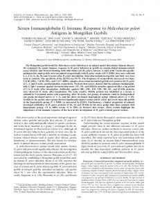
PE was diagnosed on the basis of histological examination of intestines in 1 of the 6 pigs in group A, 1 of the 12 pigs in group B, and 1 of the 6 pigs in group C. The IgG titers in the sera of PE-affected pigs were not significantly higher than those in the sera of nonaffected pigs 6 weeks after challenge. IgG titers in sera of naturally infected pigs. Seven of the 30 pigs were culled or died prior to 22 weeks of age. The sera of the remaining 23 pigs were tested by the ELISA to identify those with high IgG titers to IS intracellularis at 22 weeks of age. Fifteen pigs with high IgG titers were selected, and serum samples were tested retrospectively (Table 4). The IgG titers of 3-week-old pigs varied widely, and they declined when the pigs were between 3 and 6 weeks of age. Approximately one-third of the pigs had seroconverted by 18 weeks of age; the remaining pigs seroconverted by 24 weeks of age. To demonstrate the variability in seroconversion, four pigs were selected randomly and their IgG titers were plotted (Fig.
DISCUSSION Pigs produced IgG in their sera in response to intragastric inoculation with crude mucosal homogenate from PHE-affected pigs, and with filtrate (pore size, 0.8 ,um) containing IS intracellularis and after natural infection. In experiment 1, the antibody titers of both control and challenged pigs increased up to day 34 of the experiment. The magnitude of the immune response in challenged pigs tended to be higher than the immune response in control pigs. However, the IgG titers in the sera of challenged and unchallenged pigs were not significantly different because of variations in seroconversion among pigs. In this experiment, one control pig became infected. The IgG titer in the serum of this pig was higher than that those in the sera of the unaffected control pigs at 21 and 34 days after the trial commenced. Prior to challenge, the antibody titer in the serum of this pig was similar to those in the sera of other noninfected pigs. Cross-contamination by IS intracellularis from challenged pigs seems likely. In experiment 2, pigs inoculated with crude adenomatous intestine produced higher
ELISA FOR IgG TO INTRACELLULARIS IN PIGS
VOL. 32, 1994
1983
TABLE 2. IgG titers to IS intracellularis in sera and PE status of pigs challenged with adenomatous intestinal mucosa and noninoculated control pigs in experiment 1 IgG titer in seruma 34 days 21 days postinoculation postinoculation
38 days postinoculation
Group
Pig no.
Preinoculation
Inoculated
60 61 62 63 64 65 66 67
188 309 286 306 196 282 189 267
-C 747 452 607 446 332 343
552 849 496 691
443 507
68 69 70 71 72 73 74 75
186 206 227 155 250 177 215 221
297
318
252
223 252 243
405
205 406
366 513
Control
confirmed'
Yes Yes Yes Yes Yes No No Yes No No No No No No No Yes
369
a IgG titers in serum were measured by ELISA and were standardized by the PCF1 method. b A diagnosis of PE was made on the basis of the demonstration of adenomatous intestinal pathology and intracellular rod-shaped organisms on histological examination. c, missing values. Two pigs from each experimental group were necropsied at 2-week intervals.
antibody titers than pigs inoculated with filtered intestine. The increased antibody titer was probably the result of an increased dose of IS intracellularis, because it was estimated that approximately 80% of the IS intracellularis yield was lost through the filtering process. Pigs housed on a farm with IS intracellularis-infected pigs produced IgG after 6 weeks of age, with the highest antibody titers measured at between 18 and 24 weeks of age. The age of seroconversion is likely to affect the susceptibilities of pigs to infection and might explain the clinical variability of PE. The pathology of PHE in older pigs suggests an acute inflammatory response and is typical of other acute bacterial infections (14, 22). PHE may therefore be an acute form of intestinal adenomatosis in fully susceptible pigs. The severity of clinical signs and intestinal lesions of PHE may reflect infection in pigs with low levels of natural immunity after exposure to high numbers of IS intracellularis (27). The relatively high concentrations of IgG in the sera of these pigs at 3 weeks of age were indicative of residual levels of absorbed maternal antibody. The decay in IgG titers in pigs at between 3 and 6 weeks of age was interpreted as the decline in maternally derived IgG. In most gastrointestinal diseases, longer-lasting protection is pro-
vided by maternally derived IgA, which is superior to IgG in withstanding digestion and adhering to mucosal surfaces (7, 25). This may also be true for IS intracellularis infection. The inabilities of some naturally exposed pigs to mount an immune response to IS intracellularis before 24 weeks of age might explain the ongoing episodes of PHE in finisher pigs on the farm described here. Management strategies which involve exposure of the pigs to IS intracellularis may decrease the risk of future PE outbreaks. For example, the control strategy described by Love et al. (15) involved the removal and the subsequent replacement of medication in the diet, allowing exposure to IS intracellularis. This may allow immune development without clinical disease. Assessments of the sensitivity and specificity of the ELISA as a diagnostic test for PE were limited by small group sizes, the variability of antibody titers to IS intracellularis between groups, and the overlap in antibody titers between infected and
500
400 z 300
TABLE 3. IgG titers to IS intracellularis in sera of pigs in experiment 2 measured by ELISA Mean (SEM) IgG titer to IS
Group' A B C
No. of pigs
6 12 6
intracellularis'
P ltn Preinoculation
postinoculation
223 (15) 180 (11) 179 (17)
510 (28) 346 (34) 268 (33)
I
t ..
.......................
-
200
42 days
a Group A pigs were inoculated with intestinal homogenate, group B pigs were inoculated with filter-purified IS intracellularis, and group C pigs were uninoculated. b IgG titers to IS intracellularis were standardized by the PCF1 method.
100 0
3
6
12
18
20
24
Age (weeks) FIG. 1. Titers of IgG to IS intracellularis in the sera of four pigs housed on a farm with recurring PE. *, pig 1; *, pig 2; A, pig 3; V, pig 4.
1984
J. CLIN. MICROBIOL.
HOLYOAKE ET AL.
TABLE 4. IgG titers to IS intracellularis in sera of 15 pigs housed on a farm with endemic infection Mean (SD) Age (wk) antibody titera
60 (35.3) 34(11.8)
3. 6. 12 .64(16.9) 18 .100 (50.5) 20 .99 (37.1) 24 .212 (106.5) a Antibody titers are individual titers calculated as the mean of quadruplicate test samples measured by ELISA and corrected by the PCF1 method.
noninfected pigs. To determine antibody cutoff points for the diagnosis of PE, the sera of more pigs would need to be tested and the results would need to be correlated with the presence or absence of adenomatous pathology on histological examination. The diagnostic capacity of any ELISA is limited by the degree of variation in the immune responses of infected animals. An illustration of this is provided in humans affected with gastritis, in which the IgG titers in response to Helicobacterpylori infection are variable (20). Despite the diagnostic limitations, ELISAs used to measure H. pylon antibodies may provide epidemiological information for estimating disease prevalence. Similarly, ELISAs used to measure IS intracellularis-specific IgG may be more suited to estimating the prevalence of PE. Immunoassays based on the detection of shortlived IgM in the sera of PE-affected pigs could detect recently infected animals, but these assays have provided little epidemiological data on the prevalence of PE (9). Measurements of IgG titers by the ELISA may aid in epidemiological investigations of PE outbreaks in naturally infected herds. IS intracellularis had not been cultivated when this experiment was undertaken, so a pure source of the antigen was limited. Preliminary cultivation studies report the growth of IS intracellularis, derived from the intestines of PE-affected pigs, in a rat enterocyte cell line (8). However, Lawson et al. (8) reported that the yield of IS intracellularis from cell culture was low and was highly dependent on the multiplication cycle of the cells. Alternative methods of culturing IS intracellularis are being investigated (17) and might provide a source of antigen for retesting the ELISA. In the experiment described here, percoll gradient-purified IS intracellularis extracted from the intestines of naturally infected pigs was used to coat ELISA plates and to vaccinate pigs to produce high-positive control sera. The extraction method would not have removed immunogenic factors such as viruses or soluble antigens. Therefore, cross-reactions between nonspecific antigen components and pig sera cannot be eliminated (16). These cross-reactions were minimized by adding a solution of 1% skim milk in PBS containing 0.05% Tween 20 to the microtiter wells prior to the addition of test sera (26). It is not known whether strains of IS intracellularis with different immunogenicities exist. Other bacterial strains, including Serpulina hyodysenteriae and Actinobacillus pleuropneumoniae, vary in their immunogenicities (2, 11). The importance of strain selection for antigen when detecting antibodies to C. mucosalis was emphasized by Lawson et al. (10), who overcame the nonspecific agglutination reaction between one strain of C. mucosalis serotype B and the albumin fraction of pig sera by pooling antigen preparations from the sera of groups of pigs. In the development of the ELISA used in the present study, the problem of antigenic differences among strains was over-
come by using a pooled preparation of IS intracellularis extracted from the intestines of a number of PHE-affected pigs originating from two herds. In the present study, pigs produced low IgG titers in response to experimental IS intracellularis infection. These results, coupled with the localized intracellular nature of IS intracellularis infection, suggest that IgG plays a minor role in
preventing PE in pigs. The protection afforded by locally produced intestinal IgA against other bacterial and viral infections has been demonstrated (3, 23, 24). Experiments have been undertaken (5) to investigate the role of IgA in preventing colonization and invasion of the intestine by IS intracellularis. Preliminary investigations suggest that antibodies play a helper role with cell-mediated immune responses in the pathogenesis of IS intracellularis infection (5, 19). ACKNOWLEDGMENTS
Funding for the project was provided by the Australian Pig Research and Development Corporation. We thank the staff of the Bendigo Regional Veterinary Laboratory, especially Alison Collins and Dete Hasse for technical advice, and the staff of the farm involved in this research for their assistance. We also thank Terry Spencer and staff at the Benalla Regional Veterinary Laboratory for advice in the development of the ELISA. REFERENCES 1. Barker, I. K., and A. A. Van Dreumel. 1985. In K. V. F. Jubb, P. C. Kennedy and N. Palmer (ed.), Pathology of domestic animals, 3rd ed., vol. 2, p. 143-146. Academic Press, New York. 2. Blackall, P. J., and G. J. Storie. 1991. Studies of porcine pleuropneumonia, p. 35-46. In Proceedings of the Australian Association of Pig Veterinarians Conference, Sydney. Upjohn Australia Ltd., Rydalmere, New South Wales, Australia. 3. Crabbe, P. A., A. Carbonara, and J. Heremans. 1965. The normal human intestinal mucosa as a major source of plasma cells containing yA-immunoglobulin. Lab. Invest. 44:235-248. 4. Gebhart, C. J., S. M. Barns, S. McOrist, G. F. Lin, and G. H. K. Lawson. 1993. Ileal symbiont intracellularis, an obligate intracellular bacterium of porcine intestines showing a relationship to Desulfovibrio species. Int. J. Syst. Bacteriol. 43:533-538. 5. Holyoake, P. K. 1993. Ph.D. thesis. University of Melbourne, Melbourne, Australia. 6. Kaldor, J., H. Pritchard, A. Serpell, and W. Metcalf. 1983. Serum antibodies in Campylobacter enteritis. J. Clin. Microbiol. 18:1-4. 7. Keren, D. F., P. J. Scott, and D. Bauer. 1980. Variables affecting the local immune response in Thirty-Vella loops. II. Stability of antigen-specific IgG and secretory IgA in acute and chronic Thirty-Vella loops. J. Immunol. 124:2620-2624. 8. Lawson, G. H. K., S. McOrist, S. Jasni, and R. A. Mackie. 1993. Intracellular bacteria of proliferative enteropathy: cultivation and maintenance in vitro. J. Clin. Microbiol. 31:1136-1142. 9. Lawson, G. H. K., S. McOrist, A. C. Rowland, E. McCartney, and L. Roberts. 1988. Serological diagnosis of the porcine proliferative enteropathies: implications for aetiology and epidemiology. Vet. Rec. 122:554-557. 10. Lawson, G. H. K., L. Roberts, E. McCartney, A. C. Rowland, and A. G. Luckins. 1982. Presence of serum agglutins to Campylobacter sputorum subspecies mucosalis in pigs. Res. Vet. Sci. 32:89-94. 11. Lee, J. I., D. J. Hampson, and A. J. Lymbery. 1991. The intestinal spirochaetes: genetic diversity and disease associations, p. 140. In E. S. Batterham (ed.), Manipulating pig production III. Victorian Institute of Animal Science, Melbourne, Australia. 12. Lomax, L. G., R. D. Glock, D. L. Harris, and J. E. Hogan. 1982. Porcine proliferative enteritis: experimentally induced disease in cesarean-derived colostrum-deprived pigs. Am. J. Vet. Res. 43: 1622-1630. 13. Lomax, L. G., R. D. Glock, and J. E. Hogan. 1982. Experimentally induced porcine proliferative enteritis in specific-pathogen-free pigs. Am. J. Vet. Res. 43:1615-1621. 14. Love, D. N., and R. J. Love. 1979. Pathology of proliferative
VOL. 32, 1994 hemorrhagic enteropathy in pigs. Vet. Pathol. 16:41-48. 15. Love, R. J., D. N. Love, and M. J. Edwards. 1977. Proliferative hemorrhagic enteropathy in pigs. Vet. Rec. 100:65-68. 16. Mapother, M. E., L. A. Joens, and R. D. Glock. 1987. Experimental reproduction of porcine proliferative enteritis. Vet. Rec. 121:533536. 17. McOrist, S. (University of Edinburgh). 1993. Personal communication. 18. McOrist, S., S. Jasni, R. A. Mackie, N. MacIntyre, N. Neef, and G. H. K. Lawson. 1993. Reproduction of porcine proliferative enteropathy with pure cultures of ileal symbiont intracellularis. Infect. Immun. 61:4286-4292. 19. McOrist, S., and G. H. K. Lawson. 1989. Proliferative enteropathies: Campylobacter species in the faeces of normal and contact pigs. Vet. Rec. 124:40-41. 20. Rathbone, B. 1990. Local and systemic immune response-a comment, p. 263. In P. Malfertheimer and H. Ditschuneit (ed.), Helicobacter pylon and peptic ulcer. Springer-Verlag, Berlin. 21. Roberts, L., A. C. Rowland, and G. H. K. Lawson. 1977. Experi-
ELISA FOR IgG TO INTRACELLULARIS IN PIGS
22.
23.
24. 25.
26.
27.
1985
mental reproduction of porcine intestinal adenomatosis and necrotic enteritis. Vet. Rec. 100:12-13. Sims, L. D. The pathology of proliferative enteritis, p. 127-137. In E. S. Batterham (ed.), Manipulating pig production II. Victorian Institute of Animal Science, Melbourne, Australia. Stacey, A. R., P. R Hawtin, and D. G. Newell. 1990. Local immune responses to Helicobacter pylori infections, p. 162-165. In P. Malfertheiner and H. Ditschuneit (ed.), Helicobacterpyloni, gastritis and peptic ulcer. Springer-Verlag, Berlin. Stokes, C. R, J. F. Soothill, and M. W. Turner. 1975. Immune exclusion is a function of IgA. Nature (London) 255:74-75. Tomasi, T. B., and J. Bienenstock. 1968. Secretory immunoglobulins. Adv. Immunol. 9:1-96. Vogt, R F., D. L. Phillips, L. 0. Henderson, W. Whitfield, and F. W. Spiero. 1987. Quantitative differences among various proteins as blocking agents for ELISA microtitre plates. J. Immunol. Methods 101:43-50. Ward, G. E., and N. L Winkelman. 1990. Diagnosing, treating and controlling proliferative enteritis in swine. Vet. Med. 85:312-318.