Fermitins, the Orthologs of Mammalian Kindlins, Regulate the Development of a Functional Cardiac Syncytium in Drosophila melanogaster James H. Catterson, Margarete M. S. Heck, Paul S. Hartley* The University of Edinburgh/British Heart Foundation Centre for Cardiovascular Science, The University of Edinburgh, Edinburgh, Midlothian, United Kingdom
Abstract The vertebrate Kindlins are an evolutionarily conserved family of proteins critical for integrin signalling and cell adhesion. Kindlin-2 (KIND2) is associated with intercalated discs in mice, suggesting a role in cardiac syncytium development; however, deficiency of Kind2 leads to embryonic lethality. Morpholino knock-down of Kind2 in zebrafish has a pleiotropic effect on development that includes the heart. It therefore remains unclear whether cardiomyocyte Kind2 expression is required for cardiomyocyte junction formation and the development of normal cardiac function. To address this question, the expression of Fermitin 1 and Fermitin 2 (Fit1, Fit2), the two Drosophila orthologs of Kind2, was silenced in Drosophila cardiomyocytes. Heart development was assessed in adult flies by immunological methods and videomicroscopy. Silencing both Fit1 and Fit2 led to a severe cardiomyopathy characterised by the failure of cardiomyocytes to develop as a functional syncytium and loss of synchrony between cardiomyocytes. A null allele of Fit1 was generated but this had no impact on the heart. Similarly, the silencing of Fit2 failed to affect heart function. In contrast, the silencing of Fit2 in the cardiomyocytes of Fit1 null flies disrupted syncytium development, leading to severe cardiomyopathy. The data definitively demonstrate a role for Fermitins in the development of a functional cardiac syncytium in Drosophila. The findings also show that the Fermitins can functionally compensate for each other in order to control syncytium development. These findings support the concept that abnormalities in cardiomyocyte KIND2 expression or function may contribute to cardiomyopathies in humans. Citation: Catterson JH, Heck MMS, Hartley PS (2013) Fermitins, the Orthologs of Mammalian Kindlins, Regulate the Development of a Functional Cardiac Syncytium in Drosophila melanogaster. PLoS ONE 8(5): e62958. doi:10.1371/journal.pone.0062958 Editor: Rajesh Gopalrao Katare, University of Otago, New Zealand Received November 6, 2012; Accepted March 26, 2013; Published May 15, 2013 Copyright: ß 2013 Catterson et al. This is an open-access article distributed under the terms of the Creative Commons Attribution License, which permits unrestricted use, distribution, and reproduction in any medium, provided the original author and source are credited. Funding: This work was supported by a British Heart Foundation Centre of Research Excellence (CoRE) award to the University of Edinburgh. JHC is supported by a 4-year BHF PhD award, and PSH is supported by a BHF CoRE Transitional Fellowship Award. The funders had no role in study design, data collection and analysis, decision to publish, or preparation of the manuscript. Competing Interests: The authors have declared that no competing interests exist. * E-mail:
[email protected]
a contractile syncytium and pump haemolymph from the abdomen, through the thorax towards the head (see schematic in Figure 1A). Drosophila expresses two orthologs of human KIND2, Fermitin1 (Fit1) and Fermitin2 (Fit2) that contain a highly conserved FERM (4.1, Ezrin, Radixin, Moesin) domain which is required for binding to the cytoplasmic tail of b-integrins [18] (see Fig. 1B and Fig. S1). Both Fit1 and Fit2 message and protein are detected in adult heart tissue, consistent with a role in cardiac function [19,20]. To explore the role of Fermitins in the Drosophila heart we silenced their expression in cardiomyocytes and analysed the function of the adult heart. It was found that cardiomyocyte expression of Fermitins is critical for the development of a functional syncytium in Drosophila and that the genes can functionally compensate for each other. The findings support the hypothesis that disruption of cardiomyocyte KIND2 may lead to abnormal intercalated discs and cardiomyopathies in humans.
Introduction The mammalian Kindlins are a family of evolutionarily conserved proteins that mediate cell-cell and cell-matrix adhesion by regulating integrin function [1–3]. Findings from several animal models suggest that the formation of junctions between neighbouring cardiomyocytes (intercalated discs) may require integrins and integrin binding proteins [4–8]. It is important to understand the role of these proteins because mutations and polymorphisms that affect intercalated disc formation may be associated with the development of cardiomyopathies in humans [6,9,10]. Murine KIND2 protein is detected at intercalated discs, however genetic ablation of Kind2 in mice causes early embryonic lethality at the peri-implantation stage, before cardiogenesis [11,12]. In zebrafish, the global knock-down of Kind2 causes cardiac hyperplasia, disrupts intercalated disc formation and significantly reduces cardiac contractility [11]. These findings link Kind2 with the development of a functional cardiac syncytium; however a cardiomyocyte-specific test of this hypothesis is yet to be conducted. Drosophila melanogaster is an important model for the identification and study of genes that have relevance to mammalian cardiovascular development and function [13–17]. The abdominal heart in Drosophila consists of individual cardiomyocytes that form PLOS ONE | www.plosone.org
Methods Drosophila stocks and fly husbandry Flies were raised on standard medium at 25uC on a 12/12 hr light/dark schedule. Flies used in this paper were w1118, 1
May 2013 | Volume 8 | Issue 5 | e62958
Fermitins and the Drosophila Heart
Poole, Dorset, UK) overnight at 4uC. For fly heart RT-PCR, total RNA was extracted from 20 dissected hearts of 1-wk-old adult females using TRIzol (Invitrogen, Paisley, UK) phenol-chloroform extraction method with isopropanol precipitation. RNA was resuspended in nuclease-free DEPC-water and treated with RQ1 RNase-free DNase (Promega, Southampton, UK), then cDNA was generated with M-MLV Reverse Transcriptase (Promega, Southampton, UK) using oligo-dT primer and RNasin Plus RNase Inhibitor (Promega, Southampton, UK). PCR was performed using relevant primers, dNTP Mix (Promega, Southampton, UK), and GoTaq DNA Polymerase in 5X Green GoTaq Reaction Buffer (Promega, Southampton, UK). The primer sets were as follows: for RP49, 59-GACAATCTCCTTGCGCTTCT39 and 59-CCAGTCGGATCGATATGCTAA-39; for Fit1, 59AACAGTGAGGTCTGGGTGAGAT-39 and 59-AACAGCTCCTCCTTCTTGTGTC-39; for Fit2, 59-ACGGTATCAACAGTGAGGTGTG-39 and 59-GATGCCCGTCGAACTTAATG-39 (Eurofins MWG Operon, Acton, London, UK). PCR samples were then analysed by gel electrophoresis and images taken with Uvitec Gel Doc system (Uvitec, Cambridge, UK).
Analysis of cardiac synchrony and generation of cardiac raster plots A video of the beating heart was opened in ImageJ [22], converted to greyscale and a movement movie processed by subtracting a frame’s pixel intensity from that of the preceding frame using the ‘Image Calculator’ function. The mean pixel intensity of two, non-overlapping regions (the upper left quartile and lower right quartile) of each frame in the movement movie was then measured using the ‘Measure stack’ function. Regression analysis was then performed on the mean pixel intensity data obtained from the two regions. When the two regions are in perfect synchrony, R2 = 1; when regions are not in synchrony, R2 approaches zero. The mean (6SEM) R2 value of 7 flies for each genotype is presented. Cardiac raster plots were generated by concatamerising the image stills from the movement movies in ImageJ so that there were 12 consecutive images per row over 30 columns. The resulting raster plot corresponds to 10.8 seconds of cardiac motion. Under normal conditions a rhythmically occurring event produces a geometric pattern in a raster plot, whereas loss of rhythmicity leads to a more random pattern. This methodology of illustrating a physiological rhythm is based on the analysis of biological rhythm data in the circadian biology field.
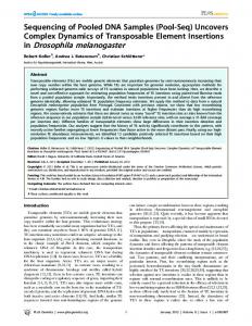
Figure 1. Overview of the adult Drosophila heart and Fermitins. (A) The schematic illustrates the position of the Drosophila heart (shaded grey) in the adult fly. The contractile portion of the heart (the ventricle) is within the abdomen and is connected directly to a noncontractile aorta which directs haemolymph (the insect equivalent of blood) from the abdomen, through the thorax and onwards to the head. The second schematic illustrates the ventricle, which consists of paired cardiomyocytes that function together as a single contractile syncytium (small arrows). Long arrows show the direction of circulation; however the heart can contract in different directions, so as to direct haemolymph to the fly’s anterior or posterior. (B) The integrin binding FERM domain of human Kind2 is present in both Fit1 and Fit2. doi:10.1371/journal.pone.0062958.g001
Fit1KG05576, and Fit2EY08530 were obtained from the Bloomington stock centre. HandC-GAL4 4.2 [21] (Hand-Gal4) flies were kindly provided by A. Paululat. HandC-GAL4; tub-GAL80ts, UAS-Fit1VDRC; UAS-Fit2VDRC, and HandC-GAL4; Fit1D161 UAS-Fit2VDRC flies were generated following standard crosses. Vienna Drosophila RNAi Centre (VDRC) RNAi lines used – Fit1 (#46494), Fit2 (#37010), and Mys (#29619). Transgenic RNAi Project (TRiP) RNAi lines used – Fit1 (#25966).
Fluorescent labelling and imaging of adult heart structures
Genomic PCR and RT-PCR
Fluorescent labelling of Drosophila heart structures was performed as described [23]. In brief, semi-intact heart preparations of four flies of each genotype were fixed in 4% formaldehyde (Fisher Scientific, Loughborough, UK) in PBS for 20 mins then 3610 min washes with PBS-Triton-X (PBST)(Sigma, Poole, Dorset, UK). Hearts were then incubated with primary antibodies and/or stained in PBST overnight at 4uC with constant agitation. Hearts were washed 3610 mins in PBST, incubated with secondary antibodies for 1-hour at room temp, washed 3610 mins in PBST plus final wash in PBS alone. Hearts were then mounted in 50% glycerol (Sigma, Poole, Dorset, UK) in PBS and visualised using a Zeiss Axioskop 2 mot plus fluorescent microscope and images were taken using Openlab imaging software. Anti-bPSintegrin (1:10 of hybridoma supernatant (100 mg/ml), Developmental Studies Hybridoma Bank) was used to stain cell junctions, AlexaFluor 546-conjugated phalloidin (13.2 nM) to stain F-actin
For genomic DNA, whole flies were homogenised in lysis buffer (1M TrisHCL pH 7.5, 500 mM EDTA, pH 8.0, 4M NaCl, 10% SDS)(Sigma, Poole, Dorset, UK) using Kontes pellet pestle (Fisher Scientific, Loughborough, UK) and incubated at 65uC for 30 mins. LiCl/KAc solution (6M LiCl, 5M KAc)(Sigma, Poole, Dorset, UK) was then added, mixed, and incubated on ice for 10 mins. Samples were centrifuged for 15 mins at 12,000 rpm at room temperature. Supernatant was transferred to fresh 1.5 mL centrifuge tubes and isopropanol (VWR, Lutterworth, Leicestershire, UK) added to precipitate DNA. Samples were then centrifuged for 20 mins at 12,000 rpm at room temperature. Pellet was washed with cold 70% EtOH (VWR, Lutterworth, Leicestershire, UK) and centrifuged for 10 mins at 12,000 rpm at room temperature. DNA pellets were left to air dry for 1-hour then resuspended in Tris-EDTA (10 mM Tris, 1 mM EDTA)(Sigma, PLOS ONE | www.plosone.org
2
May 2013 | Volume 8 | Issue 5 | e62958
Fermitins and the Drosophila Heart
(Invitrogen, Paisley, UK), and DAPI (1 ng/ml) to stain nuclei (Sigma, Poole, Dorset, UK). The hybridoma developed by [24] was obtained from the Developmental Studies Hybridoma Bank developed under the auspices of the NICHD and maintained by The University of Iowa, Department of Biology, Iowa City, IA 52242.
Results Fermitins regulate cardiac syncytium development To assess the effect of Fermitin knock-down on adult heart morphology, the cardiac Hand-GAL4 driver was used to disrupt gene expression. In control flies the cardiomyocytes display a characteristic spiral myofibrillar arrangement that can be visualised by phalloidin staining, with b-integrin staining junctions between adjacent cardiomyocytes (Figure 2, Hand-Gal4; w1118). Silencing Fit1 using the RNAi line from the Vienna Drosophila RNAi centre (VDRC [28]) led to loss of the cardiac syncytium and loss of contiguous b-integrin staining between adjacent cardiomyocytes)(Fig. 2, Hand-Gal4; UAS.Fit1VDRC). When both Fit1 and Fit2 were silenced, a more severe phenotype developed (Fig. 2, Hand-Gal4; UAS.Fit1/Fit2VDRC), with cardiomyocytes exhibiting a ‘rounded-up’ phenotype, comparable to that resulting from the silencing of the b-integrin myospheroid in cardiomyocytes (Fig. 2, Hand-Gal4; UAS.MysVDRC). In contrast, when Fit1 was silenced using the Harvard TRiP (Transgenic RNAi Project) RNAi line (Fig. 2, Hand-Gal4; UAS.Fit1TRiP), the cardiac syncytium developed normally and the cardiomyocytes exhibited a cytoskeletal morphology similar to that seen in controls. Similarly, when Fit2 was silenced on its own, the cardiac syncytium developed normally (Fig. 2, Hand-Gal4; UAS.Fit2VDRC). The discrepancy between the phenotype caused by the Fit1VDRC and Fit1TRiP RNAi lines can be explained by the VDRC line targeting both Fit1 and Fit2 message (see Fig. S2), whereas the TRiP line targets only Fit1. To confirm the effect of Fermitin knock-down on the heart the distance between neighbouring cardiomyocytes was quantified (Fig. 3A). In control flies and flies where no effect of an RNAi was observed (w1118 flies crossed with the UAS.Fit1VDRC line in the absence of the Hand-Gal4 driver), the distance between adjacent cardiomyocytes were never more than 3 mm. In contrast, the distance between cardiomyocytes when Fit1 was silenced using the VDRC line, was ten-fold greater (34.263.7 mm, (P,0.001)). When Fit2 was silenced there appeared to be a small effect on distances between cardiomyocytes, however this did not reach statistical significance.
Measurement of distance between neighbouring cardiomyocytes ImageJ (http://rsbweb.nih.gov/ij/) was used to measure the distance between neighbouring cardiomyocytes. In ImageJ, the ‘straight line’ drawing tool was selected and a line was drawn between neighbouring cardiomyocytes. The scale was calibrated (Analyse.Set Scale) to measure in microns, and distances were measured using the ‘Measure’ function (Analyse.Measure, or CTRL+M) in ImageJ. Four measurements were taken for each fly heart, and up to 7 flies were used for each genotype – resulting in 12–28 measurements from 4–7 independent flies for each genotype. Data was then grouped for each genotype and mean +/2 SEM was calculated.
Heart preparation and functional assays It is necessary to surgically expose the fly heart for detailed analysis of cardiac contractions – making them accessible for video microscopy [25]. We used the previously published heart assay and analysis methodology [26], and used between 6 and 11 female flies, aged 5–7 days old for all analyses. In brief, the ventral abdomen was dissected in artificial haemolymph (108 mM NaCl, 5 mM KCl, 2 mM CaCl2, 8 mM MgCl2, 1 mM NaH2PO4, 4 mM NaHCO3, 5 mM HEPES, 10 mM sucrose, 5 mM trehalose) to expose the fly’s heart, which lines the dorsal cuticle, and digital movies of the heart over body segments A2 and A3 were taken (video acquisition by Pinnacle Software (Pinnacle Systems, Iver Heath, UK)). Video footage was analysed using a MatLabbased image analysis program, M-modes were generated and cardiac parameters including heart rate, arrhythmias, heart periods, diastolic and systolic diameters, and fractional shortening were quantified [27]. Data was then grouped for each genotype and mean +/2 SEM was calculated.
Generation of Fit1
D161
Silencing Fermitin expression disrupts heart contractility The cardiac syncytium in vertebrates is a critical determinant of the heart’s contractility. It was therefore reasoned that loss of the syncytium caused by Fermitin knock-down in Drosophila would dramatically impact cardiac function and this hypothesis was tested in adult flies using videomicroscopy. It was found that the cardiomyocytes of Fit1VDRC silenced flies (in which both Fit1 & Fit2 are silenced) beat at the same rate as controls (w1118 and w1118 crossed with Fit1VDRC) and flies in which Fit2 had been knocked down (Figure 3C, P.0.05). However the distance the heart moved between diastole and systole (fractional shortening) was greatly reduced in Fit1VDRC silenced flies, compared to all other genotypes (see the M-mode in Figure 3B and the quantified data in Figure 3D; P,0.001). Consistent with this finding, the end systolic diameter was greater in the Fit1VDRC silenced flies than the other genotypes (Figure 3E, P,0.01); whereas diastolic diameter was similar to the controls (P.0.05). Examples of the beating hearts from the different genotypes and the disruption caused by the UAS.Fit1VDRC line can be seen in videos S1 to S4.
deletion line
The P-element inserted in Fit1KG05576 was mobilized by following standard crosses. Mobilization events were screened by PCR using different primer combinations in the genomic DNA flanking the P-element – Fit1D161 (59- CATCCGTTCCGATAAGTTCG-39 and 59- CCAACAGCTCCTCCTTCTTG-39) resulted from the imprecise excision of the P-element and creates an out-of-frame deletion which removes 1685 bp (629 bp from 59 UTR region and 1055 bp from coding sequence)(deletion corresponds to nucleotides 67–1752) and retains a 12 bp fragment (CATGATGAAATA) from the start of the 11,467 bp P-element.
Statistics One-way analysis of variance (ANOVA) followed by Tukey’s post-hoc test was used to identify differences between three or more means derived from uneven sample sizes. The student’s unpaired t-test was used to identify significant differences between two means of uneven sample size. A statistical difference of P,0.01 was regarded as significant.
Synchrony between cardiomyocytes is reduced when Fermitin expression is silenced It was clear from the functional studies that cardiomyocytes in the Fit1VDRC silenced flies were capable of beating at a frequency similar to that of controls, but that fractional shortening was
PLOS ONE | www.plosone.org
3
May 2013 | Volume 8 | Issue 5 | e62958
Fermitins and the Drosophila Heart
Figure 2. Fermitins regulate coupling between cardiomyocytes. The monochrome micrographs show the adult Drosophila heart (segments A2 to A5) stained with phalloidin, oriented with the anterior region to the left. Genes were silenced using the Hand-Gal4 enhancer which drives expression in cardiomyocytes (as well as pericardial nephrocytes and enterocytes of the gut) and abnormal cardiomyocyte/heart morphology is highlighted by the arrows. The colour panels show higher magnification micrographs of at least two cardiomyocytes stained with phalloidin (red) and antibodies to the Drosophila b-integrin, myospheroid (green); arrowheads indicate normal integrin staining between cardiomyocytes; asterisks indicate an abnormal staining pattern associated with loss of cardiomyocyte junction integrity. A wild type phenotype (Hand-Gal4; w1118), is characterised by contiguous cardiomyocytes and b-integrin staining between cardiomyocytes. doi:10.1371/journal.pone.0062958.g002
PLOS ONE | www.plosone.org
4
May 2013 | Volume 8 | Issue 5 | e62958
Fermitins and the Drosophila Heart
Figure 3. Silencing Fermitin expression prevents cardiomyocyte coupling and disrupts heart contractility. (A) The distance between nearest neighbouring cardiomyocytes was measured for four different genotypes. n = 16–28 measurements from four independent flies for each genotype. ***P,0.001, ns = not significantly different from any other genotype. (B) Representative M-modes illustrating heart contractions in the different genotypes. The white ‘D’ and ‘S’, and the black bars in the first M-mode denote the heart at diastole and systole. The arrow and arrowhead highlight reduced contractility and loss of synchrony between different regions of the heart. (C) Mean (6SEM) heart rate and (D) Fractional shortening (Diastolic diameterMAX – Systolic diameterMAX). (E) Quantification of end diastolic and systolic diameter (EDD and ESD, respectively). ***P,0.001, n = 6–7 flies per genotype. a = not significantly different from controls’ diastolic diameter; b = significantly different (P,0.01) from controls systolic diameter. Legend applies to A–E. doi:10.1371/journal.pone.0062958.g003
was significantly reduced in Hand-Gal4; UAS.Fit1VDRC flies compared to control flies (w1118, UAS.Fit1VDRC; mean R2 values, of 0.3460.11 and 0.8160.03, respectively (P,0.001, Figure 4C & D). Examples of the beating hearts and movement movies from the different genotypes can be seen in videos S5 to S8.
dramatically reduced. It was also clear that cardiomyocytes were no longer in close contact with each other and it was therefore reasoned that synchrony between cardiomyocytes had been lost. This loss of synchrony is a novel phenotype in Drosophila and was quantified by analysing movement movies from videomicroscopy footage that depict the motion of the heart during the cardiac cycle. To provide a 2-dimensional example of heart movement we explored the use of ‘actograms’, which are used to depict cyclical events over a given time period, especially in the field of biological rhythm research. In these images, a time-series of a measured or recorded parameter (e.g. behaviour, movement or gene expression) is presented as a raster plot (i.e. plotted left to right, top to bottom). A rhythmic system will produce a geometric pattern in a raster plot, whereas a random pattern is produced when a system is asynchronous. To illustrate the phenotype of the abnormal hearts in this way we concatamerised stills from the movement movies to generate raster plots of 10.8 seconds of heart activity and compared the plots from control flies (w1118, UAS.Fit1VDRC) with those from the Fermitin silenced flies (Hand-Gal4; UAS. Fit1VDRC, Fig. 4A, B). Using this method it was easy to identify the loss of rhythmicity as a transition from the geometric pattern of the control heart to the disordered plot from the Fermitin silenced fly. In order to quantify the degree of synchrony in different areas of the heart, two independent regions of the movement movie were compared to see if changes in pixel intensity (i.e. a measurement of cardiac motion) were in synchrony or not. Using simple regression analysis of two independent regions of the contracting heart in the movies, it was confirmed that synchrony PLOS ONE | www.plosone.org
Fit1 is dispensable for syncytium development The discrepancy between the phenotypes caused by the Fit1 VDRC and TRiP lines was likely due to the VDRC also targeting Fit2. This ‘double whammy’ would effectively knock-down both Fit genes and inhibit any functional compensation occurring between the two. To test this hypothesis, a Fit1 mutant was generated via D2–3 mutagenesis. The P-element in Fit1KG05576 allele was excised along with 1.7 kb of the Fit1 gene in the 39 direction to create the Fit1D161 line (Figure 5A,B). Fit1D161 flies were viable and had normal heart morphology (Figure 5). However, abnormal heart morphology was observed when Fit2 was silenced (Hand-Gal4; UAS.Fit2VDRC) in Fit1D161 mutant cardiomyocytes (Figure 5C), despite the same Fit2 RNAi line, when used on its own, having no effect on heart function (compare Figs. 2 & 3 with Fig. 5C). These findings were confirmed when the distance between neighbouring cardiomyocytes was quantified (Figure 5D). Thus, loss of Fit1 or reduced expression of Fit2 is not sufficient to cause a heart phenotype, indicating that functional redundancy exists between Fit1 and Fit2. Only when the expression of both genes is affected does a severe cardiomyopathy develop in Drosophila.
5
May 2013 | Volume 8 | Issue 5 | e62958
Fermitins and the Drosophila Heart
Figure 4. Synchrony between cardiomyocytes is reduced when Fermitin expression is silenced. Videomicroscopy of the contracting heart from control (w1118, UAS.Fit1VDRC) or Fermitin-silenced flies (Hand-Gal4; UAS.Fit1VDRC) was analysed for movement to ascertain the degree of synchronicity between the different (depicted) regions of the Drosophila heart. (A) Frames from a movement movie of the beating heart where dark pixels indicate movement relative to the previous frame. Pixel intensity in two non-overlapping regions is measured over 10 s and regression analysis performed to quantify the degree of synchrony between the two regions. (B) Concatamerised frames from the movement movie were used to develop cardiac actograms which illustrate the regular, synchronous movement of the heart in a control fly (w1118, UAS.Fit1VDRC), and asynchronous movements in the heart of a Fermitin silenced fly (Hand-Gal4; UAS.Fit1VDRC). The 360 frames run left to right and top to bottom, the entire sequence being 10.8 s long. (C) Representative XY scatter plots of pixel intensity in two regions of a 300 frame, 10 s movement movie, note the lower R2 value for the Fermitin silenced fly. (D) Synchrony (mean R2 values (6SEM)) for control (w1118, UAS.Fit1VDRC) and Fermitin silenced flies (Hand-Gal4; UAS.Fit1VDRC). n = 7 flies for each genotype, **P,0.01. doi:10.1371/journal.pone.0062958.g004
Figure 5. Cardiomyocyte Fit2 compensates for the loss of Fit1 to establish the cardiac syncytium. (A) Schematic showing the Fit1 locus with the adjacent genes (CG14995 and Ack) and the site of the P-element insertion used to create the 1.7 kb deletion mutant (Fit1D161). (B) PCR of genomic DNA from wild-type (w1118) and Fit1D161 mutant flies. (C) Micrographs of adult hearts stained with phalloidin. The arrow highlights an abnormal cardiomyocyte phenotype. (D) Mean (6SEM) distance between neighbouring cardiomyocytes. n = 12–16 measurements from four to seven independent flies for each genotype. ***P,0.001 from all other genotypes. Legend applies to C & D. doi:10.1371/journal.pone.0062958.g005
PLOS ONE | www.plosone.org
6
May 2013 | Volume 8 | Issue 5 | e62958
Fermitins and the Drosophila Heart
order to develop as a functional syncytium. When Drosophila cardiomyocytes fail to couple together to form a cardiac syncytium, synchronous contractions and fractional shortening of the adult heart are significantly reduced, despite individual cardiomyocytes remaining myogenic. Studies of vertebrate hearts suggest a role for Kind2 in cardiac development and function, however these studies are limited by the embryonic lethality of Kind2 knock-out in the mouse model, and the lack of tissue specific Kind2 silencing in the fish model [11,12]. The aberrant phenotype caused by silencing orthologs of Kind2 in the cardiomyocytes of an invertebrate demonstrates that the protein’s role in cardiac development has been evolutionarily conserved and also reiterates the validity of using Drosophila to study genes relevant to mammalian cardiac physiology. The aberrant heart phenotype caused by Fermitin knock-down in Drosophila reproduces many aspects of the cardiomyopathy caused by morpholino knock-down of Kind2 in zebrafish [11], and supports the authors’ conclusion that Kind2 regulates heart development. Morpholino-knockdown in zebrafish disrupted angiogenesis but also affected intercalated disc formation in cardiomyocytes and significantly reduced fractional shortening. Previous studies in Drosophila have identified the Fermitins as
Synchrony and contractility are disrupted when Fit2 expression is silenced in Fit1D161 mutant hearts Synchronicity between discrete regions of the Drosophila heart was quantified and control (w1118) flies displayed regular synchronous cardiac contractility. Fit1D161, and UAS.Fit2VDRC; Fit1D161 flies without Hand-Gal4 displayed comparable synchronicity to w1118 controls. In contrast, when Fit2 was silenced (Hand-Gal4; UAS.Fit2VDRC) in Fit1D161 mutant cardiomyocytes there was a significant loss of synchronicity between these regions of the heart (Figure 6A,B). There was no significant difference in heart rate between genotypes, whereas fractional shortening was significantly reduced in the Hand-Gal4; UAS.Fit2VDRC Fit1D161 mutants (Figure 6C,D). The Hand-Gal4; UAS.Fit2VDRC Fit1D161 mutants also had significantly increased end systolic and end diastolic diameters relative to the control and other genotypes (Figure 6E, P,0.01). Examples of the beating hearts and movement movies from the different genotypes can be seen in videos S9 to S16.
Discussion The current findings unequivocally demonstrate that cardiomyocytes must express members of the Fermitin/Kindlin family in
Figure 6. Cardiomyocyte Fit2 compensates for loss of Fit1 to maintain cardiac synchrony and contractility. (A) Representative cardiac actograms from four different genotypes. (B) Mean (6SEM) synchrony between different regions of the beating heart. n = 7 different flies for each genotype. ***P,0.01. (C) Representative M-modes of beating hearts showing the normal contractions in the Fit1D161 mutant flies and shallow contractions in the Fit1D161 mutant flies when Fit2 is silenced (arrow). (D) Quantification of heart rate and fractional shortening in the four genotypes confirms that heart function was normal in the Fit1D161 mutant flies and only becomes abnormal when Fit2 expression was silenced in this mutant. (E) Quantification of end diastolic and systolic diameter (EDD and ESD, respectively). n = 10–11 flies per genotype, ***P,0.001; a = P,0.01 compared to all other genotypes’ diastolic diameter; b = P,0.01 compared to all other genotypes’ systolic diameter. doi:10.1371/journal.pone.0062958.g006
PLOS ONE | www.plosone.org
7
May 2013 | Volume 8 | Issue 5 | e62958
Fermitins and the Drosophila Heart
mediators of muscle assembly [29], and the current findings extend this observation to include the assembly of a functional cardiac syncytium. Integrins and related adhesion proteins (ILK, Talin, Laminin A, Syndecan, Robo, and Slit) are necessary for heart assembly in Drosophila [30–32]. The current work is the first to demonstrate the functional consequences for the Drosophila heart when assembly of the cardiac syncytium is abnormal. Human heart diseases attributed to intercalated disc dysfunction include arrhythmogenic right ventricular cardiomyopathy caused by mutations in D proteins (e.g. Desmoplakin, Plakophilin-2, and Desmoglein-2) [9], and hypertrophic and dilated cardiomyopathy caused by a missense mutation in the integrin-binding adhesion protein, Vinculin [10]. Integrins and related binding proteins (e.g. Vinculin and Talin) were also identified at intercalated discs in adult human cardiac tissue [33,34]. Thus, there is a need to develop genetically tractable models with which to study the role of these proteins in cardiac development and function. Intercalated disc-like structures have been identified between Drosophila cardiomyocytes, however there have been no empirical studies examining the molecular components nor the functional role of this structure in the heart [35]. The current data provide evidence that the b-integrin encoded by myospheroid is localised at the boundaries between adjacent cardiomyocytes, and also indicate that the development of a functional cardiac syncytium is strictly dependent on myospheroid as well as the expression of Fermitins. It would be informative to further assess the integrity of the cardiomyocyte junctions in both wild type and mutant flies using antibodies to cadherin and b-catenin [36]. The cardiomyopathy caused by silencing Fit1 and Fit2 phenocopied myospheroid knock-down and is therefore consistent with the Fermitins regulating b-integrin signalling and promoting cardiomyocyte coupling during development [31,35]. There is evidence from mammalian models that Kindlins, via the FERM F3 subdomain, interact directly with the cytoplasmic tail of b-integrins [12,18]. The FERM domain of Drosophila Fermitins is conserved from flies to humans and it is therefore speculated that Fermitins directly interact with the b-integrin myospheroid to form a signalling complex required for cardiomyocyte coupling. No study has yet manipulated the expression of mammalian Kind2 solely in the cardiomyocytes, however it is predicted that a phenotype would develop that is similar to that seen in ventricular cardiomyocytetargeted b1-integrin knockout mice [7]. Histological studies of the b1-integrin KO hearts revealed significant disruption of myofibrils and intercalated discs, with subsequent onset of dilated cardiomyopathy. There was evidence that Fit2 expression could compensate for Fit1 in the Drosophila cardiomyocyte model. A Fit1-null mutant (Fit1D161) was generated that had normal heart morphology and function, indicating that Fit1 is not required for the development of a functional syncytium. This finding also suggested that Fit2 might be compensating for the loss of Fit1. When Fit2 was silenced in flies expressing Fit1, there was little impact on the heart. However, when the same Fit2 RNAi line was expressed in the Fit1null cardiomyocytes, it caused a severe cardiomyopathy. It can therefore be concluded that Fit2 was responsible for syncytium development in the absence of Fit1. In summary, the findings identify Fermitins as important mediators of cardiomyocyte coupling and heart function in Drosophila. Given the evolutionary conservation between invertebrate Fermitins and mammalian Kindlins, the evidence supports the hypothesis that disruption of cardiomyocyte KIND2 may lead to abnormal intercalated discs and cardiomyopathies in humans.
PLOS ONE | www.plosone.org
Supporting Information Figure S1 The Kindlin-2 FERM F3 subdomain, important for integrin-binding, is highly conserved in Drosophila Fermitins. (DOCX) Figure S2 The Fit1VDRC RNAi targets both Fit1 and Fit2 gene expression in the Drosophila heart. (DOCX) Video S1 Heart of w1118 control. Note: All videos show a
representative 10 second sequence of the beating heart or movement movie derived from the beating heart. Videos S1 to S4, correspond to the data presented in Figure 3; videos S5 to S8 correspond to the data presented in Figure 4, and videos S9 to S16 correspond to the data in Figure 6. (AVI) Heart of w1118 crossed with UAS-Fit1VDRC.
Video S2
(AVI) Heart of Hand-Gal4; UAS-Fit1VDRC.
Video S3
(AVI) Heart of Hand-Gal4; UAS-Fit2VDRC.
Video S4
(AVI) Heart of w1118 crossed with UAS-Fit1VDRC.
Video S5
(AVI) Video S6 Movement movie of w1118 crossed with UAS-
Fit1VDRC heart. (AVI) Heart of Hand-Gal4; UAS-Fit1VDRC.
Video S7
(AVI) Video S8 Movement movie of Hand-Gal4; UAS-Fit1VDRC
heart. (AVI) Heart of w1118.
Video S9
(AVI) Video S10
Movement movie of w1118 heart.
(AVI) Video S11
Heart of Fit1D161.
(AVI) Video S12
Movement movie of Fit1D161 heart.
(AVI) Video S13
Heart of Fit1D161, UAS-Fit2VDRC.
(AVI) Video S14
Movement movie of Fit1D161, UAS-Fit2VDRC
heart. (AVI) Video S15
Heart of Fit1D161, Hand-Gal4; UAS-Fit2VDRC
heart. (AVI) Video S16
UAS-Fit2 (AVI)
Movement movie of Fit1D161, Hand-Gal4; heart.
VDRC
Acknowledgments We thank the TRiP at Harvard Medical School (NIH/NIGMS R01GM084947) for providing transgenic RNAi fly stocks used in this study.
8
May 2013 | Volume 8 | Issue 5 | e62958
Fermitins and the Drosophila Heart
We would also like to thank Dr. Martin Denvir (University of Edinburgh) for providing access to the Myocam.
Author Contributions Conceived and designed the experiments: JHC PSH. Performed the experiments: JHC. Analyzed the data: PSH JHC. Contributed reagents/ materials/analysis tools: MMSH PSH. Wrote the paper: JHC MMSH PSH.
References 18. Shi X, Ma YQ, Tu Y, Chen K, Wu S, et al. (2007) The MIG-2/integrin interaction strengthens cell-matrix adhesion and modulates cell motility. J Biol Chem 282: 20455–20466. 19. Cammarato A, Ahrens CH, Alayari NN, Qeli E, Rucker J, et al. (2011) A mighty small heart: the cardiac proteome of adult Drosophila melanogaster. PLoS ONE 6: e18497. 20. Chintapalli VR, Wang J, Dow JA (2007) Using FlyAtlas to identify better Drosophila melanogaster models of human disease. Nat Genet 39: 715–720. 21. Togel M, Pass G, Paululat A (2008) The Drosophila wing hearts originate from pericardial cells and are essential for wing maturation. Dev Biol 318: 29–37. 22. Schneider CA, Rasband WS, Eliceiri KW (2012) NIH Image to ImageJ: 25 years of image analysis. Nat Methods 9: 671–675. 23. Alayari NN, Vogler G, Taghli-Lamallem O, Ocorr K, Bodmer R, et al. (2009) Fluorescent labeling of Drosophila heart structures. J Vis Exp. 24. Brower DL, Wilcox M, Piovant M, Smith RJ, Reger LA (1984) Related cellsurface antigens expressed with positional specificity in Drosophila imaginal discs. Proc Natl Acad Sci U S A 81: 7485–7489. 25. Vogler G, Ocorr K (2009) Visualizing the beating heart in Drosophila. J Vis Exp. 26. Ocorr K, Reeves NL, Wessells RJ, Fink M, Chen HS, et al. (2007) KCNQ potassium channel mutations cause cardiac arrhythmias in Drosophila that mimic the effects of aging. Proc Natl Acad Sci U S A 104: 3943–3948. 27. Ocorr K, Fink M, Cammarato A, Bernstein S, Bodmer R (2009) Semiautomated Optical Heartbeat Analysis of small hearts. J Vis Exp 32: 1423. 28. Dietzl G, Chen D, Schnorrer F, Su KC, Barinova Y, et al. (2007) A genomewide transgenic RNAi library for conditional gene inactivation in Drosophila. Nature 448: 151–156. 29. Bai J, Binari R, Ni JQ, Vijayakanthan M, Li HS, et al. (2008) RNA interference screening in Drosophila primary cells for genes involved in muscle assembly and maintenance. Development 135: 1439–1449. 30. MacMullin A, Jacobs JR (2006) Slit coordinates cardiac morphogenesis in Drosophila. Dev Biol 293: 154–164. 31. Vanderploeg J, Vazquez Paz LL, MacMullin A, Jacobs JR (2012) Integrins are required for cardioblast polarisation in Drosophila. BMC Dev Biol 12: 8. 32. Knox J, Moyer K, Yacoub N, Soldaat C, Komosa M, et al. (2011) Syndecan contributes to heart cell specification and lumen formation during Drosophila cardiogenesis. Dev Biol 356: 279–290. 33. Anastasi G, Cutroneo G, Gaeta R, Di Mauro D, Arco A, et al. (2009) Dystrophin-glycoprotein complex and vinculin-talin-integrin system in human adult cardiac muscle. Int J Mol Med 23: 149–159. 34. Di Mauro D, Gaeta R, Arco A, Milardi D, Lentini S, et al. (2009) Distribution of costameric proteins in normal human ventricular and atrial cardiac muscle. Folia Histochem Cytobiol 47: 605–608. 35. Lehmacher C, Abeln B, Paululat A (2012) The ultrastructure of Drosophila heart cells. Arthropod Struct Dev 41(5): 459–474. 36. Medioni C, Astier M, Zmojdzian M, Jagla K, Semeriva M (2008) Genetic control of cell morphogenesis during Drosophila melanogaster cardiac tube formation. J Cell Biol 182: 249–261.
1. Rogalski TM, Mullen GP, Gilbert MM, Williams BD, Moerman DG (2000) The UNC-112 gene in Caenorhabditis elegans encodes a novel component of cellmatrix adhesion structures required for integrin localization in the muscle cell membrane. J Cell Biol 150: 253–264. 2. White SJ, McLean WH (2005) Kindler surprise: mutations in a novel actinassociated protein cause Kindler syndrome. J Dermatol Sci 38: 169–175. 3. Larjava H, Plow EF, Wu C (2008) Kindlins: essential regulators of integrin signalling and cell-matrix adhesion. EMBO Rep 9: 1203–1208. 4. Ehler E, Horowits R, Zuppinger C, Price RL, Perriard E, et al. (2001) Alterations at the intercalated disk associated with the absence of muscle LIM protein. J Cell Biol 153: 763–772. 5. Knoll R, Postel R, Wang J, Kratzner R, Hennecke G, et al. (2007) Lamininalpha4 and integrin-linked kinase mutations cause human cardiomyopathy via simultaneous defects in cardiomyocytes and endothelial cells. Circulation 116: 515–525. 6. Perriard JC, Hirschy A, Ehler E (2003) Dilated cardiomyopathy: a disease of the intercalated disc? Trends Cardiovasc Med 13: 30–38. 7. Shai SY, Harpf AE, Babbitt CJ, Jordan MC, Fishbein MC, et al. (2002) Cardiac myocyte-specific excision of the beta1 integrin gene results in myocardial fibrosis and cardiac failure. Circ Res 90: 458–464. 8. White DE, Coutu P, Shi YF, Tardif JC, Nattel S, et al. (2006) Targeted ablation of ILK from the murine heart results in dilated cardiomyopathy and spontaneous heart failure. Genes Dev 20: 2355–2360. 9. Basso C, Czarnowska E, Della Barbera M, Bauce B, Beffagna G, et al. (2006) Ultrastructural evidence of intercalated disc remodelling in arrhythmogenic right ventricular cardiomyopathy: an electron microscopy investigation on endomyocardial biopsies. Eur Heart J 27: 1847–1854. 10. Vasile VC, Ommen SR, Edwards WD, Ackerman MJ (2006) A missense mutation in a ubiquitously expressed protein, vinculin, confers susceptibility to hypertrophic cardiomyopathy. Biochem Biophys Res Commun 345: 998–1003. 11. Dowling JJ, Gibbs E, Russell M, Goldman D, Minarcik J, et al. (2008) Kindlin-2 is an essential component of intercalated discs and is required for vertebrate cardiac structure and function. Circ Res 102: 423–431. 12. Montanez E, Ussar S, Schifferer M, Bosl M, Zent R, et al. (2008) Kindlin-2 controls bidirectional signaling of integrins. Genes Dev 22: 1325–1330. 13. Birse RT, Choi J, Reardon K, Rodriguez J, Graham S, et al. (2010) High-fatdiet-induced obesity and heart dysfunction are regulated by the TOR pathway in Drosophila. Cell Metab 12: 533–544. 14. Kim IM, Wolf MJ (2009) Serial examination of an inducible and reversible dilated cardiomyopathy in individual adult Drosophila. PLoS One 4: e7132. 15. Medioni C, Senatore S, Salmand PA, Lalevee N, Perrin L, et al. (2009) The fabulous destiny of the Drosophila heart. Curr Opin Genet Dev 19: 518–525. 16. Neely GG, Kuba K, Cammarato A, Isobe K, Amann S, et al. (2010) A global in vivo Drosophila RNAi screen identifies NOT3 as a conserved regulator of heart function. Cell 141: 142–153. 17. Wolf MJ, Amrein H, Izatt JA, Choma MA, Reedy MC, et al. (2006) Drosophila as a model for the identification of genes causing adult human heart disease. Proc Natl Acad Sci U S A 103: 1394–1399.
PLOS ONE | www.plosone.org
9
May 2013 | Volume 8 | Issue 5 | e62958