See discussions, stats, and author profiles for this publication at: https://www.researchgate.net/publication/243464667
Systematic revision of the genus Scaphander (Gastropoda, Cephalaspidea) in the Atlantic Ocean with a molecular phylogenetic hypothesis Article in Zoological Journal of the Linnean Society · March 2013 DOI: 10.1111/zoj.12013
CITATIONS
READS
15
300
2 authors: Mari Heggernes Eilertsen
Manuel Malaquias
University of Bergen
University of Bergen
6 PUBLICATIONS 24 CITATIONS
65 PUBLICATIONS 582 CITATIONS
SEE PROFILE
SEE PROFILE
Available from: Mari Heggernes Eilertsen Retrieved on: 03 August 2016
bs_bs_banner
Zoological Journal of the Linnean Society, 2013, 167, 389–429. With 16 figures
Systematic revision of the genus Scaphander (Gastropoda, Cephalaspidea) in the Atlantic Ocean, with a molecular phylogenetic hypothesis MARI H. EILERTSEN and MANUEL ANTÓNIO E. MALAQUIAS* Phylogenetic Systematics and Evolution Research Group, University Museum of Bergen, Natural History Collections, University of Bergen, PB 7800, 5020 Bergen, Norway Received 5 October 2012; revised 27 December 2012; accepted for publication 30 December 2012
The genus Scaphander (Gastropoda, Cephalaspidea) is a group of predominantly deep-sea, soft-bottom snails with extant species distributed worldwide from the Arctic to the Antarctic. There are approximately 45 species described worldwide, of which about 18 are considered to be valid. The systematics of Scaphander has traditionally been shell-based, but shell characters often show high intraspecific variability, and this led to a high number of nominal names available of unclear taxonomic status. The main objectives of this article are to revise the systematics of the Atlantic species of Scaphander, and to produce an identification key and a molecular phylogeny to aid with species delimitation. The validity of species was assessed following an integrative approach combining the study of type material and original descriptions, shells, morpho-anatomical characters, and molecular phylogenetics. Anatomical structures were documented by drawings, macro-photography, and scanning electron microscopy (SEM). Two mitochondrial (COI and 16S rRNA) and one nuclear (28S rRNA) genes were sequenced, and Bayesian molecular phylogenetic hypotheses were produced. Representatives of the Cephalaspidean genera Bulla and Haminoea were included to test the monophyly of Scaphander. Eight species of Scaphander were recognized in the Atlantic Ocean. Three species are restricted to the western Atlantic (Scaphander clavus, Scaphander darius, and Scaphander watsoni), one is distributed only in the eastern Atlantic (Scaphander lignarius), one is endemic to the Azores (Scaphander gracilis; this species is only known from shells), and three have amphi-Atlantic distributions (Scaphander bathymophilus, Scaphander nobilis, and Scaphander punctostriatus). Shell characters and the morphology of the male reproductive system were found to be the best diagnostic characters for species recognition. The molecular phylogeny confirms the monophyly of Scaphander, and is largely congruent with species delimitation based on morpho-anatomical characters. © 2013 The Linnean Society of London, Zoological Journal of the Linnean Society, 2013, 167, 389–429. doi: 10.1111/zoj.12013
ADDITIONAL KEYWORDS: Atlantic Ocean – biogeography – Cephalaspidea – deep-sea – Gastropoda – phylogeny – Scaphander – systematics.
INTRODUCTION OF SCAPHANDER
DEFINITION
AND
TAXONOMIC PLACEMENT
Scaphander is a genus of predominantly deep-sea gastropods, with extant species distributed worldwide from the Arctic to the Antarctic (Keen, 1971; Bouchet,
*Corresponding author. E-mail:
[email protected]
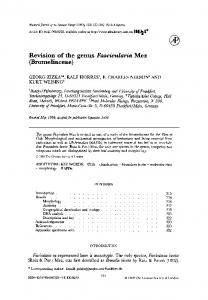
1975). There are approximately 45 species described worldwide, of which about 18 are considered to be valid (Valdés, 2008; OBIS, 2012; Rosenberg, Bouchet & Gofas, 2012). Alone, the Atlantic Ocean harbours over 50% of the global diversity, with ten recognized species (Marcus, 1974; Bouchet, 1975). Scaphander species have a strong shell and a cephalic shield that covers the mantle cavity when they burrow in soft sediments (see Fig. 1). The shell of Scaphander is pyriform to globose, with a large aperture, sunken spire, and punctuated grooves. The animal cannot
© 2013 The Linnean Society of London, Zoological Journal of the Linnean Society, 2013, 167, 389–429
389
390
M. H. EILERTSEN AND M. A. E. MALAQUIAS
Figure 1. External morphology of Scaphander; cs, cephalic shield; f, foot; m, mantle; p, parapodia; pl, pallial lobe; s, shell; sg, seminal groove. Scale bar: 5 mm.
retract completely into the shell and lacks operculum (Burn & Thompson, 1998). The genus Scaphander belongs to the clade (‘Order’) Cephalaspidea (Heterobranchia; class Gastropoda), and is often placed in the superfamily Philinoidea (including families Philinidae, Retusidae, Cylichnidae, Gasteropteridae, and Aglajidae), characterized by similarities in the radula and lack of spines in the gizzard (Burn & Thompson, 1998; Mikkelsen, 2002; Bouchet & Rocroi, 2005), but Malaquias et al. (2009a) and Göbbeler & Klussmann-Kolb (2011) did not retrieve this group as monophyletic in a molecular phylogenetic analysis of the Cephalaspidea. Sars (1878) introduced the family Scaphandridae for Scaphander only, but later Fischer (1887) assigned several other genera to it, such as Sabatia, Smaragdinella, Atys, Cylichna, and Amphisphyra. In more recent times Scaphander has often been ascribed to the family Cylichnidae, together with Cylichna, based on several morpho-anatomical potential synapomorphies (e.g. a strong external shell, shape of lateral radular teeth, and lack of distinct parapodia; Mikkelsen, 1996; Burn & Thompson, 1998; Mikkelsen, 2002; Bouchet & Rocroi, 2005). The family name Scaphandridae has seldom been used (e.g. McGinty, 1955; Thompson & Brown, 1984), but Malaquias et al. (2009a) found that Cylichnidae (represented by Cylichna and Scaphander) is not monophyletic, and reinstated Scaphandridae as the valid family name for the genus Scaphander.
BIOLOGY
OF
deeper waters (Marcus, 1974; Pequegnat, 1983). They burrow into the sediment aided by ciliary movement on the cephalic region that sends small particles over the dorsal side (Hurst, 1956). Observations of gut content indicate that Scaphander ingest mud and sand with diatom frustules, foraminifers, and small animals like bivalves, young sea urchins, polychaetes, scaphopods, gastropods, and gastropod shells containing sipunculids (Hurst, 1956; Yonge & Thompson, 1976). Scaphander are simultaneous hermaphrodites, but very little is known about their reproductive biology and larval development. This is clearly demonstrated by Schaefer (1996) in his summary of cephalaspidean reproduction, where the only data on Scaphander is the spawning period of Scaphander lignarius (January–February near Plymouth, and April near the Isle of Man; Thompson & Brown, 1984). The information available on the reproduction and development in Cephalaspidea is very uneven, with some genera and families more studied than others (e.g. Haminoea and Philine, see Schaefer, 1996), but planktotrophy appears to be most common (Schaefer, 1996). Scaphander lignarius is known to produce secondary metabolites, lignarenone A and lignarenone B, which are probably alarm pheromones used to warn conspecifics of predators, but this has not been possible to test because of the deep bathymetric distribution of these snails (Cimino, Spinella & Sodano, 1989). The biosynthetic pathways of these metabolites have been studied by Cutignano et al. (2008).
SCAPHANDER
Members of the genus Scaphander live at depths of 16–4255 m, with some species having a shallower bathymethric distribution, and some living only in
AIMS The main objective of this work is to revise the systematics of the Atlantic species of Scaphander and
© 2013 The Linnean Society of London, Zoological Journal of the Linnean Society, 2013, 167, 389–429
SYSTEMATICS OF ATLANTIC SCAPHANDER to produce a molecular phylogeny to aid in species delimitation. Furthermore, we aim to establish the morphological characters that distinguish between species, the geographical distribution of each species, and to create a dichotomous key to facilitate species identification.
MATERIAL AND METHODS SAMPLING, TYPE MATERIAL, SYNONYMIES,
AND
GEOGRAPHIC DISTRIBUTIONS
Specimens for anatomical and molecular work have been assembled from natural history museums (see list of institutional abbreviations below). Sampling cruises were undertaken along the coast of Norway under the research project MAREANO. Original descriptions for all nominal names of Atlantic Scaphander (valid names and synonyms) were assembled, as well as more recent works on Scaphander species, including re-descriptions. Type specimens for nearly all nominal species were studied and photographed. Specimens were checked against literature and types. Synonymies attempt to be as complete as possible. Geographical distributions have been inferred from the study of museum material, newly collected specimens, and reliable literature records, and were plotted using ArcMap (ESRI, 2011). When geographical coordinates were not available they were estimated from locality descriptions. If identification was uncertain or single specimens occurred outside the range set by reliable observations, the record was included, but labelled with a question mark (?). Bathymetric distributions were established using a conservative approach: for species where both empty shells and live specimens were known, depth information was collected only from the latter. When depth was known as a range, then the average between the maximum and minimum depth was used.
ABBREVIATIONS ARC, Atlantic Reference Centre, St Andrews, New Brunswick, Canada; FLMNH, Florida Museum of Natural History, Gainseville, FL, USA; H, shell height; MCZ, Museum of Comparative Zoology, Harvard University, Boston, MA, USA; MNHN, Muséum national d’Histoire naturelle, Paris, France; MZUSP, Museu de Zoologia da Universidade de São Paulo, Brazil; NHMUK, Natural History Museum, London, UK; RMNH, National Museum of Natural History (Naturalis), Leiden, the Netherlands; SEM, scanning electron microscopy; sh., shell(s); SMNH, Naturhistoriska Riksmuseet, Stockholm, Sweden; spc(s), preserved animal(s); USNM, National Museum of Natural History, Smithsonian Institution, Wash-
391
ington DC, USA; ZMBN, Natural History Collections, University Museum of Bergen, Norway.
ANATOMICAL
CHARACTERS
For the species where complete animals were available, between three and 16 specimens were dissected to establish the morphological characters that characterize the species, and to understand intraspecific variability. Drawings of the male reproductive system and digestive tract were made using a dissecting microscope fitted with a drawing tube. Shells and gizzard plates were photographed with a camera equipped with macro lens. Radulae, penial papillae, and gizzard plates were mounted for SEM. The radulae and gizzard plates were cleaned with commercial bleach and an ultrasonic machine to remove tissue prior to mounting on SEM stubs. Penial papillae were critical-point dried before mounting in order to maintain the shape of the structures. The SEM stubs were coated with a gold–palladium mixture and then photographed at low magnification to obtain a general view of the entire structures, and then at higher magnification for the details. For the species where only shells were available, species status was assessed by comparing shells with original descriptions and other relevant literature.
DNA
EXTRACTION, AMPLIFICATION, AND SEQUENCING
DNA was extracted from tissue samples from the foot or head shield using the QIAGEN DNeasy Blood and Tissue Kit, following the manufacturer’s protocol (spin-column protocol; QIAGEN). For difficult specimens that did not yield a satisfactory polymerase chain reaction (PCR) product, new extractions were performed using the EZNA Mollusc DNA Kit, following the manufacturer’s protocol. The genetic markers used were cytochrome oxidase c subunit I (COI) and 16S rRNA (two primers each, see Table 1), and nuclear 28S rRNA (four primers, see Table 1). For 28S rRNA, two primers (in bold font in Table 1) were used for amplification and an additional two internal primers were used for the sequencing. The PCR reaction volume was 50 mL for the three genes. For COI and 16S rRNA, 17.5 mL Sigma-Aldrich water, 5 mL buffer, 5 mL dNTP, 10 mL Q solution, 7 mL MgCl2, 2 mL of each of the primers, 0.5 mL TAQ, and 1 mL DNA were used, whereas for the 28S rRNA only 2 mL of MgCl2 was added, and the Sigma-Aldrich water volume used was 22.5 mL; all other items were the same for COI and 16S rRNA. For each of the genetic markers, optimization PCRs with annealing temperatures of 43–55 °C (COI) and 48–60 °C (16S and 28S) were run to establish the optimum annealing temperature. PCR thermal cycles for COI, 16S rRNA, and 28S rRNA were the same, but with specific annealing temperatures. An initial
© 2013 The Linnean Society of London, Zoological Journal of the Linnean Society, 2013, 167, 389–429
392
M. H. EILERTSEN AND M. A. E. MALAQUIAS
Table 1. Sequencing primers for polymerase chain reactions
COI 16S 28S
Name
Sequence 5′ → 3′
Source
LCO1490 (F) HCO2198 (R) 16S ar-L 16S br-H LSU5-F 900-F LSU1600-R ECD2S-R
GGTCAACAAATCATAAAGATATTGG TAAACTTCAGGGTGACCAAAAATCA CGCCTGTTTATCAAAAACAT CCGGTCTGAACTCAGATCACGT TAGGTCGACCCGCTGAAYTTAAGCA CCGTCTTGAAACACGGACCAAG AGCGCCATCCATTTTCAGG CTTGGTCCGTGTTTCAAGACGG
Folmer et al. (1994)
denaturation of 3 min at 95 °C, followed by 39 cycles with denaturation of 45 s at 94 °C, with annealing for 45 s at 45.0 °C (COI), 51.5 °C (16S), or 52.0 °C (28S), and with an extension for 2 min at 72 °C. The final extension was achieved by 10 min at 72 °C. One positive and one negative control were included in each run to check for a successful amplification reaction and to rule out contamination. The quality of PCR products was assessed using gel electrophoresis imaging. PCR product (5 mL) was mixed with 2 mL of loading buffer and run on a 1.2% agarose gel based on half-strength TAE buffer (Tris base, acetic acid, and EDTA) and containing the staining agent GelRed (Biotium, Hayward, CA, USA). pGEM marker (5 mL; Promega, Madison, WI, USA) was used to quantify and estimate the length of amplified DNA fragments. The gel was run at 80–100 V for 40–60 min and analysed under UV light. GeneSnap (SynGene) was used to capture images. In cases were gel electrophoresis imaging revealed double bands, the total PCR product was run on a new gel (same specifications as above) for 90–120 min, the individual bands were then cut out from the gel with a sterile scalpel blade and placed individually in sterile 1.5-mL tubes. Gel extraction was performed using QIAquick Gel Extraction Kit (QIAGEN) following the manufacturer’s protocol. PCR products were quantified using manual band quantification in GeneTools (SynGene). Successful PCRs were purified using Exonuclease 1 (EXO, 10 U mL–1) and shrimp alkaline phosphatase (SAP, 10 U mL–1, USB®) in 25-mL reactions (EXO 0.25 mL, SAP 2.5 mL, Sigma-Aldrich water = 2.25 mL, PCR product 20 mL). Samples were incubated at 37 °C for 15 min followed by an inactivation step at 80 °C for 15 min. The purified PCR products were sequenced using an Automatic Sequencer 3730XL.
ALIGNMENT
OF
DNA
SEQUENCES AND
PHYLOGENETIC ANALYSES
SEQUENCHER 4.10.1 (Gene Codes Corp.) was used to assemble the forward and reverse strands, and
Palumbi et al. (1991) Littlewood, Curini-Galletti & Herniou (2000) Olson et al. (2003) Williams, Reid & Littlewood (2003) Modified from Littlewood et al. (2000)
to assess the quality of the sequences, which were edited by careful examination of chromatograms, and verified by forward and reverse comparisons. The sequences were blasted in GenBank to check for potential contamination and aligned using ClustalX (Thompson et al., 1997), with a gap-opening penalty of 60 and a gap-extension penalty of 30. The alignments were then optimized by eye using MacClade 4.06 (Maddison & Maddison, 2000), trimmed to the longest sequence, and missing data at the ends were coded with question marks (?). The sequences have been deposited in GenBank (see Table 2), and the concatenated alignment and consensus tree is listed in TreeBASE (study no. 13753). Saturation was tested for each gene and for the first, second, and third codon positions of the COI gene by plotting general time-reversible (GTR) pairwise distances against total substitutions (transitions + transversions). Four individual gene analyses were performed (trees not shown): (1) COI; (2) COI (excluding the third codon positions); (3) 16S rRNA; and (4) 28S rRNA, as well as a complete concatenated data set (all taxa and all sequences included), with empty regions of the alignment coded as missing data (see Table 2 for complete list of specimens). Representatives of the genera Bulla and Haminoea were included to test the monophyly of Scaphander. All analyses were rooted with Diaphana because it represents the most basal lineage of Cephalaspidea (Malaquias et al., 2009a; Jörger et al., 2010). The best-fitting models of evolution (see Table 3) were selected using Akaike’s information criterion (Akaike, 1974) implemented in MODELTEST 3.7 (Posada & Crandall, 1998). Distances and substitutions were calculated in PAUP* (Swofford, 2003) and plotted in R (R Development Core Team, 2012). The phylogenetic analyses were performed in MRBAYES (Huelsenbeck & Ronquist, 2001; Ronquist & Huelsenbeck, 2003), with three parallel runs of 2 million generations for the single-gene data sets and 5 million generations for the concatenated data set, with sampling every 100 generations. The con-
© 2013 The Linnean Society of London, Zoological Journal of the Linnean Society, 2013, 167, 389–429
Seq no. 1 2 12 19 37 51 GB1 GB2 GB3 3 4 34 35 36 38 5 8 15 17 9 13 52 21 29 31 30 32 33
Species
Scaphander lignarius Scaphander lignarius Scaphander lignarius Scaphander lignarius Scaphander lignarius Scaphander lignarius Scaphander lignarius Scaphander lignarius Scaphander lignarius Scaphander punctostriatus Scaphander punctostriatus Scaphander punctostriatus Scaphander punctostriatus Scaphander punctostriatus Scaphander punctostriatus Scaphander watsoni Scaphander watsoni Scaphander watsoni Scaphander watsoni Scaphander nobilis Scaphander bathymophilus Scaphander bathymophilus Scaphander darius Scaphander mundus Scaphander mundus Scaphander sp. A Scaphander sp. B Scaphander subglobosus Haminoea orbignyana Haminoea orteai Bulla occidentalis Bulla striata Diaphana sp.
Spain Bergen, Norway Celtic Sea Spain Bergen, Norway Norway Algarve, Portugal Algarve, Portugal Blanes, Spain 60°09′ N, 05°19′ E Newfoundland Norway Norway Skagerrak Norway Brazil Brazil Gulf of mexico Gulf of Mexico Bay of Biscay Azores Puerto Rico Brazil Philippines Philippines New Caledonia New Caledonia Philippines Naples, Italy Faial, Azores Florida
Locality
ZMBN 88002 MNHN, IM-2009-29695 ZMBN 88006 ZMBN 88005 ZMBN 88004 ZMBN 88003 MZSP 34644 MZSP 86804 USNM 1151226 USNM 1151240 MNHN, IM-2009-29696 RMNH unnr. MZSP 75708 MZSP 29016 MNHN, IM-2009–4319 MNHN, IM-2009–4318 MNHN, IM-2009–4317 MNHN, IM-2009–4371 MNHN, IM-2009–4339 ZMBN 81714 NHMUK 20070459 NHMUK 20030779/1 NHMUK 20030784/3
ZMBN 87998 ZMBN 87999 MNHN, IM-2009-29694 MCZ 371884 ZMBN 88000 ZMBN 88001 NHMUK 20060325 NHMUK 20060114
Voucher no.
KC351572 KC351573 KC351574 KC404964 KC404963 DQ986543 DQ986566 EF489394.1
KC351560 KC351565
KC351559
KC351575 KC351576
KC351568 KC351566 KC351571 KC351570 KC351569 KC351567
KC351561 KC351563 KC351564 DQ974663 DQ974664
KC351562
COI
Table 2. Taxa set used for molecular phylogenetics with locality, voucher numbers, and GenBank accession numbers
KC351530 KC351520 KC351519 KC351521 KC351529 KC351528 KC351537 KC351538 KC351539 KC404959 KC404960 DQ986603 DQ986631 EF489325.1
KC351532 KC351531 KC351536 KC351535 KC351534 KC351533 KC351540 KC351541 KC351542
KC351524 KC351525 KC351523 KC351522 KC351526 KC351527 DQ923454
16S
KC351547 KC351546 KC351554 KC351555 KC351556 KC404961 KC404962 DQ986666 DQ986693 EF489373.1
KC351557 KC351558
DQ927221 DQ927212 EF489372 KC351549 KC351548 KC351553 KC351551 KC351552 KC351550
KC351545
KC351543 KC351544
28S
SYSTEMATICS OF ATLANTIC SCAPHANDER
© 2013 The Linnean Society of London, Zoological Journal of the Linnean Society, 2013, 167, 389–429
393
394
M. H. EILERTSEN AND M. A. E. MALAQUIAS
Table 3. Best-fit model and estimated parameters for phylogenetic analyses Parameter
COI
16S
28S
No. specimens No. characters Best-fit model Frequency A Frequency C Frequency G Frequency T Gamma shape Prop. inv. sites R-matrix [A-C] R-matrix [A-G] R-matrix [A-T] R-matrix [C-G] R-matrix [C-T] R-matrix [G-T]
25 698 TVM+I+G 0.2853 0.1504 0.1543 0.4100 0.8877 0.5582 0.1855 9.4538 0.5056 0.5378 9.4538 1.0000
28 465 TVM+I+G 0.3097 0.1564 0.2110 0.3229 0.6974 0.4077 0.3500 7.7476 4.8052 0.0000 7.7476 1.0000
23 1514 GTR+I+G 0.1922 0.3221 0.2613 0.2245 1.3875 0.7717 2.9100 17.4542 3.6316 1.6120 2.9827 1.0000
Prop. inv. sites = proportion of invariant sites.
vergence of runs was assessed using TRACER 1.5 (Rambaut & Drummond, 2007) and the burn-in was set to 10%. Consensus phylograms were generated in MRBAYES, annotated and converted to graphics in FIGTREE 1.3.1 (Morariu et al., 2008), and final adjustments were made in ADOBE ILLUSTRATOR CS6.
RESULTS SYSTEMATIC GENUS SCAPHANDER
DESCRIPTIONS DE
MONTFORT, 1810
Charta Martini, 1769: 283, 284, pl. 21, figs 194, 195. Type by monotypy Charta convoluta [ = Scaphander lignarius (Linnaeus, 1758)]. Nomen oblitum following International Commission on Zoological Nomenclature (ICZN), article 53.9. Gioeni Gioeni, 1783: 5–36, pl. 1, figs 1–13. Type by subsequent designation Tricla gioeni Philipsson, 1788 [ = Scaphander lignarius (Linnaeus, 1758)]. Nomen oblitum following ICZN article 53.9. Tricla Philipsson, 1788: 8; Winckworth, 1932: 232. Type by monotypy Tricla gioeni Philipsson, 1788 [ = Scaphander lignarius (Linnaeus, 1758)]. Suppressed by ICZN (1954: opinion 287). Gioenia Bruguière, 1792: 12, 502–504. Type by monotypy Gioenia sicula. Supressed by ICZN (1954: opinion 287). Scaphander de Montfort, 1810: 334, pl. 84. Type by monotypy Scaphander lignarius (Linnaeus, 1758). Nomen protectum following ICZN, article 53.9. Bulla (Scaphander) Adams, 1855: 574; Weinkauff, 1862: 336.
Scaphander (Sabatia) Dall, 1889a: 86, pl. 17, figs 9, 9b; Dall, 1889b: 53, 54, pl. 17, figs 9, 9b. Scaphander (Sabatina) Dall, 1908: 240, 241. Type by original designation Scaphander (Sabatina) planeticus: Dall, 1908. Assula Schumacher, 1817: 258. Type by monotypy Assula convoluta [ = Scaphander lignarius (Linnaeus, 1758)]. Sabatia (Sabatina) Dall, 1927: 25. Bulla (Bullocardia) Nordsieck, 1972: 29, pl. 7, fig. 25. Type by original designation Bulla millepunctata Locard, 1897 (= Scaphander nobilis Verill, 1884). Bucconia Dall, 1890: 16, 17, pl. 10, fig. 9; Habe, 1955: 69; Bullis, 1956: 2, 3, pl. 2, figs A,B,D,E. Type by original designation Scaphander nobilis Verill, k1884: 209, 210, pl. 32, figs 18, 18a–d. Eoscaphander Habe, 1952: 75–77, figs 7, 8. Type by monotypy Eoscaphander fragilis. Nipponoscaphander Kuroda & Habe in Kuroda, Habe & Oyama, 1971: 292, pl. 64, fig. 27. Type by original designation Scaphander japonicus Adams, 1962. Taxonomic history The first species of Scaphander to be described (S. lignarius) was originally placed in the genus Bulla, as defined by Linnaeus (1758). Bulla was redefined during the 19th century (reviewed in Malaquias & Reid, 2008), and the name Scaphander was introduced by de Montfort (1810) to include Bulla lignaria Linnaeus, 1758. Martini (1769) described a shell resembling a roll of paper (‘Das eingerollte papier’) and named it Charta convoluta. His figure shows the shell of Scaphander lignarius, and he also mentions Bulla lignaria Linnaeus, 1758 as a synonym. This overlooked publication contains the oldest available putative synonym of Scaphander de Montfort, 1810. Schumacher (1817) proposed a new genus, Assula, for the species Charta convoluta Martini, 1769, without any justification of this change. According to ICZN (1999; article 23.9), the precedence of names can be reversed if the senior synonym has not been used as valid after 1899 and the junior synonym has been used as valid in at least 25 publications, by at least ten different authors, in the immediately preceding 50 years, and encompassing a span of no less than 10 years. To our best knowledge, the name Charta has not been used as valid since its publication in 1769, and the junior synonym (Scaphander de Montfort, 1810) is used as valid in at least the following publications: Marcus & Marcus (1967), Marcus (1971), Keen (1971), Marcus (1974), Bouchet (1975), Yonge & Thompson (1976), D’Angelo & Gargiullo (1978), Pequegnat (1983), Thompson & Brown (1984), Cimino et al. (1989), Poppe & Goto (1991), Mikkelsen (1996), Schaefer
© 2013 The Linnean Society of London, Zoological Journal of the Linnean Society, 2013, 167, 389–429
SYSTEMATICS OF ATLANTIC SCAPHANDER (1996), Burn & Thompson (1998), Mikkelsen (2002), Cutignano et al. (2008), Klussmann-Kolb et al. (2008), Valdés (2008), Malaquias et al. (2009a), Malaquias, Berecibar & Reid (2009b), Segers, Swinnen & de Prins (2009), Rios (2009), Poppe (2010), Daccarett & Rossio (2011), and Gofas, Moreno & Salas (2011). Therefore, Scaphander de Montfort, 1810 is here validated as a nomen protectum and Charta Martini, 1769 is considered a nomen oblitum. Gioeni (1783) found a gizzard of S. lignarius and described it as if it was a complete animal and named the new taxon Gioeni (of unspecified rank). Philipsson (1788) regarded Gioeni as the species epithet and gave the supposed animal the binominal name Tricla gioeni, whereas Bruguière (1792) considered it the genus epithet and gave it the name Gioenia sicula. The true nature of the ‘animal’ was exposed by Draparnaud (1800), and the names Tricla and Gioenia were not used until Winckworth (1932) introduced Tricla as a generic name for S. lignarius in his ‘British Marine Mollusca’. He was followed by a few other authors until Tricla and Gioenia were suppressed by ICZN (1954: opinion 287), following an application by Lemche. Gioeni Gioeni, 1783 was never invalidated by the ICZN because it is a uninominal name, and Lemche considered it not available; however, as pointed out by Valdés (2008), ICZN (1999: article 11.4.1) states that ‘A published work containing family-group names or genus-group names without associated nominal species is accepted as consistent with the Principle of Binominal Nomenclature in the absence of evidence to the contrary’. Gioeni Gioeni, 1783 has, to the best of our knowledge, not been used as valid after 1899, and as shown above, Scaphander de Montfort, 1810 fulfils the conditions of ICZN (1999: article 23.9.1). Gioeni Gioeni, 1783 is therefore here considered a nomen oblitum, and Scaphander de Montfort, 1810 is maintained as a nomen protectum. Adams (1855) considered Scaphander a subgenus of Bulla, where he included the species Bulla lignaria Linnaeus, 1758 and Bulla puncto-striata Mighels & Adams, 1842, along with three other species later assigned to the genera Philine and Johania (CLEMAM, 2012). Dall (1890) divided the genus Scaphander into two subgenera: Scaphander and Bucconia. He defined Scaphander based on the pyriform shape of the shell of the type species S. lignarius, and Bucconia by a more globose shape of the shell and a ‘posterior pillar extended backward and supporting an expansion of the outer lip’, where he included the species Scaphander nobilis Verill, 1884. Dall (1890) also transferred the fossil species Scaphander grandis to the subgenus Bucconia. The subgenus was adopted by Bullis (1956) and Habe (1955), who gave it generic status, but Bouchet (1975) found no consistent ana-
395
tomical differences supporting the subgenus Bucconia, and Valdés (2008) came to the same conclusion from examining Pacific specimens of Scaphandridae. The species Scaphander bathymophilus (Dall, 1881) was originally described as an Atys (Dall, 1881), but a few years later Dall assigned it to Sabatia (Bellardi, 1876) and made this a subgenus of Scaphander (Dall, 1889a, 1889b). Sabatia (Bellardi, 1876) is based on a Pliocene fossil, which later led Dall (1908) to propose the name Sabatina to separate the recent species from the fossil group. Valdés (2008) suggested that Sabatina should be considered a synonym of Sabatia based on the presence of a parietal callus as a synapomorphy, but stressed that the taxonomic validity of the genera/subgenera Scaphander, Bucconia, Sabatia, and Sabatina needed further work. In the present study neither anatomical differences nor phylogenetic evidence were found to support the division of Scaphandridae in different genera (see Discussion). Habe (1952) described a new genus, Eoscaphander, and a new species, Eoscaphander fragilis. The author claimed that his species differs from the type species of Scaphander in the radular characters, but gave no details about these differences. Valdés (2008) compared the anatomy of E. fragilis with Scaphander species from the West Pacific and found no sound anatomical differences. Eoscaphander is therefore considered a synonym of Scaphander. Kuroda et al. (1971) assigned Scaphander japonicus to a new genus Nipponoscaphander defined by a smaller and more ovoid shell than typical Scaphander species. Valdés (2008) reviewed S. japonicus and found no anatomical or shell differences that separate this species from other Scaphander, and therefore Nipponoscaphander is here considered a synonym of Scaphander. Bullocardia was described as a subgenus of Bulla with Bulla millepunctata Locard, 1897 as the type species. Bulla millepunctata is a synonym of S. nobilis (see systematic description of S. nobilis), and therefore Bullocardia is considered a synonym of Scaphander. Diagnosis Shell external, usually solid, pyriform to ovoid, with only one visible whorl. Shell sculpture composed of spiral grooves, usually punctuated. Aperture as long as shell; spire sunken. Animal can only partially withdraw into shell; operculum absent. Large head shield without posterior lobes, parapodial lobes present, not concealing the shell. Eyes absent. Radular formula N ¥ 1.1.1 or N ¥ 1.0.1, rachidian teeth, when present, small and fragile, probably vestigial. Flexible, non-muscular crop, gizzard large with three calcified gizzard plates (two large paired and one smaller unpaired), bound together by muscular fibre. Reproductive system monaulic, penis armed or unarmed.
© 2013 The Linnean Society of London, Zoological Journal of the Linnean Society, 2013, 167, 389–429
396
M. H. EILERTSEN AND M. A. E. MALAQUIAS
Remarks The presence or absence of rachidian teeth in the radula of Scaphander has been a matter of debate. In several publications (e.g. Sars, 1878; Marcus & Marcus, 1967) a rachidian tooth is depicted, but Bullis (1956) stated that this must be a mistake and that he did not find rachidian teeth in the species he examined. Valdés (2008) did not find rachidian teeth in the Scaphander species from the south-west Pacific. In this study rachidian teeth were found to be very small and fragile, and easily lost during the preparation of the radula. Rachidian teeth in Scaphander are vestigial, and may not serve any function, but this study proves their presence in all of the Atlantic species studied. The fragile nature of these teeth is likely to be the reason for the misunderstanding, but careful preparation of the radula might show them to be present in all species in the genus. Valdés (2008) defined Scaphander as having a penis that can be ‘armed or not’. The penis is unarmed in all Atlantic species for which the male reproductive system is known. Based on Valdés’ (2008) diagnosis of Scaphander, the genus is here defined as potentially having an armed penis, as we could not confirm this for the Indo-Pacific species.
SCAPHANDER
BATHYMOPHILUS
(DALL, 1881)
Atys bathymophila Dall, 1881: 98 (Yucatan Strait; types seen, 2 syntypes, MCZ 6990, H = 4.9, 5.4 mm, Gulf of Mexico (24°1′N, 88°58′W); 1 syntype MCZ 6987, H = 15 mm, Yucatan; 3 syntypes, USNM 95198, H = 3.6, 4.9, 8.3 mm). Scaphander (Sabatia) bathymophila Dall, 1889a: 86, pl. 17, figs 9, 9b; Dall, 1889b: 53, 54, pl. 17, figs 9, 9b; Pilsbry, 1893: 256, pl. 32, figs 27, 28; Johnson, 1934: 147. Sabatia bathymophila Maury, 1922: 49; Thiele, 1931: 391. Roxania (Sabatia) bathymophila Thiele, 1925: 244; Zilch, 1959–60: 27. Sabatia (Sabatina) bathymophila Dall, 1927: 25. Sabatia bathymophilus: Bullis, 1956: 2; Clarke, 1962: 40. Scaphander (Sabatina) bathymophilus Dall, 1908: 241; Marcus, 1974: 341–345, figs 67–82. Scaphander punctostriatus var. clavus Dall, 1889b: 52 (in part; specimen from Barbados). Scaphander loisae Bullis, 1956: 8, figs 2G, H (Barbados, Caribbean Sea; type seen, holotype USNM 95188, H = 17.2 mm). Taxonomic history Dall (1881) first proposed the name Atys bathymophila for shells from the Yucatan Strait, casting
doubts, however, on the generic assignment. Later, Dall (1889a, 1889b) placed the species in the subgenus Sabatia Bellardi, 1876. For discussion of this subgenus, see the taxonomic history of Scaphander. The anatomy of S. bathymophilus was studied by Marcus (1974), and shows great similarity to that of other Scaphander species. This study confirms these similarities, and Atys bathymophila Dall, 1881 is here considered a member of the genus Scaphander. Bullis (1956) examined the two specimens of S. punctostriatus var. clavus described by Dall (1889b) and found them to be two distinct species. The specimen from Barbados was chosen as the type specimen of a new species, namely Scaphander loisae. This species is only known from the type specimen, and nothing is known of the anatomy. The type specimen of S. loisae was examined, and it has the characteristic tuberculate callus of S. bathymophilus. The outline of the shell is also identical to the latter species (see Fig. 2: 12). Continuous striations, as seen in S. loisae, are not an uncommon intraspecific variation (e.g. S. clavus), and can also be present in some sections of the shell in a specimen with otherwise punctuated striations. Scaphander loisae Bullis, 1956 is here considered a synonym of S. bathymophilus (Dall, 1881). Diagnosis Shell solid, ovoid, white or cream. Striations punctuated by round, interconnected depressions. Parietal wall covered by thick, tuberculate, white callus. Body white. Lateral radular teeth with complex denticulation, rachidian teeth rectangular, with two posterolateral, triangular extensions. Prostate cylindrical, rounded at the end. Prostatic duct separating prostate and penial chamber, penial chamber cylindrical, extended where prostatic duct attaches. Penial papilla short with smooth surface. Type locality: Yucatan strait. Material examined Yucatan strait, 2 sh., MCZ 66690, H = 4.9, 5.4 mm; Gulf of Mexico (24°1′N, 88°58′W), 1 sh. MCZ 6987, H = 15 mm; Cape Hatteras, 32°50′N, 77°00′W, 1 spc. dissected, H = 17.4 mm; locality unknown, 3 sh., USNM 95198, H = 3.6, 4.9, 8.3 mm; locality unknown, 1 sh., USNM 836689, H = 24.6 mm; Puerto Rico, 1 spc. dissected, MZSP 75708, H = 30.5 mm; Azores, 2 spcs dissected, RMNH unnumbered, H = 19.2, 19.8 mm. Shell (Fig. 2: 6–9, 11, 12): Maximum H = 37.5 mm (Marcus, 1974). Shell solid and ovoid, aperture very narrow posteriorly, extended anteriorly. Posterior edge of outer lip rounded, protruding beyond apex. A thin, white, or cream periostracum sometimes visible,
© 2013 The Linnean Society of London, Zoological Journal of the Linnean Society, 2013, 167, 389–429
SYSTEMATICS OF ATLANTIC SCAPHANDER
397
Figure 2. Shells and SEM images of the shell scuplture of Scaphander nobilis (1–5), Scaphander bathymophilus (6–9, 11, 12), and Scaphander gracilis (10); 1, holotype, Delaware Bay, USA (USNM 35641, H = 34.2 mm); 2, Scaphander stigmaticus, holotype, Gulf of Mexico (USNM 95196, H = 35.4 mm); 3, surface structure of shell, Georges Bank, USA (USNM 45562, H = 11.3 mm); 4, surface structure of shell, Bay of Biscay (MNHN, IM-2009-29696, H = 31.9 mm); 5, Bay of Biscay (MNHN, IM-2009-29696, H = 31.9 mm); 6, syntype, Yucatan, Mexico (MCZ 6987, H = 15.0 mm; 7, surface structure of shell, Cape Hatteras (MCZ 364990, H = 12 mm); 8, Azores (RMNH, unnumbered, H = 19.8 mm); 9, Cape Hatteras (MCZ 364990, H = 12 mm); 10, São Miguel I., Azores, syntype (NHMUK 1887.2.9.2187-8, H = 13.0 mm; image courtesy of the NHMUK photographic unit); 11, Yucatan Strait, syntype (MCZ 6990, H = 5.4 mm); 12, Barbados, holotype Scaphander loisae (USNM 95188, H = 17.2 mm). Scale bars: 500 mm. © 2013 The Linnean Society of London, Zoological Journal of the Linnean Society, 2013, 167, 389–429
398
M. H. EILERTSEN AND M. A. E. MALAQUIAS
Figure 3. Shells and SEM images of the shell scuplture of Scaphander lignarius (1–4), Scaphander punctostriatus (5–8), and Scaphander clavus (9–12); 1, Høylandssundet, Hardangerfjord, Norway (ZMBN 62008, H = 33.0 mm); 2, surface structure of shell, Norway (ZMBN 17883, H = 23.7 mm); 3, syntype, Scaphander lignarius var. alba, Shetland Islands (USNM 176422, H = 37.4 mm); 4, Dublin Bay, Ireland (RMNH, unnumbered, H = 70 mm); 5, syntype, Scaphander librarius, Finnmark, Norway (SMNH 8039, H = 16.2 mm); 6, surface structure of shell, South Newfoundland (MNHN, IM-2009-29695, H = 33.2 mm); 7, neotype, locality unknown (MCZ 177003, H = 11.0 mm); 8, surface structure of shell, East of Norfolk, Virginia, USA (USNM 757101, H = 12 mm); 9, surface structure of shell, Suriname (USNM 836676, H = 25.5 mm); 10, Panama (MZSP 75.943, H = 17.5 mm), 11, lectotype, Scaphander punctostriatus clavus, Louisiana, USA (MCZ 6981, H = 16.7 mm); 12, Suriname (USNM 836676, H = 25.5 mm). Scale bars: 2, 100 mm; 6, 8, 500 mm; 9, 200 mm. © 2013 The Linnean Society of London, Zoological Journal of the Linnean Society, 2013, 167, 389–429
SYSTEMATICS OF ATLANTIC SCAPHANDER
399
Figure 4. Shells and SEM images of the shell scuplture of Scaphander watsoni (1–5), Scaphander darius (6, 8–9), and Scaphander pilsbryi (7); 1, syntype, off Sombrero Island (MCZ 6982, H = 8.1 mm); 2, Scaphander watsoni rhederi, holotype, Gulf of Mexico (USNM 609948, H = 39.3 mm); 3, surface structure of shell, Gulf of Mexico (USNM 1151226, H = 31.5 mm); 4, surface structure of shell, off Florida, Gulf of Mexico (USNM 855168, H = 13.1 mm); 5, Santos, Brazil (USNM 34644, H = 31.9 mm); 6, holotype, off Colombia and Panama (USNM 679054, H = 15.5 mm); 7, holotype (FLMNH 174325, H = 30.6 mm); 8, Colombia, paratype (MZSP 76266, H = 12.0 mm); 9, off Florida, Gulf of Mexico (USNM 855168, H = 13.1 mm). Scale bars: 500 mm. © 2013 The Linnean Society of London, Zoological Journal of the Linnean Society, 2013, 167, 389–429
400
M. H. EILERTSEN AND M. A. E. MALAQUIAS
shell white under periostracum. Striations with round, interconnected punctuations. Parietal wall covered by a thick, tuberculate, white callus, sometimes forming an extension into the aperture (see Fig. 2: 8). Radula (Fig. 5A–D): Lateral teeth are curved, with a broad base and weak denticulation on inner edge, consisting of several rows of denticles flattened along the side of the teeth. Rachidian teeth rectangular, with two posterolateral, triangular extensions. Digestive tract (Fig. 5G, H): Salivary glands very long, flattened, with uneven, glandular surface. Paired gizzard plates kidney shaped to subtriangular. Male reproductive system (Fig. 5E, F): Prostate cylindrical, rounded at the end and filled with spongy tissue. Prostatic duct separating prostate and penial chamber, penial chamber cylindrical, extended where prostatic duct attaches. Penial papilla short with smooth surface. Ecology Feeds on 1609 m.
foraminiferans.
Depth
range:
805–
Distribution (Fig. 12) From Delaware Bay, USA (Pilsbry, 1893) to Caribbean Sea, Azores (present study). Remarks Dall (1927) reports ‘numerous young specimens’ of S. bathymophilus collected near Georgian and Fernandina (Florida, USA), and also specimens from the Yucatan Strait and around Cuba. He gives a depth range of 1342–5130 m, but does not indicate if the specimens consisted of shells only or live specimens, or what the depth ranges of individual dredgings were. Thus, it is possible that the depth range of S. bathymophila is much deeper than considered in this work (see Ecology).
SCAPHANDER
CLAVUS
DALL, 1889B
Scaphander punctostriatus var. clavus Dall, 1889b: 52; Pilsbry, 1893: 246; Dautzenberg & Fischer, 1896: 401, 402 (Gulf of Mexico, off Louisiana, USA; type seen, 1 lectotype, MZC 6981, H = 16.7 mm. Scaphander clavus Bullis, 1956: 8, fig. 2I; Marcus & Marcus, 1967: 599, figs 1–4; Marcus, 1974: 333, 334, figs 34, 81, 85; Pequegnat, 1983: 159. Taxonomic history The name ‘clavus’ was suggested by Dall (1889b) to describe two shells found in the Gulf of Mexico. He
considered them a variety of S. punctostriatus, differing from that species by a more blunt apex and a ‘more Bulla-like form’. Bullis (1956) raised this variety to specific status and gave a more detailed description of the shell morphology, and which characters separate this species from S. punctostriatus (mainly the shell shape and details of the striation). At this time no live specimens were known. Marcus & Marcus (1967) recorded the first living specimen of this species and depicted the radular teeth, apex, and male genital system, including the penial papilla. They noted that the shape of the shell and striations agreed perfectly with the description by Bullis (1956) and images of the type specimen. Marcus (1974) described the anatomy of another specimen from Surinam, and the description and figures agree with that of Marcus & Marcus (1967). Bouchet (1975) concluded that S. clavus Dall, 1889b is nothing more than a geographical variety of S. punctostriatus. Bouchet (1975) stated that there are differences in shell morphology and radular characters, but he believed these differences were not enough to justify specific status. Bouchet (1975) did not comment on the characters of the male reproductive system of S. clavus and S. punctostriatus, because he believed the specimens he examined to be sexually immature, and therefore had no material to compare with the figure in Marcus & Marcus (1967). The specimens of S. punctostriatus dissected here showed that the species has no penial papilla at any life stage, and adding this anatomical difference to the shell and radular characters it is clear that S. clavus and S. punctostriatus are two valid species. Diagnosis Shell solid, subrectangular, white to orange. Striations closely set with quadrate punctuations separated by short spaces. Body white. Lateral radular teeth with simple denticulation on outer edge, trilobed rachidian teeth with rounded lobes. Prostate tapering towards a short prostatic duct, connecting to the penial chamber. Penial papilla bulbous with a flagellate extension, the bulbous base of the papilla and the tip of the flagellum is covered with warts. Type locality: Gulf of Mexico. Material examined Gulf of Mexico, off Louisiana, USA, 1 sh., MCZ 6981, H = 16.7 mm; Colombia, Cartagena, 1 sh., MZSP 75941, H = 25 mm; Panama, 3 sh., MZSP 75943, H = 17.5, 22.3, 23 mm; Colombia, Isla Fuerte, 1 sh., MZSP 75433, H = 23 mm; Suriname, 1 spc. dissected (anterior part of digestive tract and male reproductive system were missing because of a previous dissection), USNM 836676, H = 25.50 mm.
© 2013 The Linnean Society of London, Zoological Journal of the Linnean Society, 2013, 167, 389–429
SYSTEMATICS OF ATLANTIC SCAPHANDER
401
Figure 5. Anatomical details of Scaphander bathymophilus: A, radula, arrows point to location of rachidian teeth and details of lateral teeth; B, details of lateral teeth, arrow points to location of denticulation; C, denticulation of the outer edge of lateral teeth; D, rachidian teeth; E, male reproductive system; F, penial papilla; G, anterior part of digestive tract; H, gizzard plates; bb, buccal bulb; c, crop; go, genital opening; mo, mouth; o, oesophagus; p, prostate; pc, penial chamber; pd, prostatic duct; pgp, paired gizzard plates; sg, salivary gland; ugp, unpaired gizzard plate. Scale bars: A, 200 mm; B, 50 mm; C, 25 mm; D, 50 mm; E, 1 mm; F, 500 mm; G, 1 mm; H = 5 mm. © 2013 The Linnean Society of London, Zoological Journal of the Linnean Society, 2013, 167, 389–429
402
M. H. EILERTSEN AND M. A. E. MALAQUIAS
Shell (Fig. 3: 9–12): Maximum H = 30 mm. Shell thick and subrectangular in outline. Posterior edge of outer lip rounded, protruding slightly beyond apex. Periostracum sometimes visible, colour white to orange. Shells white under periostracum. Thick, closely set striations, some specimens with coloured striations (see Fig. 3: 12). Punctuations quadrate, separated by a short space. Sometimes fused together forming a continuous line (see Fig. 3: 9). Parietal wall covered by white callus. Radula: Lateral teeth are curved with a broad base and simple denticulation on inner edge. Rachidian teeth with square base and three anterior lobes (based on Marcus & Marcus, 1967). Digestive tract: Paired gizzard plates subtriangular, with a strongly calcified, yellow centre, and more porous edges, as in the other species (present study, data not shown). Shape of anterior part of digestive tract (in front of the gizzard, including salivary glands) not known. Male reproductive system: Prostate tapering towards a short prostatic duct, connecting to the penial chamber. Penial papilla bulbous with a flagellate extension, base of papilla and the tip of flagellum is covered with warts. Walls of the penial chamber harbours a long ridge (Marcus & Marcus, 1967; Marcus, 1974). Ecology Marcus & Marcus (1967) found isopods and calcareous algae in the crop of one specimen from the Caribbean Sea. Depth range: 595–1056 m. Distribution (Fig. 14) Gulf of Mexico (Pequegnat, 1983), Caribbean Sea (Marcus, 1974), and Suriname (present study). Remarks Only one specimen was available for dissection, and it had already been dissected by someone else. The anterior part of the digestive tract (anterior to the gizzard) and the male reproductive system were missing. Dautzenberg & Fischer (1896) reported finding S. punctostriatus var. clavus Dall, 1889b around the Azores, and they described the specimens as heavy shells with less attenuation at the top. With this little information about the specimens, and no figures or pictures to confirm the identification, this record is not considered reliable. Knowing the vari-
ability of shell morphology found in S. punctostriatus, it might as well be that species, which is common around the Azores.
SCAPHANDER
DARIUS
MARCUS & MARCUS, 1967
?Scaphander pilsbryi McGinty, 1955: 82, 83, pl. 2, fig. 8 (Pensacola, Florida, USA; type images seen, FLMNH 174325, H = 30.6 mm). Scaphander darius Marcus & Marcus, 1967: 603, 604, figs 10–17; Marcus, 1971: 925; Marcus, 1974: 336–340, figs 57–66, 81, 86 (Caribbean Sea, off Colombia and Panama; type seen, holotype, USNM 679054, H = 15.5 mm; type images seen, MZSP 76266, 2 paratypes, H = 12.0, 15.3 mm. Taxonomic history Scaphander darius was described by Marcus & Marcus (1967), who commented on its morphological similarity to both the eastern Atlantic Scaphander lignarius and the western Atlantic Scaphander watsoni, with the latter being so similar that one could not separate between the two species without examining the male copulatory apparatus (Marcus, 1974). Diagnosis Shell thin or solid, pyriform, white to light brown. Striations punctuated by round interconnected punctuations. Parietal wall often covered by thin, white callus. Body white. Lateral radular teeth with simple denticulation on both edges, rachidian teeth trilobed. Prostate short, cylindrical; prostatic duct separating prostate and penial chamber. Penial chamber bulbous, penial papilla bulbous, covered with warts. Type locality: off Colombia and Panama, Caribbean Sea. Material examined Caribbean Sea, off Colombia and Panama, 1 sh., USNM 679054, H = 15.5 mm; 9°31.3′N, 76°15.4′W, 2 sh., MZSP 76266, H = 12.0, 15.3 mm; off Florida, 25°16′54″N, 83°43′11″W, 1 spc. dissected, USNM 855168, H = 13.1 mm; Guarapari, Brazil, 1 spc. dissected, MZSP 29016, H = 12.0 mm. Shell (Fig. 4: 6, 8, 9): Maximum H = 23 mm. Shell thin or solid, pyriform, anterior of aperture only very slightly extended. Posterior edge of outer lip sometimes pointed, protruding slightly beyond apex. Periostracum often visible, colour white to light brown. Shell white under periostracum. Thick punctuated striations. Punctuations are round and interconnected along the striae. Grooves are separated by gaps much wider than the grooves themselves. Radula (Fig. 6A–D): Lateral teeth curved with a broad base, one row of denticulation on both edges.
© 2013 The Linnean Society of London, Zoological Journal of the Linnean Society, 2013, 167, 389–429
SYSTEMATICS OF ATLANTIC SCAPHANDER
403
Figure 6. Anatomical details of Scaphander darius: A, radula, arrows pointing to location of rachidian teeth and details of lateral teeth; B, details of lateral teeth, arrow points to location of denticulation; C, denticulation of the outer edge of lateral teeth; D, rachidian tooth; E, male reproductive system; F, penial papilla, arrow pointing to circle of large warts; G, anterior part of digestive tract; H, gizzard plates; bb, buccal bulb; c, crop; go, genital opening; m, muscle; mo, mouth; o, oesophagus; p, prostate; pc, penial chamber; pd, penial duct; pgp, paired gizzard plates; sg, salivary gland; upg, unpaired gizzard plate. Scale bars: A, 500 mm; B, 100 mm; C, 20 mm; D, 50 mm; E, 5 mm; F, 200 mm; G, 2 mm; H, 5 mm. © 2013 The Linnean Society of London, Zoological Journal of the Linnean Society, 2013, 167, 389–429
404
M. H. EILERTSEN AND M. A. E. MALAQUIAS
Rachidian teeth wider than long, base square, anterior end trilobed with triangular lobes. Digestive tract (Fig. 6G, H): Salivary glands short with uneven surface. Paired gizzard plates subtriangular. Central area of gizzard plates often yellowish and thicker than lateral area, which is porous and thinner. Male reproductive system (Fig. 6E, F): Prostate cylindrical, rounded at distal end, prostatic duct separating prostate and penial chamber. Prostate filled with spongy tissue. Penial chamber bulbous, penial papilla bulbous, with a circle of large warts around the base, and papilla covered with small warts. Ecology Feeds on foraminiferans. Depth range: 16–97 m. Punctuations are round and interconnected along the striae. Grooves are separated by gaps much wider than the grooves themselves. Distribution (Fig. 13) Gulf of Mexico, Caribbean Sea, and Brazil (Marcus & Marcus, 1967; Marcus, 1971; Marcus, 1974). Remarks Scaphander pilsbryi McGinty, 1955 was described based on a single shell from Pensacola, Florida. Images of the type shell (FLMNH 174325, H = 31 mm) have been examined (Fig. 9: 7), and although the posterior end is more open compared with typical S. darius and S. watsoni, this feature falls within the variation of shell morphology seen in the latter two species. It is, however, impossible to say to which of these two species the shell of S. pilsbryi belongs, because, as discussed earlier, these species are only distinguishable by their internal anatomy.
SCAPHANDER
GRACILIS
WATSON, 1883
Scaphander gracilis Watson, 1883: 345, 346; Watson, 1886: 645, 646, pl. 47, figs 4a–c; Pilsbry, 1893: 247, 248, pl. 31, figs 19, 20; Dautzenberg & Fischer, 1896: 402; Locard, 1897: 47–49, pl. 1, figs 15–18; Bouchet, 1975: 338, pl. 3, fig. i (Western Azores, south of Flores Island; types seen, 4 syntypes, NHMUK 1887.2.9.2183–6, H = 8.8, 5.1, 11.3, 13.5 mm. south of São Miguel Island, Azores; 2 syntypes, NHMUK 1887.2.9.2187–8, H = 13.0, 14.4 mm). Scaphander gracilis var. minor Locard, 1897: 47–49.
Scaphander gracilis var. major Locard, 1897: 47–49. Taxonomic history The name Scaphander gracilis was first used by Watson (1883) for shells found in the Azores during the ‘Challenger’ expedition. Watson (1883) compared the shells with S. punctostriatus, but differences in the shape and form of the callus and striations separate them from the latter species. Shells of this species have later been found around the Azores (the only known locality; Locard, 1897), but complete specimens have never been observed. Locard (1897) described a variety major, larger and more pyriform than the variety minor, with a more pronounced callus on the total length of the parietal wall. This most likely represents intraspecific variability correlated with the size of the animal, but it is difficult to assess the status of these varieties without any information on the anatomical characters of the animal. They are here considered synonyms of Scaphander gracilis Watson, 1883. Diagnosis Shell thin or solid, pyriform to ovoid, and white or yellow. Striations with round punctuation separated by short spaces. Anatomical characters not known. Type locality: archipelago of the Azores (south of São Miguel and Flores islands). Material examined West of the Azores, 4 sh., NHMUK 1887.2.9.2183–6, H = 8.8, 5.1, 11.3, 13.5 mm; San Miguel, Azores, 2 sh., NHMUK 1887.2.9.2187–8, H = 13.0, 14.4 mm. Shell (Fig. 2: 10): Maximum H = 24 mm (Locard, 1897). Shell thin or solid, pyriform with anterior of aperture extended very little. Posterior edge of outer lip pointed, protruding beyond apex. Periostracum often visible, colour white to yellow. Shell white under periostracum. Medium, thick, punctuated striations. Punctuations are round, separated by short spaces. Striations are separated by gaps wider than the grooves themselves. Parietal wall covered by thick, white callus, sometimes forming a tooth-like projection in the anterior half of the aperture. Radula: Unknown. Digestive tract: Unknown. Male reproductive system: Unknown. Ecology Feeding habits unknown. Depth range: 1299–2995 m.
© 2013 The Linnean Society of London, Zoological Journal of the Linnean Society, 2013, 167, 389–429
SYSTEMATICS OF ATLANTIC SCAPHANDER Distribution (Fig. 14) Azores (Watson, 1883; Dautzenberg & Fischer, 1896; Locard, 1897; Marcus, 1974; Bouchet, 1975). Remarks Marcus (1974) reported two shells of S. gracilis from the Caribbean Sea (12°55′N, 71°55′W). The author expressed uncertainty about the taxonomic identity of the shells, and admitted they could belong to S. punctostriatus, a common species in the area. AmphiAtlantic distributions are known for several species of Scaphander (S. nobilis, S. punctotriatus, and S. bathymophilus) but because of a lack of sound evidence, S. gracilis is here considered restricted to the Azores. The depth distribution is based on shells only, and is therefore not entirely reliable because shells can be moved by currents and animals such as crustaceans.
SCAPHANDER
LIGNARIUS
(LINNAEUS, 1758)
Bulla lignaria Linnaeus, 1758: 727; da Costa, 1778: 26–28, pl. 1, fig. 9; Schröter, 1783: 175; Humphrey, 1794: 15, pl. 2, figs 1–8; Montagu, 1803: 205; Maton & Rackett, 1807: 125; Pennant, 1812: 254, pl. 63, fig. 2; Turton, 1819: 19, figs 3, 4 (gizzard plates); Blainville, 1825: 626, pl. 45, fig. 8; Payraudeau, 1826: 95; Costa, 1829: 75; Requien, 1848: 41; Petit, 1852: 81; Scacchi, 1857: 10 (Sicily and Adriatic Sea; type image seen, image reference: G-M 0010160, The Linnean Collections Online [http://www.linnean-online.org/16898/]). Scaphander lignarius de Montfort, 1810: 334; Lovèn, 1846: 142; Leach, 1852: 39; Jeffreys, 1867: 443, pl. 8, fig. 6; Hidalgo, 1870: 2, pl. 9, figs 1, 2; Monterosato, 1878: 111; Sars, 1878: 292, pl. 18, fig. 7 (shell), pl. 26, fig. 4 (whole animal), pl. 11, fig. 13a–h (digestive tract, radula); Vayssière, 1879–80: pl. 10, figs 85–94 (head shield, digestive system, radula), pl. 11, figs 99–102 (nervous system); Bucquoy, Dautzenberg & Dollfus, 1882: 536–539, pl. 63, figs 1– 3; Monterosato, 1884: 144; Locard, 1886: 69; Pilsbry, 1893: 245, pl. 31, figs 17, 21–23, pl. 32, figs 24 (whole animal), 25 (gizzard), pl. 61, figs 33–37 (gizzard), 39, 40 (radula); Locard, 1896: 207; Locard, 1897: 42; Friele & Grieg, 1901: 112; Sykes, 1904: 34; Perrier & Fisher, 1911: 72–126; Lemche, 1948: 59, 86, 87; Marcus, 1974: 325, fig. 31; Bouchet, 1975: 340; Yonge & Thompson, 1976: 116; Thompson & Brown, 1984: 125, figs 63, 64; Cutignano et al., 2008; Valdés, 2008: 667–668. Bulla (Scaphander) lignaria Adams, 1855: 574, pl. 121, fig. 47; Weinkauff, 1862: 336. Scaphander lignarius var. alba Jeffreys, 1867: 444; Pilsbry, 1893: 245 (Scotland; types seen, five syntypes USNM 176422, H = 31.7, 37.4, 7.6, 6.1, 3.6 mm).
405
Scaphander lignarius var. curta Jeffreys, 1867: 444; Pilsbry, 1893: 245 (Scotland; types seen, 2 syntypes, USNM 176430, H = 18.2, 17.7 mm). Scaphader lignarius var. hidalgoi Bucquoy, Dautzenberg & Dollfus, 1882: 536–539; Hidalgo, 1870: 2, pl. 9, fig. 3. Scaphander lignarius var. brittanica Monterosato, 1884: 144; Pilsbry, 1893: 245, pl. 31, fig. 17. Scaphander lignarius var. minuscula Monterosato, 1884: 144; Pilsbry, 1893: 245. Scaphander lignarius var. targionia Monterosato, 1884: 144; Pilsbry, 1893: 243. Tricla lignaria Winckworth, 1932: 232. Charta convoluta Martini, 1769: 283, pl. 21, figs 194, 195. Assula convoluta Schumacher, 1817: 258. Tricla gioeni Philipsson, 1788: 8. Gioenia sicula Bruguière, 1792: 12, 502–504. Scaphander giganteus Risso, 1826: 51, pl. 2, fig. 12. Scaphander targionius Risso, 1826: 51, pl. 2, fig. 13. Bulla zonata Turton, 1834: 352, 353. Scaphander librarius Lovèn, 1846: 142 (in part; 1 syntype, SMNH 8039, H = 9.5 mm, remaining syntypes are S. punctostriatus). Scaphander brownii Leach, 1852: 40. Tricla lignaria brownii Fisher, 1935: 172. Taxonomic history This species was first described as Bulla lignaria by Linnaeus (1758). Linnaeus (1758) does not refer to a type locality, but Gmelin (1791), in the 13th edition of Systema Naturae, places the type locality in the western Mediterranean Sea (Sicily and Adriatic Sea). Charta convoluta Martini, 1769 is a senior synonym of S. lignarius, but precedence is here reversed in favour of Scaphander de Montfort, 1810 (see the taxonomic history of Scaphander). Assula convoluta Schumacher, 1817 is a synonym of Charta convoluta Martini, 1769, and therefore a synonym of S. lignarius. Gioeni (1783) described the gizzard of S. lignarius as a new animal, but the names Gioeni Gioeni, 1783, Tricla lignaria Philipsson, 1788, and Gioenia sicula Bruguière, 1792 given to this ‘stomach-animal’ are not valid today (see the taxonomic history of Scaphander). de Montfort (1810) introduced the genus Scaphander to include the species S. lignarius (Linnaeus, 1758). Adams (1855) placed Scaphander as a subgenus of Bulla, but by the end of the 1800s the genus Scaphander was widely accepted. Towards the end of the 19th century several variations of S. lignarius were described (e.g. Jeffreys, 1867; Monterosato, 1884), based on shell characters like variations in size, colour, and striations. Scaphander lignarius var. hidalgoi was described by Bucquoy, Dautzenberg & Dollfus, 1882 based on a figure by Hidalgo (1870) of a small S. lignarius with a uniform
© 2013 The Linnean Society of London, Zoological Journal of the Linnean Society, 2013, 167, 389–429
406
M. H. EILERTSEN AND M. A. E. MALAQUIAS
brown colour. In the present study none of these shell variations have correlated with variations in the anatomy of the animal, and they should be considered nothing more than part of the intraspecific variability of the species. Scaphander brownii Leach, 1852 and Bulla zonata Turton, 1834 were described from young S. lignarius. Lovèn (1846) described S. librarius from Finnmark, Norway, but this is a synonym of S. punctostriatus (Mighels & Adams, 1842). However, examination of the syntypes revealed that one of them is S. lignarius (SMNH 8039, H = 9.5 mm). Scaphander lignarius is a well-known species in both scientific and amateur malacological/ conchological communities, and the literature available is vast. It was therefore not feasible to provide a comprehensive list of literature for this species, and only a selection of considered key works have been included. For a more complete list of literature prior to 1948 see Lemche (1948). Diagnosis Shell solid, pyriform, and white to brown. Striations punctuated with round, usually interconnected depressions. Body white, yellow, or brown. Lateral radular teeth with simple denticulation on outer edge, trilobed rachidian teeth. Prostate tapering toward penial chamber, no penial papilla, inside of penial chamber covered with soft, wart-like structures. Type locality: Sicily and Adriatic Sea (Mediterranean Sea). Material examined Scotland, UK, 5 sh., USNM 176422, H = 3.6, 6.1, 7.6, 31.7, 37.4 mm; Scotland, UK, 2 sh., USNM 176430, H = 18.2, 17.7 mm; Bergenfjorden, Norway, 2 spcs dissected, ZMBN 28436, H = 35.5, 36.1 mm; Høylandssundet, Norway, 2 spcs dissected, MZBN 62008, H = 18.2, 33 mm; Halsnøy, Hardangerfjorden, Norway, 2 spc. dissected, ZMBN 62007, H = 20.1, 21.3 mm; Barcelona, Spain, 1 spc. dissected, MCZ 371885, H = 40.0 mm; Celtic Sea (48°39′N, 09°46′S), 1 spc. dissected, MNHN, IM-2009-29694, H = 30 mm. Shell (Fig. 3: 1–4): Maximum H = 70 mm. Shell solid, pyriform with anterior of aperture extended. Posterior edge of outer lip rounded, usually not protruding beyond apex. Periostracum often visible, colour white to brown. Shell white under periostracum. Thick punctuated striations, some specimens with coloured striations (see Fig. 3: 1). Punctuations are round and usually interconnected along the striae. Groves are separated by gaps much wider than the grooves themselves. Major growth lines present and common. Radula (Fig. 7A–D): Lateral teeth curved with broad base and simple denticulation (one row) on outer edge or on both edges. Rachidian teeth with quadrate base,
anterior end trilobed with two rounded lobes and one pointed lobe in the centre. Digestive tract (Fig. 7G, H): Salivary glands short with uneven surface. Paired gizzard plates subtriangular to trilobed. Central area of gizzard plates often yellowish and thicker than lateral area, which is porous and thinner. Male reproductive system (Fig. 7E, F): Prostate cylindrical, rounded at the end, and tapering towards penial chamber. Prostate filled with spongy tissue. Penial chamber cylindrical. No penial papilla, the inside of the penial chamber is covered with soft warts. Ecology Feeds on foraminiferans, bivalves, gastropods, scaphopods, and occasionally sipunculids. Produces secondary metabolites, probably alarm pheromones (Cimino et al., 1989). Depth range: 70–630 m. Distribution (Fig. 14) East Atlantic; from Finnmark (Norway) to the British Isles, Europe, and the Mediterranean Sea (Gmelin, 1791; Locard, 1897; Hidalgo, 1917; Bouchet, 1975; present study). Canaries (Hernàndez et al., 2011) and Madeira (Nordsieck & Garcia-Talavera, 1979). Remarks Vayssière (1879–80) described the presence of a nipple-shaped protrusion – ‘mammillae’ – covered with cartilaginous cones in the penial chamber of S. lignarius located around the opening of the prostate. A swelling similar to the structure described by Vayssière was found in some specimens during this study; however, the cone-shaped warts covering the inner walls of the penial chamber are not cartilaginous, but are instead made of soft tissue. Marcus & Marcus (1967) mentioned the unpublished thesis of Lloyd (1952), who described the penial papilla of S. lignarius as hammer-shaped, but no distinct papilla has been found in any of the specimens dissected in this study.
SCAPHANDER
NOBILIS
VERILL, 1884
Scaphander nobilis Verill, 1884: 209, 210, pl. 32, fig. 18a–d; Dall, 1889a: 86; Dall, 1889b: 53, pl. 64., fig. 106; Pilsbry, 1893: 249, 250, pl. 32, figs 31, 32; Maury, 1922: 49; Dall, 1927: 26; Johnson, 1934: 147; Clarke, 1962: 40; Bouchet, 1975: 335, 336, figs 7A–C, pl. 3, figs a–c, map 5 (Delaware Bay; type seen, holotype, USNM 35641, H = 34.2 mm).
© 2013 The Linnean Society of London, Zoological Journal of the Linnean Society, 2013, 167, 389–429
SYSTEMATICS OF ATLANTIC SCAPHANDER
407
Figure 7. Anatomical details of Scaphander lignarius: A, radula, arrows point to location of rachidian teeth and details of lateral teeth; B, details of lateral teeth, arrow points to location of denticulation; C, denticulation of the outer edge of lateral teeth; D, rachidian teeth; E, male reproductive system; F, penial chamber turned inside out, arrow pointing to warts covering the inside of the chamber; G, anterior part of digestive tract; H, gizzard plates; bb, buccal bulb; c, crop; go, genital opening; mo, mouth; o, oesophagus; p, prostate; pc, penial chamber; pgp, paired gizzard plates; sg, salivary gland; ugp, unpaired gizzard plate. Scale bars: A, 500 mm; B, 100 mm; C, 50 mm; D, 50 mm; E, 5 mm; F, 500 mm; G, 5 mm; H, 1 mm. © 2013 The Linnean Society of London, Zoological Journal of the Linnean Society, 2013, 167, 389–429
408
M. H. EILERTSEN AND M. A. E. MALAQUIAS
Scaphander (Bucconia) nobilis Dall, 1890: 16, 17, pl. 10, fig. 9; Thiele, 1925: 319; Bullis, 1956: 6, figs 2A,B. Scaphander stigmatica Dall, 1927: 26 (Gulf of Mexico; type seen, holotype, USNM 95196, H = 35.4 mm). Scaphander (Bucconia) stigmatica Bullis, 1956: 6, figs 2D,E. Scaphander stigmaticus Marcus, 1974: 334, figs 51– 56. Bulla millepunctata Locard, 1897: 52, 53. Atys millepunctatus Martens & Thiele, 1903: 15, pl. 5, fig. 20. Taxonomic history This species was first described by Verill (1884), based on specimens from the New England coast (USA). Dall (1889a, 1889b) reported finding S. nobilis in the Gulf of Mexico, but later described this shell as a new species, namely Scaphander stigmatica (Dall, 1927). Locard (1896) described the species Bulla millepunctata and reported its occurrence on the west coast of Africa and the Azores (Locard, 1897). Martens & Thiele (1903) transferred this species to the genus Atys. Bouchet (1975) compared the types of B. millepunctata Locard, 1896 and S. stigmatica Dall, 1927 with the figures of the type of S. nobilis Verill, 1884 in Bullis (1956), and concluded that they represent variation in the shell morphology of S. nobilis. In the present study the types of S. nobilis and S. stigmatica were studied and compared with the figure of Atys millepunctatus in Martens & Thiele (1903), and the same conclusion was reached. Diagnosis Shell thin, ovoid, and white. Striations with round punctuations separated by short spaces or interconnected. Body white. Lateral radular teeth with weak denticulation on the inner edge, rachidian teeth rectangular. Prostate tapering toward penial chamber, penial papilla flagellate, covered with warts. Type locality: Delaware Bay, USA. Material examined Delaware Bay, USA, 1 sh., USNM 35641, H = 34.5 mm; South of Cuba, 1 sh., USNM 95196, H = 35.4 mm; Georges Bank, USA, 1 spc. dissected, USNM 45562, H = 11.3 mm; Bay of Biscay, 2 spcs dissected, MNHN, IM-2009-29696, H = 26.2, 31.9 mm. Shell (Fig. 2: 1–5): Maximum H = 40 mm. Shell thin and ovoid, aperture wide. Posterior edge of outer lip pointed, protruding well beyond apex. A thin, translucent periostracum visible, shell white. Striations punctuated by small, round punctuations separated by short spaces or interconnected (see Fig. 2: 3, 4)
Radula (Fig. 8A–D): Lateral teeth curved with broad base and weak denticulation on inner edge. Rachidian teeth rectangular, longer than wide, anterior end pointed in the centre. Digestive tract (Fig. 8G, H): Salivary glands medium long, uneven surface. Paired gizzard plates kidney shaped. Central area of gizzard plates orange in specimens from Bay of Biscay, thicker than lateral area, which is porous and thinner. Male reproductive system (Fig. 8E, F): Prostate round, tapering towards penial chamber, and filled with spongy tissue. Penial chamber cylindrical and covered with soft warts on the inside, also in juvenile specimens without a developed penial papilla. Penial papilla with bulbous base and flagellum covered with warts. Ecology Feeds mainly on foraminiferans ingested along with sand. One specimen from the Bay of Biscay had empty polychaete tubes, coccoliths, and faecal pellets containing small foraminiferans in the crop. Depth range: 1493–4255 m. Distribution (Fig. 15) From Martha’s Vineyard, MA, USA (Dall, 1927), the Gulf of Mexico (Bullis, 1956), and from the Caribbean Sea to south Brazil (Marcus, 1974). Bay of Biscay, the Azores (Bouchet, 1975), and the north-west coast of Africa from the Canaries to Senegal (Locard, 1897; Marcus, 1974). Remarks Marcus (1974) reported finding a small specimen of S. nobilis in Brazil. The author pointed out that the rachidian tooth of the specimen was different from the drawings by Verill (1884). Verill’s drawings have very few details, and it is unclear if the markings in the centre of the radula refer to rachidian teeth. Nevertheless, the rachidian teeth found in this study are of a different shape than the one drawn by Marcus (1974), but are similar to the tooth depicted by Bouchet (1975; see Fig. 6D). Variability in the shape of rachidian teeth is, however, not uncommon in Scaphander species (see Fig. 5). Yet, the description by Marcus (1974) of the male reproductive system of the Brazilian specimen, with a penial papilla covered with soft warts, coincides with our observations of S. nobilis specimens. Several reports on the occurrence of Scaphander mundus Watson, 1883 in the Atlantic Ocean have been published (Locard, 1897; Marcus & Marcus, 1966; Pequegnat, 1983), even though S. mundus is an Indo-West Pacific species, originally described from Aru Island,
© 2013 The Linnean Society of London, Zoological Journal of the Linnean Society, 2013, 167, 389–429
SYSTEMATICS OF ATLANTIC SCAPHANDER
409
Figure 8. Anatomical details of Scaphander nobilis: A, radula, arrows point to location of rachidian teeth and details of lateral teeth; B, details of lateral teeth, arrow points to location of denticulation; C, denticulation of the outer edge of lateral teeth; D, rachidian teeth; E, male reproductive system; F, penial papilla, arrow pointing to flagellum; G, anterior part of digestive tract; H, gizzard plates; bb, buccal bulb; c, crop; f, flagellum; go, genital opening; mo, mouth; o, oesophagus; p, prostate; pc, penial chamber; pd, prostatic duct; pgp, paired gizzard plates; sg, salivary gland; ugp, unpaired gizzard plate. Scale bars: A, 500 mm; B, 100 mm; C, 20 mm; D, 50 mm; E, 5 mm; F, 500 mm, G, 5 mm; H, 5 mm. © 2013 The Linnean Society of London, Zoological Journal of the Linnean Society, 2013, 167, 389–429
410
M. H. EILERTSEN AND M. A. E. MALAQUIAS
West of Papua, Indonesia (Watson, 1883; Valdés, 2008). These observations in the Atlantic Ocean are probably based on misidentifications of S. nobilis, which is very similar in shell morphology to S. mundus. To test for this, one specimen of S. mundus from the Philippines was dissected (illustrations not shown), and the anatomy of the male reproductive system was confirmed to differ. Moreover, two specimens of S. mundus from the Philippines were included in the molecular phylogenetic analyses, which corroborated the morpho-anatomical results. Therefore, the records of S. mundus for the western Atlantic are here included in the distribution of S. nobilis (Fig. 13).
SCAPHANDER PUNCTOSTRIATUS (MIGHELS & ADAMS, 1842) Bulla puncto-striata Mighels & Adams, 1842: 43, 44, pl. 4, fig. 10; Stimpson, 1851: 17, 18 (no locality; type seen, neotype, MCZ 177003, H = 11 mm). Bulla (Scaphander) puncto-striata Adams, 1855: 575, pl. 121, fig. 50. Scaphander puncto-striatus Gould, 1870: 215, fig. 505; Sars, 1878: 292, pl. 18, fig. 7, pl. 26, fig. 4; Friele & Grieg, 1901: 113. Scaphander punctostriatus Watson, 1886: 642, 643; Locard, 1886: 70; Dall, 1889a: 86, pl. 72, fig. 4; Dall, 1889b: 52; Pilsbry, 1893: 246, pl. 31, fig. 16; Dautzenberg & Fischer, 1896: 401, 402; Locard, 1896: 208; Locard, 1897: 45; Sykes, 1904: 34; Maury, 1922: 49; Johnson, 1934: 146; Lemche, 1948: 59, 60, 87, 88; Bullis, 1956: 5, 6, figs 2 C, F; Marcus, 1974: 325, 326, fig. 33; Bouchet, 1975: 338–340, figs 8A–F, pl. 3, figs d–f, map 6; Thompson & Brown, 1984: 126, fig. 65. Scaphander librarius Lovén, 1846: 142; Jeffreys, 1867: 445, 446; Monterosato, 1878: 111 (Finnmark, Norway; types seen, 1 syntype, SMNH 8039, H = 16.2 mm, 2 syntypes, SMNH 8040, H = 7.6,14.8 mm). Cryptaxis crebripunctatus Jeffreys, 1883: 298, pl. XLIV, figs 11, 11a–c; Pilsbry, 1893: 293, pl. 27, Scaphander punctostriatus var. intermedia Locard, 1897: 45. Scaphander punctostriatus var. inflata Locard, 1897: 45. Scaphander punctostriatus var. elongata Locard, 1897: 45. Clistaxis crebripunctatus Cossmann, 1895: 90. Brocktonia crebripunctatus Iredale, 1915: 340. Taxonomic history The name Bulla puncto-striata was introduced by Mighels & Adams (1842) for a shell found in the stomach of a haddock in Casco Bay, Maine, USA. The original types were lost in a fire at the Portland
Society of Natural History and a neotype was selected from C.B. Adams’ collection (Johnson, 1949). A few years later Lovén (1846) described some shells from Finnmark, Norway, as S. librarius. The type specimens of the latter species were studied and found to correspond to S. punctostriatus, except one (SMNH 8039, H = 9.5 mm), which is S. lignarius. The species Cryptaxis crebripunctatus Jeffreys, 1883 was described based on small shells (H = 5 mm) found at around 1000 m depth between the Hebrides and the Faroe Islands. He grouped these shells together with Cylichna parvula in a new genus Cryptaxis based on the partial concealment of the spire. Cryptaxis Jeffreys, 1883 is, however, a junior homonym of Cryptaxis Lowe, 1854, and the new name Clistaxis Cossmann, 1895 was proposed for the genus. Iredale (1915) created another genus, Brocktonia Iredale, 1915, for the species Clistaxis crebripunctatus, with the argument that ‘This species does not really fall into Cryptaxis Jeffreys, 1883’. From the original description by Jeffreys and the plates, it looks like the animals described are young S. punctostriatus, which was also the conclusion of Friele & Grieg (1901) and Lemche (1948). Diagnosis Shell usually solid, ovoid to pyriform, and white or yellow. Striations narrow, rounded or oblong punctuations separated by short spaces. Body white or yellow. Lateral radular teeth with or without denticulation, bilobed or trilobed rachidian teeth. Prostatic duct separating prostate and penial chamber, penial chamber usually with small blind sac near entrance of prostatic duct. No penial papilla. Original type locality: Casco Bay, Maine, USA; neotype locality: unknown. Material examined Locality unknown, 1 sh., MCZ 177003, H = 11 mm; locality unknown, 1 sh., MCZ 156451, H = 5,4 mm; Mudheim, Norway, 4 spcs dissected, ZMBN 62015, H = 26.1, 26.7, 27.3, 28.5 mm; South Newfoundland, 3 spcs dissected, MNHN, IM-2009-29695, H = 26.7, 28.9, 33.2 mm; Eidsfjorden, Norway, 1 spc. dissected, ZMBN 62022, H = 24.1; Samlafjord, Norway, 2 spcs dissected, ZMBN 62014, H = 21.0, 26.4 mm; Cape Cod, USA, 1 spc. dissected, MCZ 304991, H = 24 mm; Between Canary Is and Morocco (27°89′–27°85′N, 13°91′–13°88′W), north-west Africa, 1 spc. dissected, NHMUK 19980349, H = 32 mm; Between Canary Islands and Morocco (27°90′N, 13°90′W), northwest Africa, 1 spc. dissected, NHMUK 19980249, H = 41.2 mm; Cape Sable, Nova Scotia, 1 spc. dissected, MCZ 200802, H = 28 mm; Gulf of St Lawrence, 1 spec dissected, ARC 9460084, H = 26 mm.
© 2013 The Linnean Society of London, Zoological Journal of the Linnean Society, 2013, 167, 389–429
SYSTEMATICS OF ATLANTIC SCAPHANDER Shell (Fig. 3: 5–8): Maximum H = 41.2 mm. Shell usually solid, pyriform to ovoid with anterior of aperture extended. Posterior edge of outer lip curves towards apex, forming a cup, usually not protruding much beyond apex. Periostracum often visible, colour white to yellow. Shell white under periostracum. Narrow, punctuated striations. Punctuations are oblong or round, with a short space between. Grooves are separated by gaps much wider than the grooves themselves. Growth lines present, but usually not major; thin to medium in thickness, white callus on parietal wall sometimes present. Radula (Figs 9A–D, 10A–D): Lateral teeth curved with a broad base; denticulation present or absent, simple (one row of denticles) or in multiple rows (Fig. 9C, D). Base of rachidian teeth with posterolateral triangular extensions, anterior end bilobed or trilobed, with triangular or rounded lobes (see Fig. 10). Digestive tract (Fig. 9G, H): Salivary glands medium long, surface uneven. Paired gizzard plates kidneyshaped to subquadrate. Male reproductive system (Fig. 9E, F): Prostate cylindrical, rounded at the end and tapering towards a long prostatic duct. Prostate filled with spongy tissue; often positioned on the left side of the digestive tract with the prostatic duct running under the digestive tract, connecting to the penial chamber on the right side. Penial chamber cylindrical, usually with small, rounded blind sac where the prostatic duct opens into the penial chamber. Interior walls of penial chamber harbours soft, longitudinal ridges running lengthwise from prostatic duct to genital opening. Ridges fill up most of the space inside penial chamber. Ecology Feeds on foraminiferans, often ingested along with sand and mud. Depth range: 264–2730 m. Distribution (Fig. 15) East Atlantic: from Norway, Iceland, Faroe Islands, southwards to the British Isles and Mediterranean Sea (Dautzenberg & Fischer, 1896; Locard, 1897; Sykes, 1904; Lemche, 1929; Bouchet, 1975; Marcus, 1977). West Atlantic: from Greenland, east coast of North America, Antilles, and Sargasso Sea (Locard, 1897; Lemche, 1929; Marcus, 1974; present study). Remarks The radula of S. punctostriatus shows extensive variability in denticulation of the lateral teeth and shape of rachidians. In a specimen from Cape Cod (MCZ 304991, H = 24 mm) and one from Gulf of St
411
Lawrence (ARC 9460084, H = 26 mm) the denticulation on the lateral teeth was formed by one row of long denticles on the inner edge of the teeth and a band of short denticles at the base (see Fig. 4C). One specimen from Norway (ZMBN 62015, H = 26.1 mm) has one row of denticles on the outer edge, and one specimen from East of Norfolk, Virginia (USNM 757101, H = 12 mm) has one row of denticles on the inner edge. Specimens from Nova Scotia (MCZ 200802, H = 41.2 mm), Norway (ZMBN 62015, H = 27.3, 26.76 mm), and Newfoundland (MNHN, IM-2009-29695, H = 26.7, 28.9, 33.2 mm) show no denticulation on the lateral teeth. The variability of the rachidian teeth of S. punctostriatus was depicted by Bouchet (1975), and can also be seen in Figure 5. This intraspecific variation does not follow a geographical pattern.
SCAPHANDER
WATSONI
DALL, 1881
Scaphander watsoni Dall, 1881: 99, 100; Dall, 1889a: 86, pl. 17, fig. 10; Dall, 1889b: 52, 53, pl. 17, fig. 10; Pilsbry, 1893: 248, 249, pl. 31, fig. 18; Maury, 1922: 49; Johnson, 1934: 147; Marcus, 1974: 326–328, figs 35–41, 81, 87; Pequegnat, 1983: 159 (Caribbean Sea, Sombrero Island; type seen, 1 syntype, USNM 95190, H = 8.8 mm, 2 syntypes, MCZ 6982, H = 8.1, 8.1 mm). Scaphander (Scaphander) watsoni watsoni Bullis, 1956: 10, figs 3C,D. Scaphander (Scaphander) watsoni rhederi Bullis, 1956: 13–15, figs 3A–D [Gulf of Mexico, off Mobile; type seen, holotype, USNM 609948, H = 39.3 mm. (29°10′N, 88°03′W); type seen, 1 paratype, MCZ 214372, H = 40.8 mm]. Scaphander watsoni rhederi Marcus & Marcus, 1967: 602, 603, figs 5–9. ?Scaphander pilsbryi McGinty, 1955: 82, 83, pl. 2, fig. 8 (Pensacola, Florida, USA; type images seen, FLMNH 174325, H = 30.6 mm). Taxonomic history Scaphander watsoni was first described by Dall (1881), based on a very small shell (H = 8.8 mm) from Sombrero Island, Caribbean Sea, probably a juvenile specimen, but adult shells (measuring up to 38 mm) were found later by Dall (1889a, 1889b). Bullis (1956) placed S. watsoni in the subgenus Scaphander, related in shape to the type species S. lignarius, and described a new subspecies, Scaphander watsoni rhederi, differing from Scaphander watsoni watsoni by a slightly higher width/height ratio and slightly more striae per length of shell surface. Bullis (1956) also mentioned a more ‘pinched together’ appearance of the unpaired gizzard plate. In the present study none of these characters have shown any correlation
© 2013 The Linnean Society of London, Zoological Journal of the Linnean Society, 2013, 167, 389–429
412
M. H. EILERTSEN AND M. A. E. MALAQUIAS
Figure 9. Anatomical details of Scaphander punctostriatus: A, radula, arrows point to location of rachidian teeth and details of lateral teeth; B, details of lateral teeth, arrow points to location of denticulation; C, denticulation of the outer edge of lateral teeth; D, variation of denticulation found in S. punctostriatus; E, male reproductive system; F, penial chamber opened, arrow pointing to ridges on the inside; G, anterior part of digestive tract; H, gizzard plates; bb, buccal bulb; c, crop; go, genital opening; mo, mouth; o, oesophagus; p, prostate; pc, penial chamber; pd, prostatic duct; pgp, paired gizzard plates; sg, salivary gland; ugp, unpaired gizzard plate. Scale bars: A, 500 mm; B, 100 mm; C, 5 mm; D, 5 mm; E, 5 mm; F, 500 mm; G, H, 5 mm. © 2013 The Linnean Society of London, Zoological Journal of the Linnean Society, 2013, 167, 389–429
SYSTEMATICS OF ATLANTIC SCAPHANDER
413
Figure 10. Variation in the rachidian teeth of Scaphander punctostriatus. A, Cape Cod, Massachusets, USA, MCZ 304991, H = 24 mm; B, off north-west Africa, 27°9′N, 13°9′W, NHMUK 19980249, H = 41.2 mm; C, East of Norfolk, Virginia, USA, USNM 757101, H = 12 mm; D, Mundheim, Norway, ZMBN 62015, H = 27.3 mm. Scale bars: 50 mm.
with variation in other anatomical features, and they are considered part of the intraspecific variability of S. watsoni. Marcus & Marcus (1967) depicted the penial papilla of S. watsoni rhederi, and this is identical to the papilla found in S. watsoni in the present study, supporting conspecificity.
Diagnosis Shell solid, pyriform, white to brown. Striations with round interconnected punctuations. Parietal wall often covered by thin, white callus. Body white. Lateral radular teeth without denticulation, rachidian teeth trilobed. Prostate disc-shaped, prostatic duct separating prostate and penial chamber. Penial
chamber cylindrical, penial papilla bulbous with flagellate extension. Type locality: Sombrero Island, Caribbean Sea.
Material examined Sombrero Island, Caribbean Sea, 1 sh., USNM 95190, H = 8.8 mm; Gulf of Mexico, off Mobile, 1 sh., USNM 609948, H = 39.3 mm; off Louisiana, Gulf of Mexico (29°10′N, 88°03′W), 1 sh., MCZ 214372, H = 16.7 mm; Cabo Frio, Rio de Janeiro, Brazil, 2 spc. dissected, MZSP 86804, H = 26.2, 30.7 mm; Cabo Frio, Rio de Janeiro, Brazil, 1 spc. dissected, MZSP 32986, H = 39.5 mm; Santos, São Paulo, Brazil, 3 spc. dissected, MZSP 34644, H = 31.3, 31.4, 31.9 mm; Gulf of
© 2013 The Linnean Society of London, Zoological Journal of the Linnean Society, 2013, 167, 389–429
414
M. H. EILERTSEN AND M. A. E. MALAQUIAS
Mexico, 1 spc. dissected, USNM 1151226, H = 31.5 m; Gulf of Mexico, 1 spc. dissected, USNM 1151240, H = 33.8 mm; Caribbean Sea, 1 spc. dissected, USNM 836700, H = 32 mm. Shell (Fig. 4: 1–5): Maximum H = 39.5 mm. Shell solid, pyriform, anterior of aperture only very slightly extended. Posterior edge of outer lip often pointed, protruding well beyond apex. Periostracum often visible, colour white to brown. Shell white under periostracum. Thick, punctuated striations, some specimens with coloured striations (see Fig. 4: 2). Punctuations are round and interconnected along the striae. Groves are separated by gaps much wider than the grooves themselves. Major growth lines common. Radula (Fig. 11A–D): Lateral teeth curved with a broad base, inner edge with a ridge, but no denticulation. Base of rachidian teeth square, anterior end trilobed with triangular lobes. Digestive tract (Fig. 11G, H): Salivary glands short with uneven surface. Paired gizzard plates subtriangular to kidney-shaped. Central area of gizzard plates often yellowish and thicker than lateral area, which is porous and thinner. Male reproductive system (Fig. 11E, F): Prostate discshaped, prostatic duct separating prostate and penial chamber. Prostate filled with spongy tissue; often positioned on the left side of the digestive tract, with the prostatic duct running under the digestive tract, connecting to the penial chamber on the right side. Penial chamber cylindrical, penial papilla bulbous, with flagellate extension, surface smooth. Ecology Feeds on foraminiferans, often very large species. Depth range: 110–476 m. Distribution (Fig. 13) Caribbean Sea (Marcus & Marcus, 1967; Marcus, 1974), Gulf of Mexico (Bullis, 1956; Pequegnat, 1983), and Brazil (present study). Remarks Marcus (1974) pointed out that it is impossible to distinguish between the shells of S. watsoni and S. darius: these species can only be separated by examining the male reproductive system, and this is confirmed by our results. This implies that records of S. watsoni in which identification is based on shells only are unreliable. Caution should be applied when identifying specimens of these species; our work shows that reliable identifications can only be achieved by either anatomical dissections or DNA
barcoding. Scaphander pilsbryi is a synonym of either S. watsoni or S. darius (this is discussed under remarks for S. darius).
DNA
SEQUENCE ANALYSES
Our data set included representatives of six Atlantic and four Pacific species (for a complete specimen list, see Table 2). Seven sequences from S. lignarius were downloaded from GenBank and included in the data set. The COI alignment was 704 bp, the 16S alignment was 466 bp, and the 28S alignment was 1514 bp in length. Most gaps were caused by the out-groups, and none of the alignments contained indel-rich regions, which made determination of homology problematic. Uncorrected p-distances for COI ranged between 0.3–1.2% for species of Scaphander and 11.2–19.7% for all species (see Table 4); however, one specimen of S. lignarius from Barcelona, Spain (spc. 19) showed unusually high divergence from conspecific specimens (9.7–10.3%). The lineages recognized with the COI phylogeny confirmed the species identifications based on morpho-anatomical data. The sequencing success was highest for 16S, and this data set was the most complete, with 25 Scaphander specimens representing six species (one species more than in the COI data set, and three more than in the 28S data set). For the species where COI sequences were not available, molecular species status was assessed by comparison of 16S distances between the already established species (based on COI data) and the remaining specimens. Uncorrected p-distances varied in the 16S gene between 0–1.6% within species and 1.0–5.3% between species (see Table 5). Clearly the separation of species based on 16S is more difficult, as there is overlap between the ranges of distances observed within and between species. The overlap, however, was caused by just a few specimens: S. lignarius from the Celtic Sea (seq. 12), and from Barcelona, Spain (seq. 19), that showed high divergence (0.46–1.9%, Table 5) from the conspecifics; and S. darius from Brazil that shows low divergence from S. watsoni from Brazil (1–1.2%, Table 5). Scaphander bathymophilus from Puerto Rico (not included in the COI analysis) differs from S. bathymophilus from the Azores by 0.23% in 16S, which supports conspecificity of these specimens and an amphi-Atlantic distribution of S. bathymophilus. The Atlantic species S. nobilis and the Indo-Pacific S. mundus showed very low divergence in 16S (0.23%), but the anatomy of the male reproductive system is different, supporting that they are different species. The Incongruence Length Difference (ILD) test (Farris et al., 1995) was performed on the concatenated data set, and indicated no incongruence
© 2013 The Linnean Society of London, Zoological Journal of the Linnean Society, 2013, 167, 389–429
SYSTEMATICS OF ATLANTIC SCAPHANDER
415
Figure 11. Anatomical details of Scaphander watsoni: A, radula, arrows pointing to location of rachidian teeth and details of lateral teeth; B, details of lateral teeth, arrow pointing to edge of lateral teeth; C, edge of lateral teeth with no denticulation; D, rachidian teeth; E, male reproductive system; F, penial papilla, arrow pointing to flagellum; G, anterior part of digestive tract; H, gizzard plates; bb, buccal bulb; c, crop; go, genital opening; m, muscle; mo, mouth; o, oesophagus; p, prostate; pc, penial chamber; pd, penial duct; pgp, paired gizzard plates; sg, salivary gland; upg, unpaired gizzard plate. Scale bars: A, 800 mm; B, 100 mm; C, 50 mm; D, 20 mm; E, F, G, 5 mm; H, 50 mm. © 2013 The Linnean Society of London, Zoological Journal of the Linnean Society, 2013, 167, 389–429
Figure 12. Distributions of Scaphander bathymophilus and Scaphander clavus. Distributions are based on studied material and reliable literature records. Only records of live specimens are included.
416 M. H. EILERTSEN AND M. A. E. MALAQUIAS
© 2013 The Linnean Society of London, Zoological Journal of the Linnean Society, 2013, 167, 389–429
Figure 13. Distributions of Scaphander darius and Scaphander watsoni. Distributions are based on studied material and reliable literature records. Only records of live specimens are included.
SYSTEMATICS OF ATLANTIC SCAPHANDER
© 2013 The Linnean Society of London, Zoological Journal of the Linnean Society, 2013, 167, 389–429
417
Figure 14. Distributions of Scaphander gracilis and Scaphander lignarius. Distributions are based on studied material and reliable literature records. For S. lignarius, only records of live specimens are included. Live specimens of S. gracilis are not known, and the distribution is based on shells.
418 M. H. EILERTSEN AND M. A. E. MALAQUIAS
© 2013 The Linnean Society of London, Zoological Journal of the Linnean Society, 2013, 167, 389–429
Figure 15. Distributions of Scaphander nobilis and Scaphander punctostriatus. Distributions are based on studied material and reliable literature records. Only records of live specimens are included.
SYSTEMATICS OF ATLANTIC SCAPHANDER
© 2013 The Linnean Society of London, Zoological Journal of the Linnean Society, 2013, 167, 389–429
419
0.1863 0.1622 0.1422 0.1209 0.1846 0.1591 0.0000 0.0132 0.0090 0.1000 0.1544 0.1499 0.1585 0.1495 0.1529 0.1650 0.1706 0.1667 0.1631 0.1614 0.1608 0.1938 0.1741 Scaphander lignarius Norway 2 Scaphander punctostriatus Norway 3 Scaphander lignarius GenBank 2 Scaphander lignarius GenBank 1 Scaphander lignarius Norway 51 Scaphander lignarius Norway 37 Scaphander lignarius Spain 19 Scaphander punctostriatus Norway 35 Scaphander punctostriatus Norway 34 Scaphander punctostriatus Norway 36 Scaphander punctostriatus Norway 38 Scaphander punctostriatus Newfoundland 4 Scaphander bathymophilus Azores 13 Scaphander darius Brazil 21 Scaphander watsoni Gulf of Mexico 17 Scaphander watsoni Gulf of Mexico 15 Scaphander mundus Philippines 29 Scaphander subglobosus Philippines 33 Scaphander sp. A New Caledonia 30 Scaphander sp. B New Caledonia 32
0.1523 0.0117 0.0079 0.0044 0.0060 0.0983 0.1536 0.1507 0.1592 0.1518 0.1523 0.1650 0.1733 0.1676 0.1641 0.1564 0.1615 0.1924 0.1739
0.1577 0.1499 0.1508 0.1578 0.1646 0.0045 0.0060 0.0075 0.0088 0.0015 0.1385 0.1618 0.1514 0.1532 0.1124 0.1310 0.1861 0.1515
0.0079 0.0080 0.0965 0.1508 0.1456 0.1565 0.1472 0.1494 0.1626 0.1644 0.1623 0.1576 0.1619 0.1534 0.1968 0.1723
0.0045 0.0970 0.1522 0.1493 0.1579 0.1504 0.1509 0.1637 0.1707 0.1647 0.1626 0.1550 0.1617 0.1893 0.1724
0.1028 0.1561 0.1536 0.1624 0.1548 0.1562 0.1695 0.1739 0.1677 0.1654 0.1591 0.1637 0.1952 0.1754
0.1703 0.1701 0.1743 0.1695 0.1672 0.1549 0.1592 0.1378 0.1353 0.1684 0.1529 0.1788 0.1620
0.0045 0.0090 0.0059 0.0030 0.1389 0.1633 0.1527 0.1528 0.1152 0.1316 0.1879 0.1531
0.0106 0.0015 0.0045 0.1406 0.1601 0.1531 0.1526 0.1156 0.1330 0.1897 0.1534
0.0119 0.0060 0.1474 0.1713 0.1559 0.1598 0.1206 0.1371 0.1952 0.1561
0.0074 0.1416 0.1622 0.1543 0.1551 0.1169 0.1326 0.1876 0.1559
0.1408 0.1646 0.1528 0.1528 0.1139 0.1302 0.1882 0.1531
0.1457 0.1466 0.1473 0.1451 0.1489 0.1729 0.1632
0.1221 0.1232 0.1511 0.1488 0.1668 0.1848
0.0120 0.1438 0.1245 0.1856 0.1593
0.0901 0.1882 0.1576
33 29 15 17 21 13 4 38 36 34 35 19 37 51 GB1 GB2 3 2
Table 4. COI uncorrected p-distance matrix
0.1594
M. H. EILERTSEN AND M. A. E. MALAQUIAS
30
420
between the gene markers (P > 0.05). Substitution saturation analysis for the COI gene showed signs of saturation in the third codon position, and therefore a data set was created including only first and second codon positions for comparison. This data set was run in MrBayes with the same settings as the one including the third codon position, but the resulting consensus tree and node support (posterior probability, PP) was the same as for the complete data set (data not shown).
PHYLOGENETIC
HYPOTHESIS
Scaphander was shown to be monophyletic, with maximum branch support (see Fig. 16). All individual gene trees were congruent, recognizing the monophyly of the species that have been analysed in common. The only exception is the pair S. darius/S. watsoni in the 16S gene tree analysis, where reciprocal monophyly was not achieved. The concatenated data set was analysed twice. The first analysis (data not shown) rendered S. watsoni paraphyletic, very likely influenced by the fact that the two specimens from Brazil only had data from the 16S gene, which already rendered paraphyly of this species in its own individual analysis. The two specimens from Brazil were removed, and the data set was analysed again, retrieving monophyly of S. watsoni and maximum support for a sister relationship between S. watsoni and S. darius (see Fig. 16). Specimens from the two best-sampled morphospecies, S. lignarius and S. punctostriatus, rendered monophyletic groups with maximum node support (Fig. 16), and the monophyly of S. punctostriatus, including specimens from both sides of the Atlantic, is confirmed. The two specimens of S. bathymophilus form a monophyletic clade with maximum node support. The sister relationship between S. nobilis and S. mundus is not supported in the phylogeny (PP = 0.58), but morpho-anatomical similarities between these two species suggest a potential close relationship (see remarks for S. nobilis).
MONOPHYLY
DISCUSSION OF SCAPHANDER
AND GENERIC
CLASSIFICATION
Previous molecular phylogenies have included sequences of Scaphander, but not more than a single species (Tholleson, 1999; Dayrat et al., 2001; Klussmann-Kolb et al., 2008; Malaquias et al., 2009a; Jörger et al., 2010); therefore, the monophyly of the genus has never been tested in a molecular phylogenetic framework, and is here confirmed for the first time (Fig. 16). Several authors have advocated the separation of Scaphander in several genera/subgenera based on
© 2013 The Linnean Society of London, Zoological Journal of the Linnean Society, 2013, 167, 389–429
S. nobilis Bay of Biscay 9 S. punctostriatus Nowfoundland 4 S. punctostriats Norway 38 S. punctostriatus Norway 34 S. punctostriatus Norway 35 S. punctostriatus Norway 3 S. bathymophilus Azores 13 S. punctostriatus Skagerrak 36 S. watsoni Gulf of Mexico 15 S. watsoni Brazil 8 S. watsoni Brazil 5 S. darius Brazil 21 S. bathymophilus Brazil 52 S. lignarius Spain 19 S. lignarius Celtic Sea 12 S. lignarius Spain 1 S. lignarius GenBank 1 S. lignarius Norway 51 S. lignarius Norway 37 S. lignarius Norway 2 S. mundus Philippines 31 S. mundus Philippines 29 S. subglobosus Philippines 33 Scaphander sp. A New Caledonia 30 Scaphander sp. B New Caledonia 32
S. nobilis Bay of Biscay 9 S. punctostriatus Nowfoundland 4 S. punctostriats Norway 38 S. punctostriatus Norway 34 S. punctostriatus Norway 35 S. punctostriatus Norway 3 S. bathymophilus Azores 13 S. punctostriatus Skagerrak 36 S. watsoni Gulf of Mexico 15 S. watsoni Brazil 8 S. watsoni Brazil 5 S. darius Brazil 21 S. bathymophilus Brazil 52 S. lignarius Spain 19 S. lignarius Celtic Sea 12 S. lignarius Spain 1 S. lignarius GenBank 1 S. lignarius Norway 51 S. lignarius Norway 37 S. lignarius Norway 2 S. mundus Philippines 31 S. mundus Philippines 29 S. subglobosus Philippines 33 Scaphander sp. A New Caledonia 30 Scaphander sp. B New Caledonia 32
0.0046 0.0069 0.0069 0.0189 0.0167 0.0164 0.0381 0.0380 0.0379 0.0516 0.0524
19
52
0.0484 0.0488 0.0483 0.0454 0.0450 0.0430 0.0420 0.0263 0.0262 0.0357 0.0469 0.0429
0.0023 0.0000 0.0024 0.0023 0.0285 0.0023 0.0234 0.0207 0.0163 0.0225 0.0302 0.0301 0.0302 0.0277 0.0302 0.0258 0.0238 0.0233 0.0071 0.0071 0.0166 0.0376 0.0476
4
0.0047 0.0069 0.0049 0.0072 0.0071 0.0232 0.0068 0.0231 0.0247 0.0209 0.0270 0.0246 0.0344 0.0347 0.0320 0.0340 0.0259 0.0238 0.0232 0.0023 0.0023 0.0166 0.0374 0.0426
9
Table 5. 16S rRNA uncorrected p-distance matrix
0.0069 0.0069 0.0165 0.0143 0.0163 0.0357 0.0356 0.0356 0.0519 0.0501
12
0.0000 0.0024 0.0024 0.0301 0.0000 0.0208 0.0183 0.0166 0.0195 0.0275 0.0306 0.0284 0.0303 0.0278 0.0259 0.0237 0.0236 0.0071 0.0071 0.0165 0.0378 0.0475
38
0.0023 0.0118 0.0095 0.0093 0.0357 0.0355 0.0355 0.0539 0.0500
1
0.0000 0.0000 0.0288 0.0000 0.0214 0.0189 0.0166 0.0201 0.0286 0.0308 0.0285 0.0284 0.0285 0.0238 0.0239 0.0238 0.0072 0.0071 0.0167 0.0381 0.0477
34
0.0118 0.0095 0.0093 0.0357 0.0356 0.0356 0.0541 0.0499
GB1
0.0047 0.0310 0.0024 0.0237 0.0212 0.0166 0.0225 0.0308 0.0330 0.0285 0.0306 0.0308 0.0261 0.0239 0.0261 0.0072 0.0072 0.0166 0.0403 0.0478
35
0.0000 0.0024 0.0262 0.0262 0.0262 0.0471 0.0452
51
0.0311 0.0023 0.0237 0.0207 0.0161 0.0226 0.0301 0.0321 0.0301 0.0298 0.0300 0.0236 0.0239 0.0257 0.0072 0.0071 0.0166 0.0399 0.0477
3
0.0000 0.0263 0.0263 0.0263 0.0454 0.0454
37
0.0302 0.0302 0.0324 0.0289 0.0370 0.0023 0.0498 0.0479 0.0473 0.0488 0.0451 0.0431 0.0427 0.0264 0.0263 0.0359 0.0473 0.0429
13
0.0263 0.0262 0.0261 0.0447 0.0453
2
0.0208 0.0180 0.0162 0.0196 0.0270 0.0298 0.0300 0.0296 0.0272 0.0259 0.0238 0.0232 0.0071 0.0071 0.0165 0.0374 0.0476
36
0.0000 0.0143 0.0406 0.0454
31
0.0069 0.0047 0.0147 0.0276 0.0424 0.0403 0.0422 0.0394 0.0378 0.0357 0.0353 0.0238 0.0237 0.0284 0.0471 0.0453
15
0.0143 0.0405 0.0453
29
0.0024 0.0121 0.0291 0.0391 0.0393 0.0389 0.0362 0.0400 0.0380 0.0371 0.0261 0.0260 0.0307 0.0491 0.0476
8
0.0451 0.0548
33
0.0099 0.0280 0.0369 0.0370 0.0347 0.0349 0.0355 0.0358 0.0351 0.0238 0.0237 0.0284 0.0472 0.0453
5
0.0428
30
0.0345 0.0379 0.0357 0.0377 0.0347 0.0403 0.0381 0.0378 0.0277 0.0276 0.0327 0.0501 0.0529
21
SYSTEMATICS OF ATLANTIC SCAPHANDER
© 2013 The Linnean Society of London, Zoological Journal of the Linnean Society, 2013, 167, 389–429
421
422
M. H. EILERTSEN AND M. A. E. MALAQUIAS
Figure 16. Phylogram generated by Bayesian inference analysis of the concatenated data set (COI + 16S + 28S) in MrBayes. Lables on branches represent node support as posterior probabilities (PPs). Scaphander species are delimited by the grey box. A representative of the genus Diaphana was used as the out-group. Grey branches refer to western Pacific species.
shell characters (see the taxonomic history of Scaphander). In our phylogeny we included representatives of three of these genera/subgenera, namely Bucconia [S. nobilis, type species, and S. mundus (e.g. Bullis, 1956)], Sabatina [S. bathymophilus; also ascribed to the genus Sabatia (e.g. Dall, 1927; Bullis, 1956)], and Scaphander (S. lignarius, type species). Recognition of Bucconia and Sabatina/Sabatia as natural groups would imply the paraphyly of Scaphander proper (Fig. 16). Moreover, our anatomical studies did not find any correlation between differences in shell morphology and anatomy (see systematic descriptions). Eventually a revision and phylogeny of the worldwide species of Scaphander may produce evidence for the division of Scaphandridae in several genera, but present results suggest the inclusion of all species in the genus Scaphander.
SPECIES
RECOGNITION
After revising the genus Scaphander in the Atlantic Ocean, eight species are recognized as valid, one of which is only known from shells (S. gracilis). Most species can be identified based on shells only, even though shell characters can be variable within species. The dichotomous key presented here (Table 6) is mainly based on shell characters, as these can be studied easily and non-destructively with a stereomicroscope. Additional diagnostic characters for species delimitation are included in Table 7. Only two species (that are reciprocally monophyletic; see Fig. 16) could not be separated based on shells alone, namely S. darius and S. watsoni, but these can be differentiated by examining the male copulatory apparatus.
© 2013 The Linnean Society of London, Zoological Journal of the Linnean Society, 2013, 167, 389–429
SYSTEMATICS OF ATLANTIC SCAPHANDER
423
Table 6. Identification key to the Atlantic species of Scaphander, based on shells and anatomical characters Entry
Description
Species
1
Outline of shell subrectangular. Punctuations square, sometimes interconnected forming a continuous line. Medium-thick, white callus on parietal wall usually present. Outline of shell pyriform. Outline of shell ovoid. Thick, white callus on parietal wall of shell present. Forms tooth-like projection into aperture. Shell with round, separated punctuations. If callus present in shell; not forming tooth-like extension. Shell striations punctuated with round or elongate punctuations, separated by short spaces. Posterior edge of outer lip curved towards the apex, forming a small cup. Shell with round punctuations, usually interconnected. Posterior edge of outer lip not curved to form cup. Anterior of shell aperture extended, posterior edge of outer lip rounded. Prostate tapering towards penial chamber. Walls of penial chamber covered in soft warts. Anterior of shell aperture only very slightly extended, or not at all. Prostate set of from penial chamber by prostatic duct. Prostate disc-shaped; prostatic duct long. Penial papilla smooth, flagellate.
Scaphander clavus 2 6 Scaphander gracilis 3 Scaphander punctostriatus 4
2
3
4
5
6
7
Prostate cylindrical, rounded at distal end. Penial papilla bulbous, covered with soft warts. Parietal wall of shell covered by a thick, tuberculate callus, sometimes forming a projection into aperture. Punctuations round and interconnected. Callus, if present, not tuberculate. Posterior edge of outer lip extended well beyond apex, pointed. Shell punctuations round, sometimes interconnected. Posterior edge of outer lip extending slightly beyond apex, curving towards apex to form a cup. Shell striations punctuated with round or elongate punctuations, separated by short spaces.
The male copulatory apparatus provides the most reliable set of characters, and shows a high degree of consistency within species, and consistent variations between species, of Scaphander. Penial shape is believed to play a role in mate recognition and function simultaneously, as an isolating mechanism that prevents interspecific mating (Reid, 1996). The penial papilla is frequently not developed in immature specimens, and S. lignarius and S. punctostriatus do not possess distinct papillae, but in both cases the presence of other structures in the penial chamber (e.g. warts and ridges) can be used to diagnose species in combination with other characters (e.g. shell shape and microstructure). The radula is very similar between species, the only differences are in the denticulation of the lateral teeth and shape of the rachidian teeth, and both of these characters can be variable within species. In combination with other characters, however, the radula can provide additional information to help identify specimens. The definition of a species (the ‘species concept’) is a matter of debate (e.g. Wheeler & Meier, 2000; Coyne & Orr, 2004). We here apply the phylogenetic species
Scaphander lignarius 5 Scaphander watsoni Scaphander darius Scaphander bathymophilus 7 Scaphander nobilis Scaphander punctostriatus
concept, where species are recognized as reciprocally monophyletic divergent lineages (Wheeler & Meier, 2000). In sympatry, this overlaps with the biological species concept because reproductive isolation is required to maintain distinct lineages. In allopatry one cannot say if populations might interbreed if given the opportunity, but with sufficient time in isolation reproductive barriers are likely to evolve (Coyne & Orr, 2004). The principle applied here to recognize species in allopatry is that if congruent patterns are observed in two or more independent gene phylogenies (different loci), it is assumed that reproductive barriers have evolved (Avise & Wollenberg, 1997; Reid et al., 2006). Molecular evidence supports the species identification based on morpho-anatomical characters (see Fig. 16), but two species (S. clavus and S. gracilis) were not sequenced because of a lack of material suitable for DNA extraction, and the species status of both needs to be re-evaluated when suitable material becomes available. The minimum genetic distance detected between different species was 11% (uncorrected p-distance for COI gene), but two specimens of S. lignarius [seq. 12
© 2013 The Linnean Society of London, Zoological Journal of the Linnean Society, 2013, 167, 389–429
Both edges West Atlantic 16–97 m
Usually present, smooth
Cylindrical Present Present Warts
Short Inner edge West Atlantic 595–1056 m
White
Present, tuberculate
Cylindrical Present Present Smooth
Long
Weak, on inner edge Amphi-Atlantic 805–1609 m
– Callus on parietal wall 2. Male reproductive system – Prostate – Prostatic duct – Penial papilla – Ornamentation of papilla/chamber 3. Digestive tract Salivary glands 4. Radula – Denticulation of lateral teeth 5. Geographical range 6. Depth range Short
Cylindrical Absent Present Warts
Sometimes present, smooth
White/yellow/orange
Pyriform Continuous
– Colour
Subrectangular Interrupted/ continuous White
Ovoid Continuous
Scaphander darius
1. Shell – Shape – Striations
Scaphander clavus
Scaphander bathymophilus
Character
Azores 1299–2995 m
NA
NA
NA NA NA NA
Present, smooth
White/yellow
Pyriform Interrupted
Scaphander gracilis
North-East Atlantic 70–630 m
Outer or both edges
Short
Cylindrical Absent Absent Warts
Sometimes present, smooth
White/orange/brown
Pyriform Continuous
Scaphander lignarius
Table 7. Summary of the most useful characters for diagnosis of Atlantic species of Scaphander
Weak, on inner edge Amphi-Atlantic 1493–4255 m
Short
Round Absent Present Warts
Absent
Ovoid Interrupted/ continuous White
Scaphander nobilis
Present/absent on inner edge Amphi-Atlantic 264–2730 m
Short
Cylindrical Present Absent Ridges
Sometimes present, smooth
White/yellow
Ovoid/pyriform Interrupted
Scaphander punctostriatus
West Atlantic 110–476 m
Absent
Short
Round Present Present Smooth
White/orange/ brown Sometimes present, smooth
Pyriform Continuous
Scaphander watsoni
424 M. H. EILERTSEN AND M. A. E. MALAQUIAS
© 2013 The Linnean Society of London, Zoological Journal of the Linnean Society, 2013, 167, 389–429
SYSTEMATICS OF ATLANTIC SCAPHANDER (16S) and seq. 19 (COI and 16S) from the Celtic Sea and Barcelona, Spain, respectively] showed high genetic divergence from the conspecific specimens. This variability is here hypothesized to result from stochastic mutations, probably related to intrinsic biological factors. No relation between these divergent specimens and geography was detected, rejecting the hypothesis of occurrence of phylogeographic structure in the species; however, the dimension and geographical coverage of the data set was not ideal to address these questions.
PATTERNS
OF DIVERSITY AND BIOGEOGRAPHY
Establishing geographical distributions for Scaphander species in the Atlantic Ocean is hampered by sampling bias. The South Atlantic is broadly undersampled (McClain & Hardy, 2010), and even in the North Atlantic sampling has mostly been focused on coastal areas (Ramirez-Llodra et al., 2010). Sampling of the open ocean seafloor has been largely restricted to areas around islands, hydrothermal vents and other chemosynthetic habitats, whereas the abyssal plains have received less attention compared with the size of this habitat (Ramirez-Llodra et al., 2010). Out of the eight Atlantic species of Scaphander, three have amphi-Atlantic distributions, whereas five are restricted to one side of the Atlantic. The highest diversity of Scaphander species in the Atlantic is found in the tropical West Atlantic, where the geographic ranges of six of the eight species overlap. None of these are truly sympatric, however, because species have different bathymetric ranges. The percentage of amphi-Atlantic species in opisthobranch gastropods were originally estimated to be 29% (Marcus & Marcus, 1966), but more recently have been reported to be only 12.5% (Garcìa & Bertsch, 2009). These numbers are, however, based on morphospecies, and recent research using molecular phylogenetic methods have revealed that many amphi-Atlantic ‘species’ are in fact a complex of cryptic species restricted to one of the sides of the Atlantic (Malaquias & Reid, 2008; Reid, Dyal & Williams, 2010; Carmona et al., 2011; Claremont et al., 2011; Ornelas-Gatdula et al., 2012). These works focused on littoral and sublittoral species that are restricted to specific coastal habitats. The present study, however, shows the presence of amphi-Atlantic species in the deep-sea genus Scaphander supported by molecular data.
ACKNOWLEDGEMENTS We are very grateful to A. Baldinger and P. Benson (Museum of Comparative Zoology, Harvard University), E. Strong, R. Hershler, and T. Nickens (National
425
Museum of Natural History, Smithsonian Institution), A. Salvador, H. Taylor, K. Way, and J. Ablett (Natural History Museum, London), P. Bouchet (Muséum National d’Histoire Naturelle, Paris), C. Mangenta and L. Simone (Museu de Zoologia, University of São Paulo, Brazil), J. Pohle (Atlantic Reference Centre, Canada), J. Goud [National Museum of Natural History (Naturalis), Leiden, the Netherlands], J. Slapcinsky (Florida Museum of Natural History, USA), and A. Wáren (Naturhistoriska Riksmuseet, Stockholm, Sweden) for their help with specimens and images of type material. Without the collaboration of these colleagues and institutions this study would not have been possible. At the University of Bergen, we give special thanks to E. Willassen for valuable discussions on phylogenetic analysis, K. Kongshavn for help with image editing and the production of distribution maps, J. Kongsrud and L. Ohnheiser for facilitating access to specimens, E. Erichsen for help with SEM, and L. Lindblom and K. Meland for help with DNA work. This work was funded by Artsdatabanken (project number: 56-10, prosjekt 70184219) and a student grant from the Meltzer Foundation awarded to the first author, and also benefited from material collected by the Norwegian National programme MAREANO.
REFERENCES Adams A. 1855. Monograph of the family Bullidae. In: Sowerby GB II, ed. Thesaurus conchyliorum or monographs of genera of shells, Vol. 2. London: Sowerby, 553–608. Adams A. 1962. On some new species of Cylichnidae, Bullidae, and Philinidae from the seas of China and Japan. Annales and Magazine of Natural History 9: 1–12. Akaike H. 1974. A new look at the statistical model identifications. IEEE Transactions on Automatic Control 19: 716– 723. Avise JC, Wollenberg K. 1997. Phylogenetics and the origin of species. Proceedings of the National Academy of Sciences of the United States of America 94: 7748–7755. Bellardi L. 1876. Descrizione di un nuovo genera della famiglia delle Bullide fossile del terreno pliocenico inferiore del Piemonte e della Liguria. Bullettino della Società Malacologica Italiana 2: 207–210. Blainville HMDd. 1825. Manuel de Malacologie et Conchyliologie. Librarie: F. G. Levrault. Bouchet P. 1975. Opisthobranches de profondeur de l’Océan Atlantique. I.- Cephaslaspidea. Cahiers de Biologie Marine 16: 317–365. Bouchet P, Rocroi J. 2005. Classification and nomenclator of gastropod families. Malacologia 47: 1–397. Bruguière JG. 1792. Encyclopédie Méthodique. Histoire naturelle des vers. Paris: Panckoucke. Bucquoy E, Dautzenberg P, Dollfus GF. 1882. Les mollusques marins du Roussillon. J.-B. Baillière.
© 2013 The Linnean Society of London, Zoological Journal of the Linnean Society, 2013, 167, 389–429
426
M. H. EILERTSEN AND M. A. E. MALAQUIAS
Bullis HR. 1956. The genus Scaphander in the Gulf of Mexico and notes on the Western Atlantic species. Bulletin of Marine Science of the Gulf and Carribean 6: 1–17. Burn R, Thompson TE. 1998. Order Cephalaspidea. In: Beesley PL, Ross GJB, Wells A, eds. Mollusca: the southern synthesis, Part B. Melbourne: CSIRO Publishing, 943–959. Carmona L, Malaquias MAE, Gosliner TM, Pola M, Cervera JL. 2011. Amphi-Atlantic distributions and cryptic species in Sacoglossan sea slugs. Journal of Molluscan Studies 77: 401–412. Cimino G, Spinella A, Sodano G. 1989. Potential alarm pheromones from the Mediterrainan opisthobranch Scaphander lignarius. Tetrahedron letters 30: 5003–5004. Claremont M, Williams ST, Barraclough TG, Reid DG. 2011. The geographic scale of speciation in a marine snail with high dispersal potential. Journal of Biogeography 38: 1016–1032. Clarke AH. 1962. Annotated checklist and bibliography of the abyssal marine molluscs of the world. Bulletin (National Museum Canada) 181: 1–114. CLEMAM. 2012. Taxonomic Database on European Marine Mollusca. Available from: http://www.somali.asso.fr/clemam/ index.php?lang_=en. Cossmann M. 1895. Essais de paléoconchologie comparée. Volume 1. Paris. da Costa EM. 1778. Historia naturalis testaceorum Britanniæ, or, The British conchology. Millan: B. White, Elmsley and Robson, Bookfellers. Costa OG. 1829. Catalogo sistematico e regiònato de’ testacei delle due Sicilie. Dalla Tipografia delle Minerva. Coyne JA, Orr HA. 2004. Speciation. Sunderland, MA: Sinauer Associates. Cutignano A, Avila C, Domenech-Coll A, d’Ippolito G, Cimino G, Fontana A. 2008. First biosynthetic evidence on the phenyl-containing polyketides of the marine mollusc Scaphander lignarius. Organic letters 10: 2963–2966. Daccarett EY, Rossio VS. 2011. Colombian Seashells from the Caribbean Sea. Hackenheim: ConchBooks. Dall WH. 1881. Reports on the Results of Dredging by the United States Coast Survey Steamer ‘Blake’. XV Preliminary Report on the Mollusca. Bulletin of the museum of Comparative Zoology 9: 33–144. Dall WH. 1889a. Preliminary report on the shell-bearing mollusks and brachiopods of the southeaster coast of the United States, with illustrations of many of the species. Bulletin of the National Museum 37: 1–232. Dall WH. 1889b. Reports on the results of dredging, under the supervision of Alexander Agassiz, in the Gulf of Mexico (1877–78) and in the Caribbean Sea (1879–80), by the U. S. Coast Survey steamer ‘Blake’. Bulletin of the Museum of Comparative Zoology 18: 433–439. Dall WH. 1890. Contributions to the Tertiary fauna of Florida, with special reference to the Miocene Silex-beds of Tampa and the Pliocene beds of the Caloosahatchie River. Part I. Pulmonate, Opisthobranchiate, and orthodont gastropods. Transactions of the Wagner Free Institute of Science of Philadelphia 3: 1–200. Dall WH. 1908. Reports on the scientific results, U.S. steamer
‘Albatross’. Reports on the Mollusca and Brachiopoda. Bulletin of the Museum of Comparative Zoology Harvard 43: 205–487. Dall WH. 1927. Small shells from dredgings off the southeast coast of the United States by the United States Fisheries Steamer ‘Albatross’ in 1885 and 1886. Proceedings of the United States National Museum 70: 1–134. D’Angelo G, Gargiullo S. 1978. Guida alle conchiglie mediterranee: conoscerle cercarle collezionarle. Ancona: L’Informatore Piceno. Dautzenberg P, Fischer H. 1896. Dragages effectués par l’Hirondelle et par la Princesse Alice 1888–1895. 1. Mollusques Gastropodes. Mémoires de la Société zoologique de France 9: 395–498. Dayrat B, Tillier A, Lecointre G, Tillier S. 2001. New clades of euthyneuran gastropods (Mollusca) from 28S rRNA sequences. Molecular Phylogenetics and Evolution 19: 225–235. Draparnaud JPR. 1800. Observations sur la Gioenia. Journal de Physique, de Chimie, d’Histoire Naturelle et des Arts 50: 146–147. ESRI. 2011. ArcGIS desktop: release 10. Redlands, CA: Environmental Systems Research Institute. Farris JS, Källesjö M, Kluge AG, Bult C. 1995. Testing significance of incongruence. Cladistics 10: 315–319. Fischer P. 1887. Manuel de Conchyliologie et de Paléontologie Conchyliologique ou Histoire Naturelle des Mollusques Vivants et Fossiles. Paris: Libraire F. Savy. Fisher N. 1935. The marine mollusca of Magilligan, Co. Derry. Journal of Conchology 20: 168–175. Folmer O, Black M, Hoeh W, Lutz R, Vrijenhoek R. 1994. DNA primers for amplification of mitochondrial cytochrome c oxidase subunit I from diverse metazoan invertebrates. Molecular Marine Biology and Biotechnology 3: 294– 299. Friele H, Grieg JA. 1901. Mollusca III in: Den Norske Nordhavs-expedition, 1876–1878. Grøndahl og søns bogtrykkeri. Garcìa FJ, Bertsch H. 2009. Diversity and distribution of the Gastropoda Opisthobranchia from the Atlantic Ocean: a global biogeographic approach. Scientia Marina 73: 153–160. Gioeni G. 1783. Descrizione di una nuova famiglia e di un nuova genere di testacei trovati nel littorale di Catania, con qualche osservazione sopra una spezie di ostriche, per servire alle conchiologia generale. Napoli. Gmelin FJ. 1791. Caroli a Linné systema naturae per regna tria naturae, secundum classes, ordines, genera, species cum characteribus, differentis, synonymus, locis. Edition XIII aucta reformata. Lipsiae. Göbbeler K, Klussmann-Kolb A. 2011. Molecular phylogeny of the Euthyneura (Mollusca, Gastropoda) with special focus on Opisthobranchia as a framework for reconstruction of evolution of diet. Thalassas 27: 121–154. Gofas S, Moreno D, Salas C. 2011. Moluscos Marinos de Andalucía (Vol. II). Málaga: Universidad de Málaga, Junta de Andalucía. Gould AA. 1870. Report on the Invertebrata of Massachusetts. Wright and Potter, State Printers.
© 2013 The Linnean Society of London, Zoological Journal of the Linnean Society, 2013, 167, 389–429
SYSTEMATICS OF ATLANTIC SCAPHANDER Habe T. 1952. Descriptions of new genera and species of the shell-bearing opisthobranchiate molluscs from Japan (Cephalaspidea, Tectibranchia). Venus 17: 69–77. Habe T. 1955. A list of the cephalaspid Opisthobranchia of Japan. Bulletin of the Biogeographical Society of Japan 16–19: 54–79. Hernàndez JM, Rolàn E, Swinnen F, Gàmez R, Pèrez JM. 2011. Moluscos y conchas marinas de Canarias. Hackenheim: ConchBooks. Hidalgo JG. 1870. Moluscos marinos de España, Portugal y las Baleares. Imprenta de Miguel Ginesta. Hidalgo JG. 1917. Fauna malacologica de España, Portugal y las Baleares – Moluscos testaceos marinos. Trabajos del Museo Nacional de Ciencias Naturales 30: 1–752. Huelsenbeck JP, Ronquist F. 2001. MRBAYES: Bayesian inference of phylogeny. Bioinformatics 17: 754–755. Humphrey G. 1794. Account of the Shell called by Linnaeus Bulla lignaria, adressed to the President. Transactions of the Linnean Society of London 2: 15–18. Hurst A. 1956. Studies of the structure and function of the feeding aparatus of Philine aperta with a comparative consideration of some other opisthobranchs. Malacologia 2: 281–347. ICZN. 1954. Opinion 287. Validation, under the plenary powers, of the generic name ‘Scaphander’ Montfort, 1810 (class Gastropoda, order Tectibranchiata). Opinions and Declarations rendered by the International Commission on Zoological Nomenclature 8: 4962. ICZN. 1999. International code of zoological nomenclature, 4th edn. London: The International Trust for Zoological Nomenclature, 49–62. Iredale T. 1915. Notes on the names of some British marine mollusca. Proceedings of the Malacological Society of London 11: 329–342. Jeffreys JG. 1867. British Conchology, or an account of the Mollusca which now inhabit the British Isles and the surrounding seas Vol. IV Marine shells, in continuation of the Gastropoda as far as the Bulla family. J. van Voorst. Jeffreys JG. 1883. On the Mollusca procured during the Cruise of H.M.S. ‘Triton’ between the Hebrides and Faroes in 1882. Proceedings of the Zoological Society of London 51: 88–116. Johnson CW. 1934. List of the marine Mollusca of the Atlantic coast from Labrador to Texas. Proceedings of the Boston Society of Natural History 40: 1–204. Johnson RI. 1949. Occational papers on Mollusks, published by the Department of Mollusks, Museum of Comparative Zoology, Harvard University Cambridge, Massachusets. 1: 213–231. Jörger KM, Stöger I, Kano Y, Fukuda H, Knebelsberger T, Schrödl M. 2010. On the origin of Acochlidia and other enigmatic euthyneuran gastropods, with implications for the systematics of Heterobranchia. BMC Evolutionary Biolog 10: 323. Keen M. 1971. Sea shells of tropical West America. Marine Mollusks from Baja California to Peru, 2nd edn. Stanford, CA: Stanford University Press.
427
Klussmann-Kolb A, Dinapoli A, Kuhn K, Streit B, Albrecht C. 2008. From sea to land and beyond – New insights into the evolution of euthyneuran Gastropoda (Mollusca). BMC Evolutionary Biology 8: 57. Kuroda T, Habe T, Oyama K. 1971. The sea shells of Sagami Bay collected by his Majesty the Emperor of Japan. Tokyo: Maruzen. Leach WE. 1852. A synopsis of the mollusca of Great Britain, arranged according to their natural affinities and anatomical structure. Van Voorst. Lemche H. 1929. Gastropoda Opisthobranchiata. The Zoology of the Faroes 3: 1–35. Andr. Fred. Høst og søn. Lemche H. 1948. Northern and Arctic Tectibranch Gastropods. II. A revision of the Cephalaspid species. Det Kongelige Danske Videnskabernes Selskab Biologiske Skrifter 5: 1–136. Linnaeus C. 1758. Systema Naturae per regna tria naturae, secundum classes, ordines, genera, species, cum characteribus, differentiis, synonymis, locis, 10th edn. Holmiae: Laurentii Salviae. Littlewood DTJ, Curini-Galletti M, Herniou E. 2000. The interrelationships of Proseriata (Platyhelminthes: Seriata) tested with molecules and morphology. Molecular Phylogenetics and Evolution 16: 449–466. Lloyd HM. 1952. A study of the reproductive system of some molluscs. Ph.D. Thesis. University of London. Locard A. 1886. Prodrome de malacologie française: Catalogue général des mollusques vivants de France. Mollusque Marins. Lyon: H. Georg. Locard A. 1896. Catalogue des Mollusques et Brachiopodes dragués dans le golfe de Gascogne par M. le professeur Koehler (campagne du ‘Caudan’, août 1895). Annales de la Société d’Agriculture, Sciences et Industrie de Lyon 3: 205– 222. Locard A. 1897. Mollusces Testacés. In: Milne-Edwards A, ed. Expédition scientifique du Travailleur et du Talisman pendant les années. Paris: Masson et Cie, 1880, 1881, 1882, 1883. Lovén SW. 1846. Om Nordens Hafs-Mollusker. Öfversikt af Kongl. Vetenskaps-Akademiens Förhandlingar 3: 134– 160. Lowe RT. 1854. Catalogus molluscorum pneumonatorum insularum Maderensium. Proceedings of the Zoological Society of London 22: 161–218. Maddison DR, Maddison WP. 2000. MacClade 4: analysis of phylogeny and character evolution, Version 4.0. Malaquias MAE, Berecibar E, Reid DG. 2009b. Reassessment of the trophic position of Bullidae (Gastropoda: Cephalaspidea) and the importance of diet in the evolution of cephalaspidean gastropods. Journal of Zoology 277: 88–97. Malaquias MAE, Mackenzie-Dodds J, Bouchet P, Gosliner T, Reid DG. 2009a. A molecular phylogeny of the Cephalaspidea sensu lato (Gastropoda: Euthyneura): architectibranchia redefined and Runcinacea reinstated. Zoologica Scripta 38: 23–41. Malaquias MAE, Reid DG. 2008. Systematic revision of the living species of Bullidae (Mollusca: Gastropoda: Cepha-
© 2013 The Linnean Society of London, Zoological Journal of the Linnean Society, 2013, 167, 389–429
428
M. H. EILERTSEN AND M. A. E. MALAQUIAS
laspidea), with a molecular phylogenetic analysis. Zoological Journal of the Linnean Society 153: 453–543. Marcus EdBR. 1971. Opisthobranchs from northern Brazil. Bulletin of Marine Science 20: 922–951. Marcus EdBR. 1974. On some Cephalaspidea (Gastropoda: Opisthobranchia) from the western and middle atlantic warm waters. Bulletin of Marine Science 24: 300– 371. Marcus EdBR. 1977. An annotated checklist of the western Atlantic warm water opisthobranchs. Journal of Molluscan Studies (Suppl. 4) 43: 1–23. Marcus EdBR, Marcus EG. 1966. The R/V Pillsbury deepsea biological expedition to the gulf of Guinea, 1964–65, Opisthobranchs from tropical west Africa. Studies in Tropical Oceanography 4: 152–208. Marcus EdBR, Marcus EG. 1967. Opisthobranchs of the Southwestern Carribean Sea. Bulletine of Marine Science 17: 597–628. Martens E, Thiele J. 1903. Die beschalten Gasteropoden der deutschen Tiefsee-Expedition, 1898–1899. In: Chun C, ed. Wissenschaftliche Ergebnisse der deutschen TiefseeExpedition auf dem Dampfer ‘Valdivia’, 1898–1899. Jena: Gustav Fischer, 3–179. Martini FHW. 1769. Neues systematisches ConchylienCabinet. G. N. Raspe. Maton WG, Rackett T. 1807. A descriptive catalouge of the British Testacea. Transactions of the Linnean Society 8: 17–250. Maury CJ. 1922. Recent species from the Gulf of Mexico and Pleistocene and Pliocene species from the Gulf states. Bulletins of American Paleontology 9: 34–172. McClain CR, Hardy SM. 2010. The dynamics of biogeographic ranges in the deep sea. Proceedings of the Royal Society B-Biological Sciences 277: 3533–3546. McGinty TL. 1955. New Marine Mollusks from Florida. Proceedings of the Academy of Natural Sciences of Philadelphia 107: 75–85. Mighels JW, Adams CB. 1842. Descriptions of twenty-four Species of the Shells of New England. Boston Journal of Natural History 4: 37–54. Mikkelsen PM. 1996. The evolutionary relationships of Cephalaspidea s.l. (Gastropoda:Opisthobranchia): a phylogenetic analysis. Malacologia 37: 375–442. Mikkelsen PM. 2002. Shelled Opisthobranchs. Advances in Marine Biology 42: 67–136. Montagu G. 1803. Testacea Britannica, or, Natural history of British shells, marine, land, and fresh-water, including the most minute: systematically arranged and embellished with figures. J.S. Hollis. Monterosato TA. 1878. Enumerazione e sinonimia delle conchiglie mediterranee. Giornale di Scienze Naturali ed Economiche 13: 61–115. Monterosato TA. 1884. Nomenclatura Generica e Specifica di Alcune Conchiglie Mediterranee. Palermo: Stabilimento Tipografico Virz. de Montfort PD. 1810. Conchyliologie systématique, et classification méthodique des coquilles. Paris. Morariu VI, Srinivasan BV, Raykar VC, Duraiswami R,
Davis LS. 2008. Automatic online tuning for fast Gaussian summation. Advances in Neural Information Processing Systems (NIPS) 21: 1113–1120. Nordsieck F. 1972. Die Europäischen Meeresschnecken (Opisthobranchia mit Pyramidellidae; Rissoacea). Vom Eismeer bis Kapverden, Mittelmeer und Schwarzes Meer. Stuttgart: Gustav Fischer Verlag. Nordsieck F, Garcia-Talavera F. 1979. Molluscos Marianos de Canarias y Madera (Gastropoda). Madrid: Aula de Cultura de Tenerife. OBIS. 2012. Indo-Pacific Molluscan database. Philadelphia: The Academy of Natural Sciences. Available at: http:// clade.ansp.org/obis/ Olson PD, Cribb TH, Tkach VV, Bray RA, Littlewood DT. 2003. Phylogeny and classification of the Digenea (Platyhelminthes: Trematoda). International Journal of Parasitology 33: 733–755. Ornelas-Gatdula E, Camacho-García Y, Schrödl M, Padula V, Hooker Y, Gosliner T, Valdés A. 2012. Molecular systematics of the ‘Navanax aenigmaticus’ species complex (Mollusca, Cephalaspidea): coming full circle. Zoologica Scripta 41: 374–385. Palumbi S, Martin A, Roman S, McMillan WO, Stice L, Grabowski G. 1991. The simple fool’s guide to PCR. Honolulu: Special Publication, Department of Zoology, University of Hawaii. Payraudeau BC. 1826. Catalogue descriptif et méthodique des annelides et des mollusques de l’ile de Corse. Bechet, Levrault, Paschoud, Treuttel et Wurtz. Pennant T. 1812. British zoology. Wilkie and Robinson. Pequegnat WE. 1983. The ecological communities of the continental slope and ajacent regimes of the northern Gulf of Mexico. Contr. No. AA851-CT1-12. United States department of the interior minerals management services. Perrier R, Fisher H. 1911. Recherches anatomiques et histologiques sur la cavitè pallèale et ses dèpendances chez les Bullèens. Annales des Sciences Naturelles. Zoologie 14: 1–190. Petit S. 1852. Suite du catalogue des coquilles marines des côtes de France. Journal de conchyliologie 3: 70–96. Philipsson LM. 1788. Dissertatio historico naturalis sistens testaceorum genera. Lundae, Berlingianis. Pilsbry HA. 1893. Manual of Conchology, structural and systematic: with illustrations of the species. Vol. 15. Philadelphia: Conchological Section of the Academy of Natural Sciences of Philadelphia. Poppe GT. 2010. Philippine marine mollusks, volume 3: gastropoda. Hackenheim: ConchBooks. Poppe GT, Goto Y. 1991. European seashells, volume 1: polyplacophora, caudofoveata, solenogastra, gastropoda. Wiesbaden: Christa Hemmen. Posada D, Crandall KA. 1998. Modeltest: testing the model of DNA substitution. Bioinformatics 14: 817–818. R Development Core Team. 2012. R: a language and environment for statistical computing. Vienna, Austria: R Development Core Team. Available at: http://www.R-project.org Rambaut A, Drummond AJ. 2007. Tracer v1.4. Available at: http://beast.bio.ed.ac.uk/Tracer
© 2013 The Linnean Society of London, Zoological Journal of the Linnean Society, 2013, 167, 389–429
SYSTEMATICS OF ATLANTIC SCAPHANDER Ramirez-Llodra E, Brandt A, Danovaro R, De Mol B, Escobar E, German CR, Levin LA, Martinez Arbizu P, Menot L, Buhl-Mortensen P, Narayanaswamy BE, Smith CR, Tittensor DP, Tyler PA, Vanreusel A, Vecchione M. 2010. Deep, diverse and definitely different: unique attributes of the world’s largest ecosystem. Biogeosciences 7: 2851–2899. Reid DG. 1996. The Systematics and Evolution of Littorina. Dorset: The Ray Society. Reid DG, Dyal P, Williams ST. 2010. Global diversification of mangrove fauna: a molecular phylogeny of Littoraria (Gastropoda: Littorinidae). Molecular Phylogenetics and Evolution 55: 185–201. Reid DG, Lal K, Mackenzie-Dodds J, Kaligis F, Littlewood DTJ, Williams ST. 2006. Comparative phylogeography and species boundaries in Echinolittorina snails in the central Indo-West Pacific. Journal of Biogeography 33: 990– 1006. Requien E. 1848. Catalogue des coquilles de l’ile de Corse. Fr. Seguin Ainè, Imprimeur-Librarie. Rios EC. 2009. Compendium of Brazilian sea shells. Rio Grande, Brasil: Femorale Ltda. Risso PA. 1826. Histoire naturelle des principales productions de l’Europe Méridionale et particulièrement de celles des environs de Nice et des Alpes Maritimes. Paris: Chez F.-G. Levrault, Libraire. Ronquist F, Huelsenbeck JP. 2003. MRBAYES 3: Bayesian phylogenetic inference under mixed models. Bioinformatics 19: 1572–1574. Rosenberg G, Bouchet P, Gofas S. 2012. Scaphander Montfort, 1810. Accessed through: World Register of Marine Species at: http://www.marinespecies.org/aphia.php?p= taxdetails&id=137871 on 2012-09-28. Sars GO. 1878. Bidrag til kundskaben om Norges Arktiske fauna. I. Mollusca Regionis Arcticae Norvegiae. Christiania. Scacchi A. 1857. Catalogus conchyliorum Regni Neapolitani quae usque adhuc reperit. Typis F. Xaverii Tornese. Schaefer K. 1996. Review of data on cephalaspid reproduction, with special reference to the genus Haminaea (Gastropoda, Opisthobranchia). Ophelia 45: 17–37. Schröter JS. 1783. Einleitung in die Concylienkenntniss nach Linné. Johann Jacob Gebauer. Schumacher CF. 1817. Essai d’un nouveau système des habitations des vers testacés. Schultz. Segers W, Swinnen F, de Prins R. 2009. Marine Molluscs of Madeira: (Madeira and Selvagens Archipelago). Zwijndrecht: Snoeck Publishers. Stimpson W. 1851. Notices of several species new to Massachusets Bay, including new species. Proceedings of the Boston Society of Natural History 4: 12–18. Swofford DL. 2003. PAUP*. Phylogenetic analysis using parsimony (*and other methods). Version 4. Sykes ER. 1904. On the mollusca procured during the ‘Porcupine’ expiditions, 1869–1870. Supplemental notes, part 1. Proceedings of the Malacological Society of London 6: 23–40. Thiele J. 1925. Gastropoda der Deutschen TiefseeExpedition. II Teil. In: Chun C, ed. Wissenschaftliche ergeb-
429
nisse der Deutschen Tiefsee- expedition auf dem Dampfer ‘Valdivia’ 1898–1899. Vol. 17. Jena: Gustav Fischer, 35–382. Thiele J. 1931. Handbuch der systematischen Weichtierkunde. Jena: Gustav Fischer. Tholleson M. 1999. Phylogenetic analysis of Euthyneura (Gastropoda) by means of the 16S rRNA gene: use of a ‘fast’ gene for ‘higher-level’ phylogenies. Proceedings of the Royal Society B 266: 75–83. Thompson JD, Gibson TJ, Plewniak F, Jeanmougin F, Higgins DG. 1997. The CLUSTAL_X windows interface: flexible strategies for multiple sequence alignment aided by quality analysis tools. Nucleic Acids Research 25: 4876– 4882. Thompson TE, Brown GH. 1984. Biology of opisthobranch molluscs, Volume I. London: Ray Society. Turton W. 1819. A conchological dictionary of the British Islands. John Booth. Turton W. 1834. Description of some nondescript and rare British species of shells. Magazine of Natural History and Journal of Zoology, Botany, Mineralogy, Geology and Meteorology 7: 350–353. Valdés A. 2008. Deep-sea ‘cephalaspidean’ heterobranchs (Gastropoda) from the tropical southwest Pacific. In: Héros V, Cowie RH, Bouchet P, eds. Tropical Deep-Sea Benthos. vol. 25. Vol. 196. Paris: Mémoires du Muséum national d’ Histoire naturelle, 1–806. Vayssière MA. 1879–80. Recherches anatomiques sur les Mollusques de la famille des Bullides. Annales des Sciences Naturelles, Zoologie et Paleontologie 4: 1–123. Verill AE. 1884. Second catalogue of Mollusca recently added to the fauna of the New England coast and the adjacent parts of the Atlantic, consisting mostly of deep sea species, with notes on others previously recorded. Transactions of the Connecticut Academy of Arts and Sciences 6: 139–294. Watson RB. 1883. Mollusca of the ‘Challenger’ Expedition. Part XX. The Journal of the Linnean Society 17: 341–346. Watson RB. 1886. Report on the Scaphopoda and Gasteropoda collected by H.M.S. Challenger during the years 1873– 76. Report of the scientific results of the Voyage of H.M.S. Challenger during the years 1873–76, Zoology 15: 1–756. Weinkauff HC. 1862. Catalogue des coquilles marines recueillies sur les côtes de l’Algerie. Journal de Conchyliologie, Paris 10: 301–371. Wheeler QD, Meier R. 2000. Species concepts and phylogenetic theory: a debate. New York: Columbia University Press. Williams ST, Reid DG, Littlewood DTJ. 2003. A molecular phylogeny of the Littorininae (Gastropoda: Littorinidae): unequal evolutionary rates, morphological parallelism and biogeography of the Southern Ocean. Molecular Phylogenetics and Evolution 28: 60–86. Winckworth R. 1932. The British marine mollusca. Journal of Conchology 19: 211–252. Yonge CM, Thompson TE. 1976. Living marine molluscs. London: Collins, St James’s place. Zilch A. 1959–60. Gastropoda Teil 2, Euthyneura. In: Schindewolf OH, ed. Handbuch der Paläozoologie Vol. 6. Berlin: Gebrüder Borntraeger, 1–200.
© 2013 The Linnean Society of London, Zoological Journal of the Linnean Society, 2013, 167, 389–429