Tropical Natural History 12(1): 55-73, April 2012 2012 by Chulalongkorn University
Temporal and Spatial Variations of Heterotrophic Bacteria, Picoand Nano-phytoplankton along the Bangpakong Estuary of Thailand VICHAYA GUNBUA1,2, NITTHARATANA PAPHAVASIT1 1* AND AJCHARAPORN PIUMSOMBOON 1
Department of Marine Science, Faculty of Science, Chulalongkorn University, 254 Phayathai Road, Bangkok 10330, THAILAND 2 Present address: Department of Aquatic Science, Faculty of Science, Burapha University, Chonburi 20131, THAILAND * Corresponding author. E-mail:
[email protected] Received: 28 August 2011; Accepted: 4 January 2012
ABSTRACT.– The densities and distribution of heterotrophic bacteria and pico- and nano-phytoplankton were determined along the Bangpakong River during the dry (February and April) and wet (July and September) seasons of 2004. Heterotrophic bacteria, with average densities ranging from 1.38 x 10 6 to 1.87 x 10⁸ cells/ml, were 10 to 100 times more abundant than autotrophic picoplankton (picophytoplankton). The communities of nanophytoplankton were dominated by nanoflagellates, with average densities ranging from 2.54 x 10⁶ to 1.33 x 10⁹ cells/l. Other nanophytoplankton included diatoms, dinoflagellates, coccolithophorids and cyanobacteria, with average densities ranging from < 1 to 10 8 cells/l. The abundances of pico- and nano-plankton were higher in the wet season than in the dry season. Higher densities of both groups were also recorded in the upper estuarine region and the densities tended to decrease towards the sea. Heterotrophic bacteria and nanoflagellates dominated the pico- and nano-plankton communities throughout the study periods, while the densities of the other groups showed an increasing trend from the lower estuarine region of the river (salinity > 25 psu) towards the sea. The densities of the pico- and nanoplankton were statistically related to the temperature, salinity and dissolved inorganic nutrients in the Bangpakong River. KEY WORDS: Picophytoplankton, Nanophytoplankton, heterotrophic bacteria, Bangpakong estuary, Thailand
INTRODUCTION The importance of small-sized phytoplankton, the pico- (0.2 – 2 µm) and nanophytoplankton (2 – 20 µm), as primary producers in marine food webs, as well as their roles in biogeochemical cycles of elements, are widely recognized in temperate oceans (Detmer and Bathmann, 1997; Tarran et al., 2001; Not et al., 2002; Jochem, 2003) but not in the tropical ones (Nielsen et al., 2004; Pan et al., 2005). The current information on the role of the smallsized phytoplankton in the coastal areas of tropical regions is sporadic due to the turbid
nature of the water bodies, which interferes with the quantitative measurements of either phytoplankton abundance or biomass. In Thai waters, the determination of extracted chlorophyll-a level has been used as a measure to assess the phytoplankton biomass in both coastal and offshore environments. However, this then places a strong emphasis accordingly on the microphytoplankton (large-sized phytoplankton of 20 – 200 µm in diameter), that is the diatoms, filamentous cyanobacteria and dinoflagellates (Piumsomboon, 2002). The measurement of size-fractionated chlorophyll-a containing organisms was
56
TROPICAL NATURAL HISTORY. 12(1), APRIL 2012
used as a part of the study into the marine food web structure in the coastal and estuarine ecosystems to assess the rehabilitation and/or reforestation of degraded mangrove ecosystem in the Gulf of Thailand (Piumsomboon et al., 2001) as well as the ecosystem health and stability of the tsunami-disturbed mangrove and coastal ecosystems of the Andaman Sea (Piumsomboon et al., 2007). The contributions of phytoplankton of smaller than 20 μm in size (pico- and nano-phytoplankton) were estimated to be 41% - 91% of the total chlorophyll-a biomass in mangrove estuaries and river mouths in the Gulf of Thailand (Piumsomboon, 2002), and this proportion increased to 82% in the less turbid seawater of the near-shore waters of the Andaman Sea (Paphavasit et al., 2009). A high percentage of pico- and nanophytoplankton biomass results in a different structure of the pelagic food webs than those starting with microphytoplankton as the important producers, and so will alter the biological productivities of marine ecosystems. The variation of water quality, and especially the salinity and nutrient levels, may play an important role in affecting the distribution and densities of small phytoplankton. High densities and biomass of picophytoplankton have been found in oligotrophic waters of high salinity and low nutrient levels, whilst lower densities and biomass are observed in coastal waters with a low salinity and high nutrient concentration. Nanophytoplankton, on the other hand, had abundant densities and biomass in coastal waters rather than in the oligotrophic waters (Sieburth et al., 1978; Gobler et al., 2005; Pan et al., 2007). Besides the small-sized phytoplankton as important photosynthesis primary producers in marine ecosystem, heterotrophic bacteria
are present that consume dissolved organic matters and serve as a major biomass and carbon food source among the plankton community (Fuhrman et al., 1989; Cho and Azam, 1990). In the microbial loop food web, heterotrophic bacteria play an important role as an intermediate group between phytoplankton and zooplankton in transforming dissolved organic matter into biomass (Azam et al., 1983; Sherr and Sherr, 1994), contributing some 9 – 48% of the total carbon food source (Jochem, 2003). The Bangpakong River locates in the eastern part of the Gulf of Thailand. The river is strongly influenced by the monsoon system (Buranapratheprat et al., 2002) as well as by intense human activities, including irrigation systems, industry, aquaculture, animal farms, municipal supply and waste water discharge, which control the variations of the physico-chemical characteristics of this river, and in particular the salinity and nutrients (Wattayakorn, 2001; Bordalo et al., 2001). Indeed, the fluctuations in the salinity and nutrient concentrations will primarily alter the community structure of microphytoplankton, as has been reported in the eastern part of the Gulf of Thailand (NRCT-JSPS, 1998; Buranapratheprat et al., 2002), and hence will affect the higher tropic levels of the food chains including the fish productivity. However, there are no report concerning the community structure of the small-sized phytoplankton and heterotrophic bacteria in this area. Therefore, this study into the structure of the phytoplankton community, and in particular the pico- and nano-phytoplankton plus the heterotrophic bacteria was designed to determine the variability in the community of small-sized plankton along the Bangpakong estuary, as a baseline data
GUNBUA ET AL. –VARIATIONS OF PICO- AND NANO-PHYTOPLANKTON
57
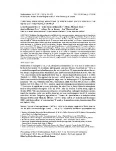
FIGURE 1. The 13 sampling stations (ST 1 – ST 13) located within the Bangpakong River (ST 1 – ST 9), estuary (ST 10 and ST 11) and sea in the river mouth (ST 12 and ST 13).
to serve for future assessment of the planktonic food webs in this ecosystem. MATERIALS AND METHODS Study Area and Period.– Bangpakong River is the most important river in the eastern part of Thailand with its 122 km length covering a watershed of 7,988 km2 in the Prachinburi and Chachoengsao provinces (NRCT-JSPS, 1998). It is under the influence of the monsoon system; the dry northeast (November to March) and the wet southwest (May to October) monsoons. Thus, the discharge controlled by the river displays seasonal variations, including
variations in the nutrient concentrations that may affect the phytoplankton biomass in this area (NRCT – JSPS, 1998; Buranapratheprat et al., 2002). Thirteen sampling stations were set up along the Bangpakong River from Baan Srang district in Prachinburi province to Bangpakong district in Chachoengsao province, which were comprised of nine, two and two sampling stations along the river, in the estuary and in the sea off the river mouth respectively (Table 1 and Fig. 1). This study was carried out during the daytime in February, April, July and September of 2004. The first two sampling months represented the dry season while
58
TROPICAL NATURAL HISTORY. 12(1), APRIL 2012
TABLE 1. Location of each sampling station and any associated human activity in the proximity of that station.
Easting 732222
Northing 1534220
Upstream Distance (km) (0 km)
737194
1518099
(30 km)
Urban
730404
1516321
(40 km)
Animal farming
724784
1513547
(50 km)
Urban
ST 5
Amphoe Bang Khla, Chachoengsao Lower Dam, Amphoe Mueang, Chachoengsao Na Mueang, Amphoe Mueang, Chachoengsao Amphoe Ban Pho, Chachoengsao
724647
1507213
(65 km)
Urban
ST 6
Amphoe Ban Pho, Chachoengsao
725051
1504103
(70 km)
Urban
ST 7
716355
1497161
(85 km)
Urban
717000
1491500
(95 km)
Urban
711014
1487408
(105 km)
ST 10
Tha Sa-an, Amphoe Bangpakong, Chachoengsao Tha Kham, Amphoe Bangpakong, Chachoengsao Amphoe Bangpakong, Chachoengsao Bangpakong estuary, Chonburi
708716
1485977
(110 km)
Industry, Aquaculture Aquaculture
ST 11
Bangpakong estuary, Chonburi
705819
1484246
(115 km)
Aquaculture
ST 12
Bangpakong estuary, Chonburi
700056
1477925
(125 km)
Aquaculture
ST 13
Bangpakong estuary, Chonburi
698150
1473581
(130 km)
Aquaculture
Station
Location
ST 1
Amphoe Ban Srang, Prachinburi
ST 2 ST 3 ST 4
ST 8 ST 9
UTM
other two months represented the wet season. Study Methods.– At each station, the presampling measurement of four physicochemical parameters, the water temperature, salinity, dissolved oxygen and pH, were carried out 0.5 m below the water surface and 1.0 m above the river / estuary bottom. Note that at all sampling sites the water depth was greater than 1.5 m. Water samples were collected for subsequent analysis of the size-fractionated chlorophylla biomass. Each sample was separated into three fractions; 20 – 200 µm, 3 – 20 µm and 0.7 – 3 µm fractions as representing the of micro-, nanoand pico-plankton, respectively. These fractions have been successfully used before in studying the estuarine plankton in The Gulf of Thailand (Piumsomboon et al., 2004). Chlorophyll-a
Activity Agriculture
from algal cells retained on the GF/F filter paper was extracted in 90% (v/v) acetone solution and the concentration was measured fluorometrically (Arar and Collins, 1992). Filtrates, after passing through GF/F paper, were also preserved for the analyses of the dissolved inorganic nutrients (NH4+-N, NO2--N, NO3--N, PO43--P and SiO32--Si) according to Parsons et al. (1984). In order to estimate the picoplankton community structure, water samples collected with a cleaned stainless water sampler were immediately filtered through a 200 µm-meshed net and then a 20 µmmeshed net. The filtrate was collected in a 150 ml cleaned bottle and preserved with buffered formalin to give a final formalin concentration of 2% (v/v). An aliquot of 1 – 10 ml (depending on the turbidity) of each filtrate was then filtered onto a 0.2 µm black
GUNBUA ET AL. –VARIATIONS OF PICO- AND NANO-PHYTOPLANKTON
polycarbonate membrane filter. Picophytoplankton were separated from heterotrophic bacteria based on their red chlorophyll-fluorescence under blue light while heterotrophic bacteria exhibited a bright blue color under UV excitation after DAPI (4’6-diamidino-2-phenylindoli) – staining due to the DNA fluorescence (Porter and Feig, 1980). At least 400 cells of each group were counted with an accuracy of ± 10% (Venrick, 1978) and the cell density were calculated (cells/ml). The nanophytoplankton community structure was determined by the FTF (filtertransfer-freeze) technique (Hewes and Holm – Hansen, 1983). Between 1 to 10 ml of the filtrate sample was filtered onto a 1.2-µm polycarbonate membrane filter under low vacuum. The filter containing the nanophytoplankton cells was placed upside down onto a clean glass slide with a small drop of water. The slide was then frozen by placing onto a cold surface or in the freezer (-20 °C) for 5 – 10 minutes. After the sample was frozen, the membrane filter was carefully peeled of leaving the nanophytoplankton cells on the glass slide. The slide was mounted with glycerol solution and examined under a compound microscope for classification at the higher taxonomic level. Cell volumes of picoplankton and nanoplankton were calculated from measuring the cell size using approximate cell geometry. Conversion factors from the literatures were used to estimate the carbon biomass of different groups of picoplankton: heterotrophic bacteria: 105 fgC/µm3 (Theil – Nielsen and Søndergaard, 1998) cyanobacteria Synechococcus: 205 fgC/cell (Kana and Glibert, 1987). The nanophytoplankton carbon contents were also calculated from their cell volumes using the following empirical relationships;
59
log C = -0.422+0.758 (log v) for diatoms (Strathmann, 1967), log C= -0.363+0.863 (log v) for flagellates and cyanobacteria (Verity et al., 1992) and log C = 0.760+0.819 (log v) for dinoflagellates (Menden-Deuer and Lessard, 2000). Data Analysis.– The Bray-Curtis similarity coefficient was calculated to explain the degree of similarity of plankton assemblages on the temporal scale of sampling months. Prior to analyses, the abundance data were transformed to log (x+1) to normalize the distributions and stabilize variances. The output similarity matrix was subjected to cluster analysis (group – average mean linkage). An ordination technique, principal component analysis (PCA) was used to explain the spatial differences in plankton communities. These analyses were performed using the PRIMER computer package version 5.0 (Clarke and Warwick, 1994). Correlation analysis was also performed upon the data to analyze the relationships between the physico-chemical and biological parameters. The test of significance of water quality parameters as well as plankton abundances from different stations and seasons were performed with ANOVA model and Duncan’s MMT for parametric analysis and two independent samples T test. RESULTS Temporal and spatial variations in the physico-chemical parameters.– Temporal variations in the average physical-chemical parameters along the Bangpakong River were clearly noticed with a higher salinity, pH and dissolved oxygen (DO) content being observed in the dry season than in the wet season (Table 2). Concentrations of dissolved inorganic nitrogen and
60
TROPICAL NATURAL HISTORY. 12(1), APRIL 2012
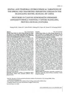
FIGURE 2. Spatial variations in the physical parameters of temperature, salinity, dissolved oxygen and pH for the dry and wet seasons. The sampling sites (ST 1 – ST 13), shown as the distance upstream from the sea, are as detailed and shown in Table 1 and Figure 1, respectively. Data are shown as the mean ± 1 SE. Means with a different lowercase letter are significantly different (p < 0.05; Duncan’s MMT).
phosphorus (DIN and DIP) in the dry season were also higher than those in wet season, although, in contrast, the silicate-silicon concentrations were significantly higher in the wet season. Likewise, in the wet season, the phytoplankton biomass, in terms of total chlorophyll-a, was also significantly higher than in the dry season (Table 2). The temperature, however, showed no significant differences. The water salinity and pH showed clear spatial variations with the salinity (especially) and the pH trending to increase towards the sea with significant and strong differences between the seasons for the salinity, being higher at all sampling stations in the dry season, and for the pH values, which were lower in the wet season
at the river sampling sites 1 – 8 (Fig. 2). The amount of DO was above 4 mg/l in either upstream stations or in the lower estuary in the dry season, but levels were lower in the wet season, when they were regularly lower than 4 mg/l at all sample sites and with the lowest values in the downstream stations of the dam, some 60 - 70 km from the uppermost sampling station. In contrast, the temperature was not significantly different at any sampling station in both seasons. (Fig. 2). The maximum value of chlorophyll-a biomass was recorded from the uppermost sampling station in the Ban Srang district, Prachinburi province, in the dry season (Fig. 3), which was accompanied by a bloom of the diatom, Cylindrotheca sp. (not shown),
GUNBUA ET AL. –VARIATIONS OF PICO- AND NANO-PHYTOPLANKTON
61
FIGURE 3. Spatial variations in the concentrations of chlorophyll-a and dissolved inorganic nutrients in the Bangpakong River estuary in the dry and wet seasons. The sampling sites (ST 1 – ST 13), shown as the distance upstream from the sea, are as detailed and shown in Table 1 and Figure 1, respectively. Data are shown as the mean ± 1 SE. Means with a different lowercase letter are significantly different (p < 0.05; Duncan’s MMT).
the highest DO content (Fig. 2) and the lowest silicate-silicon concentration (Fig. 3). In addition, spatial variations in the concentration of the other dissolved inorganic nitrogen and phosphorus were observed, such as the high nitrate and
phosphate levels upstream and the high concentrations of ammonium and nitrite downstream in the estuarine area of the river mouth (Fig. 3). These appear to correlate with the land use of farming and industry at site 9.
62
TROPICAL NATURAL HISTORY. 12(1), APRIL 2012
TABLE 2. Temporal variations in the average physico-chemical parameters across the 13 sampling sites in the Bangpakong River in both seasons.
Temperature (°C)
Dry season Mean ± SE Range 30.02 ± 0.36a 28.35 – 32.28
Wet season Mean ± SE Range 30.36 ± 0.15a 29.78 – 31.95
Salinity (psu)
23.01 ± 1.91a
7.47 ± 2.41b
Parameters
8.80 – 30.90
4.84 ± 0.23
a
pH
7.43 ± 0.08
a
7.03 – 8.12
7.17 ± 0.15
Total chlorophyll-a (mg/m3)
1.67 ± 0.50a
0.56 – 8.16
3.21 ± 0.36b b
Dissolved oxygen (mg/l)
a
3.54 – 6.55
0.15 – 25.10
3.26 ± 0.15
b
2.33 – 4.21
b
6.61 – 8.23 1.71 – 6.66
Ammonium-nitrogen (µM)
3.49 ± 0.82
N.D. – 9.14
2.39 ± 0.39
Nitrate-nitrogen (µM)
52.30 ± 6.64a
1.01 – 79.23
14.99 ± 2.17b
3.05 – 25.13
Nitrite-nitrogen (µM)
1.92 ± 0.59a
N.D. – 7.68
1.04 ± 0.16b
0.40 – 2.78
0.27 – 3.60
1.50 ± 0.16
b
3.89 – 93.04
95.23 ± 8.64b
a
Phosphate-phosphorus (µM)
2.28 ± 0.25
Silicate-silicon (µM)
60.78 ± 7.25a
0.61 – 5.52
0.76 – 3.00 31.71 – 137.36
Data are shown as the mean ± 1 SE. Means within a row that are followed by a different letter are significantly different (p < 0.05; Two Independent samples T Test). N.D. = non detected.
Temporal and spatial variations in the plankton abundances.– The communities of picophytoplankton in the Bangpakong River and estuary were composed solely of cyanobacteria cells of 0.83 – 1.77 µm in diameter, recognized as Synechococcus type-cells due to their fluorescent characteristic. Densities of this picophytoplankton were in the range of 104 - 105 cells/ml with the peak abundance in the wet season (1.48 x 10⁵ cells/ml) being some five-fold higher than that in dry season (2.06 x 10⁴ cells/ml). High densities of picophytoplankton were noticed in the peak of the rainy season (July), decreased in the late rainy season (September) and then increased slightly again in the dry season (February) during the northeast monsoon season before falling to the lowest level in April (Fig. 4). Spatially, the picophytoplankton tended to be more abundant in the river mouth and in the sea (St. 9 – 13) in both seasons (although far more abundant in the wet season) than in the upstream stations, with the highest densities at a
distance of 110 km from the inner most upstream station (Fig. 4). The communities of nanophytoplankton were composed of nanoflagellates, diatoms, dinoflagellates, coccolithophorids and cyanobacteria. Nanophytoplankton densities were higher in the wet season, 6.55 x 10⁷ – 1.33 x 10⁹ cells/l, than in the dry season, 2.54 x 10⁶ – 1.78 x 10⁸ cells/l (Fig. 5), and were dominated by nanoflagellates and cyanobacteria, while dinoflagellates, coccolithophorids and diatoms made up only small fractions. The average densities of nanophytoplankton were highest (8.95 x 10⁸ cells/l) at the peak of the rainy season (July), the same period as the picophytoplankton peak, and lowest in the dry season (February). High densities of nanoflagellates were usually found in the upstream area and ranged from 106 – 108 cells/l, and were the main component of the nanophytoplankton community along the Bangpakong River in summer (April). In contrast, cyanobacteria were the major constituent of the nanophytoplankton communities in the wet
GUNBUA ET AL. –VARIATIONS OF PICO- AND NANO-PHYTOPLANKTON
63
FIGURE 4. Temporal and spatial variations in the picophytoplankton densities. The sampling sites, shown as the distance upstream from the sea, are as detailed and shown in Table 1 and Figure 1, respectively. Data are shown as the mean ± 1 SE. Means with a different lowercase letter are significantly different (p < 0.05; Duncan’s MMT).
FIGURE 5. Temporal and spatial variations in the nanophytoplankton densities. The sampling sites, shown as the distance upstream from the sea, are as detailed and shown in Table 1 and Figure 1, respectively. Data are shown as the mean ± 1 SE. Means with a different lowercase letter are significantly different (p < 0.05; Duncan’s MMT).
FIGURE 6. Temporal and spatial variations in the heterotrophic bacterial densities. The sampling sites, shown as the distance upstream from the sea, are as detailed and shown in Table 1 and Figure 1, respectively. Data are shown as the mean ± 1 SE. Means with a different lowercase letter are significantly different (p < 0.05; Duncan’s MMT).
64
TROPICAL NATURAL HISTORY. 12(1), APRIL 2012
FIGURE 7. Dendrogram of the similarity index from phytoplankton communities in the wet and dry seasons.
season, where they were comprised of cyanobacteria at 86% to 97% of the total nanophytoplankton along the river. Heterotrophic picoplankton (heterotrophic bacteria or bacterioplankton), with an average density of 106 – 108 cells/ml, were 10 – 100 times more abundant than picophytoplankton. Their densities ranged from 1.38 x 10⁶ - 1.31 x 10⁸ and 3.29 x 10⁶ 1.87 x 10⁸ cells/ml in the dry and wet seasons, respectively (Fig. 6), although this is some 10-fold lower than that for nanophytoplankton. High densities of bacteria were found in the upper estuarine areas in the dry, and especially in the wet seasons. The communities of bacterioplankton in the lower estuary were always lower than those of the upstream areas (Fig. 6). Note that although the highest levels of heterotrophic bacteria were seen in the peak of the wet season (July), as per the nanoand pico-plankton, in contrast it did not decline much in the latter part of the wet season (September). Communities of pico- and nano-plankton in the Bangpakong River showed temporal variation between the wet and dry seasons with a similarity of 80%. The communities in the wet season showed a higher similarity, of approximately 88%, between
the early and late wet season (Fig. 7). A PCA of the abundance of picoplankton and nanophytoplankton communities (Figs 8 and 9, respectively) indicated the separation between communities in the sampling stations along the river to the upper estuary (stations 1 - 9) with those of the lower estuary (stations 10 and 11) and the sea (stations 12 & 13) due to the differences in salinity (Table 3). The abundance of heterotrophic bacteria tended to be higher in the river and upper estuarine regions rather than the lower estuary and the sea without any significant difference between seasons (Fig. 8). This result was in agreement with the significant negative relationships between heterotrophic bacteria and salinity as well as the positive relationships of heterotrophic bacteria to temperature and phosphate concentrations. The communities of picophytoplankton with a higher abundance in the sea and in the lower estuary than in the upper estuary and river showed a high positive correlation with the salinity level but an inverse relationship with the temperature, nitrate and phosphate levels (Figs 8 and 9; Table 4). On the other hand, nanoplanktonicdiatoms, which exhibited the same distribution pattern as the picophytoplankton, showed a significant relationship only with the salinity (Table 4). The distribution patterns of nanophytoplankton showed a spatial variation between the river and upper estuarine, lower estuary and the sea without any significant difference between seasons (Fig. 9). Nanoflagellates densities also indicated an inverse relationship with the salinity level, while their densities correlated well with the temperature and, ammonia, nitrate and phosphate concentrations (Table 4).
GUNBUA ET AL. –VARIATIONS OF PICO- AND NANO-PHYTOPLANKTON
65
TABLE 3. Average physico-chemical parameters from the different zones in the Bangpakong River and estuary. Parameters Temperature (°C) Salinity (psu) Dissolved oxygen (mg/l) pH
Range Mean Range Mean Range Mean Range Mean
Zone I (ST 1 – 9) Zone II (ST 10 – 11) Zone III (ST 12 – 13) Dry Wet Dry Wet Dry Wet 28.35-32.28 29.98-31.95 28.55-28.95 30.13-30.23 28.40-30.50 29.78-29.85 30.48 ± 0.26a 29.46 ± 0.42a 29.63 ± 0.44a 8.80-27.65 0.15-9.28 28.88-29.43 16.15-16.55 29.45-30.90 24.03-25.10 10.88 ± 2.58a 22.75 ± 3.70b 27.37 ± 1.66c 3.54-5.84 2.33-4.21 5.05-5.15 3.39-4.16 6.27-6.55 2.92-4.05 3.76 ± 0.21a 4.44 ± 0.41b 4.94 ± 0.88b 7.03-7.45 6.61-7.39 7.59-7.61 7.72-7.82 8.07-8.12 8.05-8.13 7.03 ± 0.07a 7.68 ± 0.05b 8.12 ± 0.04c
Data are shown as the mean ± 1 SE. Means within a row that are followed by a different letter are significantly different (p < 0.05; Duncan’s MMT). Sampling sites (ST 1 – ST 13) are as described and shown in Table 1 and Figure 1, respectively). TABLE 4. Correlations of the plankton community with the physico-chemical parameters. Heterotrophic picoplankton (log10x+1) -.462** .389** Temperature .501** -.717** Salinity ns ns Ammonia -.274* ns Nitrate ns ns Nitrite -.310* .337** Phosphate * Correlation is significant at the 0.05 level (2-tailed). ** Correlation is significant at the 0.01 level (2-tailed). ns = not significant. Pico phytoplankton (log10x+1)
Carbon biomass of the plankton community.– The carbon biomass, calculated from the biovolumes of the phytoplankton and heterotrophic bacteria, in the Bangpakong River and estuary varied from 17.1 – 1,870.7 µgC/l in the dry season to 157.9 – 4,132.8 µgC/l in the wet season (Fig. 10). Nanophytoplankton dominated the plankton communities of Bangpakong estuary in terms of the proportion of the carbon biomass ranging from 72.3% to 98.0% of the total carbon biomass in the dry season and 58.7% to 90.0% in the wet season. Heterotrophic bacteria contributed up to 28% of the total carbon biomass throughout the study period, while carbon derived from picophytoplankton was almost negligible, except in the river mouth area.
Nanoflagellates (log10x+1)
Cyanobacteria (log10x+1)
.419** -.354** .428** .597** ns .567**
ns -.597** ns -.581** ns -.293*
DISCUSSION Structure of plankton communities in the Bangpakong River estuary.– The communities of picoplankton in the Bangpakong River estuary showed a broad resemblance to that seen in the Klongkone mangrove creek in the Upper Gulf of Thailand, particularly in terms of picophytoplankton densities (Tarangkoon, 2002). However, the Bangpakong River and estuary communities were dominated by heterotrophic bacteria rather than by the autotrophic picoplankton found in Klongkone mangrove creek of Samut Songkram province. The abundance of heterotrophic bacteria from our study was 100-fold higher than that that reported for the Klongkone creek (Tarangkoon, 2002), a
66
TROPICAL NATURAL HISTORY. 12(1), APRIL 2012
FIGURE 8. Principal component analysis (PCA) of the log (x+1) transformed density of picoplankton. Sampling sites (ST 1 – ST 13) are as detailed and shown in Table 1 and Figure 1, respectively.
difference that may due to the significant basin characteristics and activities of the Bangpakong River in comparison to the Klongkone creek (Tarangkoon, 2002; Paphavasit et al., 2005). For example, the concentrations of dissolved organic carbon were in the range of 2 - 4 mg/l in the Bangpakong River and river mouth (Paphavasit et al., 2005), levels that can support the bacterial production in this estuary. The dominance of heterotrophic bacteria over picophytoplankton has also been reported before in tropical and temperate estuaries (Garrison et al., 2000; Hare et al., 2005; Pan et al., 2006, 2007). However, the abundance of heterotrophic bacteria in the Bangpakong River / estuary was much higher than those previously reported in the temperate regions of Asia and America, such as in the Hiroshima, Mutsu and Isu Bays in Japan, the Changjiang estuary in China, the Arabian Sea in the middle-east
and the St. Lucie river estuary in Florida, USA (Table 5). In contrast, the abundance of local picophytoplankton at less than 50x104 cells/ml was in the same range as that previously reported in the above estuaries (Table 5). The abundance of nanophytoplankton in the Bangpakong River / estuary found in this study was 10-fold higher than the communities reported in the Tha Chin estuary in the western part and Pak Poon estuary in the southern part of the Gulf of Thailand (Table 5), although the nanophytoplankton community structure (mostly composed of nanoflagellates and cyanobacteria with diatoms, dinoflagellates, and coccolithophorids as minor constituents) was similar to that reported for the Thachin estuary and the Pak Poon estuary in the Gulf of Thailand (Phromthong, 1999; Piumsomboon et al., 2000). Thus, the nanophytoplankton community structure is supported by the
GUNBUA ET AL. –VARIATIONS OF PICO- AND NANO-PHYTOPLANKTON
67
FIGURE 9. Principal component analysis (PCA) of the log (x+1) transformed density of nanophytoplankton. Sampling sites (ST 1 – ST 13) are as detailed and shown in Table 1 and Figure 1, respectively.
68
TROPICAL NATURAL HISTORY. 12(1), APRIL 2012
TABLE 5. Reported abundances and biomass of heterotrophic bacteria, pico- and nanophytoplankton in estuaries and coastal areas of the tropical-subtropical regions. Organisms/Abundance Environment, location, area Bangpakong estuary Gulf of Thailand Tha Chin estuary, Samut Sakhon Pak Poon estuary, Nakorn Si Thammarat Klong Kone mangrove swamp, Samut Songkhram Andaman Sea The Andaman Sea, Indian Ocean, Thailand Other locations Hiroshima Bay Mutsu Bay Ise Bay The Changjiang estuary and adjacent coastal regions The northern Arabian Sea
The Arabian Sea
The northern Gulf of Mexico The Mississippi river plume
Biomass (µgC/l)
Methods
References
2.54 – 1,330
11 – 4,132
LM
This study
-
3.41 – 24.80
-
LM
Phromthong (1999)
-
-
1 – 100
-
LM
Piumsomboon et al. (2000)
1 – 1.8
6.4 – 21.8
-
-
LM
Tarangkoon (2002)
0.2 – 0.3
-
-
20 – 150
FM
Nielsen et al. (2004)
-
0.15 – 2 0.3 – 0.9 0.02 – 9 0.2 – 27 (Syn) 0 – 21 (Pro) 0.01 – 0.9 (Euk)
0.35 – 150 0.10 – 2.10 0.23 – 710
-
FM
Nishitani et al. (2005)
0.01 – 21.40
-
FCM
Pan et al. (2007)
0.02 – 120
0.02 – 11.6
-
Garrison et al. (1998)
FCM
Garrison et al. (2000)
Heterotrophic bacteria (x106 cells/ml)
Picophytoplankton (x104 cells/ml)
Nanophytoplankton (x106 cells/l)
1.4 – 187
0 – 14.8
-
0.4 – 2.5 -
-
0.7 – 1
3 – 17 (Pro) 3 – 9.7 (Syn) 0.1 – 0.2 (Euk)
0.30 – 7.60
0.2 – 34.7 0.0 – 47.2 0.0 – 112.9 0.0 – 5.8 0.1 – 36.3
0.9 – 1.3
-
-
-
FCM
Jochem (2001)
0.1 – 2
-
-
-
FCM
Jochem (2003)
FCM
Jochem (2003)
FM
Millie et al. (2004)
The Mississippi River
1
0.1 – 4.3 (Syn and Pro)
-
5.1 – 28.3 0 – 6.1 (Syn) 0.1 – 0.8 (Pro)
The North Fork of the St. Lucie river estuary, Florida (USA)
-
0.7 – 42.5 (Syn and Synt)
-
-
FM: Fluorescence microscopy, FCM: Flow cytometry, LM: Light microscopy, Syn: Synecococcus, Synt: Synechocystis, Pro: Prochorococcus, Euk: Eukaryotic forms.
results of other studies in the subtropical northwestern Philippine Sea (Tsuji and Adachi, 1979) and the Arabian Sea (Garrison et al., 1998), and so may be a more general ecosystem characteristic. In general, the successful distribution of nanoflagellates and cyanobacteria in various
aquatic environments has been attributed to efficient light harvesting mechanisms by pigments, adaptations and mechanisms to live in diverse habitats, and their efficiency in nutrient uptake (Sigee, 2005).
GUNBUA ET AL. –VARIATIONS OF PICO- AND NANO-PHYTOPLANKTON
69
FIGURE 10. Temporal and spatial variations of the carbon biomass in the plankton community. The sampling sites, shown as the distance upstream from the sea, are as detailed and shown in Table 1 and Figure 1, respectively.
Factors influencing temporal and spatial variations in plankton communities.– Communities of pico- and nano-plankton in the Bangpakong River were characterized by the dominance of heterotrophic bacteria over autotrophic picoplankton and by the cyanobacteria dominated nanophytoplankton communities in the water with a salinity range from 0.15 to 25.1 psu. The resemblance between the plankton communities in the early and late dry season was about 85% (Fig. 7) with the average salinity ranged from 8.80 to 30.90 psu. These communities also had heterotrophic bacteria dominating the picoplankton community but the nanophytoplankton one was dominated by nanoflagellates rather than by cyanobacteria. Cyanobacteria may take advantage of their thick cell wall or mucilaginous sheath (Jeffrey et al., 1997) to
overcome the problem of the changing salinity in the wet season and hence supports the high tolerance of cyanobacteria in an extreme environment (Sigee, 2005). The situation in the dry season where the concentrations of nutrients are scarce, therefore, favors the nanoflagellates with their smaller sizes and high surface area: volume ratio which offers a high nutrient uptake rate (Safi and Hall, 1997) in comparison to larger sized phytoplankton. Our result also indicates that salinity is the major environmental factor shaping the plankton community structure in the Bangpakong River and estuary, with the concentration of dissolved inorganic nutrients as the minor controlling factors. The distribution pattern of diatoms reflected the dominance of different species inhibited the river and upper estuary in comparison to
70
TROPICAL NATURAL HISTORY. 12(1), APRIL 2012
the species in the lower upper estuary and the sea. The importance of salinity and dissolved inorganic nutrients as factors controlling picoplankton abundance in the Bangpakong River are also in agreement of the results reported for the Arabian sea (Brown et al., 1999), equatorial Pacific (Blanchot et al., 2001), Gulf of Venice (Aubry et al., 2006), Andean-Patagonian lakes (Callieri et al., 2007), subtropical coastal lagoons of Uruguay (Vidal et al., 2007) and South Australian coastal lagoons (Schapira et al., 2010). Carbon biomass of plankton community.– The estimated carbon biomass levels in this study at the Bangpakong River and estuary are higher than those reported from the temperate bays and estuaries (Table 5). The high carbon biomass of nanophytoplankton may due to the application of the microphytoplankton carbon-biovolume conversion values to calculate the carbon biomass of nanophytoplankton, since there is currently no conversion value specific for nanophytoplankton. Therefore, the biomass for nanophytoplankton is one to two orders of magnitude higher in the upstream regions than the sea, while pico-phytoplankton biomass increases in the sea, indicating their different responses to these different changing environments. Nanophytoplankton seemingly make a higher contribution to the phytoplankton biomass in the upstream regions while picophytoplankton become more important in the sea.
study period with a density of about 10-fold to 100-fold higher than that for picophytoplankton, which were composed of prokaryotic coccoid cyanobacteria or Synechococcus-like species. The nanophytoplankton were mostly comprised of nanoflagellates and cyanobacteria as the dominant groups, together with diatoms, dinoflagellates and coccolithophorids. High densities of heterotrophic picoplankton and nanophytoplankton were recorded in upstream regions (upper and lower estuary) while picophytoplankton dominated in the seaward area. Nanophytoplankton accounts for a large proportion of the biomass in terms of chlorophyll-a and make up an important contribution to the primary production in the Bangpakong River and estuary. This also suggested further investigation on the structure of the food chain/ food web based on small-size producers in this area. ACKNOWLEDGEMENTS We are grateful to members of the Marine Ecology Laboratory, Department of Marine Science for help in the field-work. Financial support for this research was provided by the Department of Marine and Coastal Resources, the Ministry of Natural Resources and Environment. Partial financial support from The Graduate School, Chulalongkorn University, to the first author’s dissertation is also appreciated. The authors also thank the anonymous reviewers for their fruitful suggestion.
CONCLUSION
LITERATURE CITED
Among the communities of picoplankton in the Bangpakong River and estuary, heterotrphic picoplankton (bacteria) were the most abundant group throughout the
Arar, E.J. and Collins, G.B. 1992. Methods 445.0: In vitro determination of chlorophyll a and phaeophytin a in marine and freshwater algae by fluorescence. In USEPA methods for the determination of chemical substances in Marine
GUNBUA ET AL. –VARIATIONS OF PICO- AND NANO-PHYTOPLANKTON and estuarine environmental samples. EPA/600/R92/121. U.S. Environmental protection Agency. Ohio. Aubry, F.B., Acri, F., Bastianini, M., Pugnetti, A. and Socal, G. 2006. Picophytoplankton contribution to phytoplankton community structure in the Gulf of Venice (NW Adriatic Sea). International Review of Hydrobiology, 91: 51-70. Azam, F., Fenchel, T., Field, J.G., Gray, J.S., MeyerReil, L.A. and Thingstad, F. 1983. The ecological role of water-colume microbes in the sea. Marine Ecology Progress Series, 10: 257-263. Blanchot, J., Andre, J.M., Navarette, C., Neveux, J. and Radenac, M.H. 2001. Picophytoplankton in the equatorial Pacific vertical distributions in the warm pool and in the high nutrient low chlorophyll conditions. Deep-Sea Research I, 48: 297-314. Bordalo, A.A., Nilsumranchit, W. and Chalermwat, K. 2001. Water quality and uses of the Bangpakong river (eastern Thailand). Water Research, 35: 36353642. Buranapratheprat, A., Yanagi., T., Boonphakdee T. and Sawangwong, P. 2002. Seasonal variations in inorganic nutrient budgets of the Bangpakong estuary, Thailand. Journal of Ocenaography, 58: 557-564. Brown, S.L., Landry, M.R., Barber, R.T., Campbell, L., Garrison, D.L. and Gowing, M.M. 1999. Picophytoplankton dynamics and production in the Arabian sea during the 1995 southwest monsoon. Deep-Sea Research II, 46: 1745-1768. Callieri, C., Modenutti, B., Queimalinos, C., Bertoni, R. and Balseiro, E. 2007. Production and biomass of picophytoplankton and larger autotrophs in Andean ultraoligotrophic lakes: differences in light harvesting efficiency in deep layers. Aquatic Ecology, 41: 511-543. Cho, B.C. and Azam, F. 1990. Biogeochemical significance of bacterial biomass in the ocean's euphotic zone. Marine Ecology Progress Series, 63: 253-259. Clarke, K.R. and Warwick, R.M. 1994. Change in marine communities approach to statistical analysis and interpretation. Plymouth Marine Laboratory, UK, 144 pp. Detmer, A.E. and Bathmann, U.V. 1997. Distribution patterns of autotrophic pico- and nanoplankton and their relative contribution to algae biomass during spring in the Atlantic sector of the southern ocean. Deep-Sea Research II, 44: 299-320.
71
Fuhrman, J.A., Sleeter, T.D., Carlson, C.A., and Proctor, L.M. 1989. Dominance of bacterial biomass in the Sargasso Sea and its ecological significance. Marine Ecology Progress Series, 57: 207-217. Garrison, D.L., Gowing, M.M. and Hughes, M.P. 1998. Nano- and microplankton in the northern Arabian Sea during the southwest monsoon, AugustSeptember 1995, A US-JGOFS study. Deep-Sea Research II, 45: 2269-2299. Garrison, D.L., Gowing, M.M., Hughes, M.P., Campbell, L., Caron, D.A., Dennett, M.R., Shalapyonok, A., Olson, R.J., Landry, M.R., Brown, S.L., Liu, H., Azam, F., Steward, G.F., Ducklow, H.W. and Smith, D.C. 2000. Microbial food web structure in the Arabian Sea: a US JGOFS study. Deep-Sea Research II, 47: 1387-1422. Gobler, C.J., Cullison, L.A., Koch, F., Harder, T.M. and Krause, J.W. 2005. Influence of freshwater flow, ocean exchange, and seasonal cycles on phytoplankton - nutrient dynamics in a temporarily open estuary. Estuarine, Coastal and Shelf Science, 65: 275-288. Hare, C.E., DiTullio, G.R., Trick, C.G., Wihelm, S.W., Bruland, K.W., Rue, E.L. and Hutchins, D.A. 2005. Phytoplankton community structure changes following simulated upwelled iron inputs in the Peru upwelling region. Aquatic Microbial Ecology, 38: 269-282. Hewes, C.D. and Holm-Hansen, O. 1983. A method for recovering nanoplankton from filters for identification with the microscope: The filtertransfer-freeze (FTF) technique. Limnology Oceanography, 28: 389-394. Jeffrey, S.W., Mantoura, R.F.C. and Wright, S.W. 1997. Phytoplankton pigments in oceanography. UNESCO, 661 pp. Jochem, F.J. 2001. Morphology and DNA content of bacterioplankton in the northern Gulf of Mexico: analysis by epifluorescence microscopy and flow cytometry. Aquatic Microbial Ecology, 25: 179194. Jochem, F.J. 2003. Photo- and heterotrophic pico- and nanoplankton in the Mississippi River plume and distribution and grazing activity. Journal of Plankton Research, 25: 1201-1214. Kana, T.M. and Glibert, P.M. 1987. Effect of irradiances up to 2000 Em-2 s-1 on marine Synechococcus WH7803.1. Growth, pigmentation, and cell composition. Deep-Sea Research, 34: 479495.
72
TROPICAL NATURAL HISTORY. 12(1), APRIL 2012
Menden-Deuer, S. and Lessard, E.J. 2000. Carbon to volume relationships for dinoflagellates, diatom, and other protest plankton. Limnology Oceanography, 45: 569-579. Millie, D.F., Carrick, H.J., Doering, P.H. and Steidinger, K.A. 2004. Intra-annual variability of water quality and phytoplankton in the North Fork of the St. Lucie River estuary, Florida (USA): a quantitative assessment. Eatuarine, Coastal and Shelf Science, 61: 137-149. Nielsen, T.G., BjØrnsen, P.K., Boonruang, P., Fryd, M., Hansen, P.J., Janekarn, V., Limtrakulvong, V., Munk, P., Hansen, O.S., Satapoomin, S., Sawangarreruks, S., Thomsen, H.A. and Østergaard, J.B. 2004. Hydrography, bacteria and protist communities across the continental shelf and shelf slope of the Andaman Sea (NE Indian Ocean). Marine Ecology Progress Series, 274: 69-86. Nishitani, G., Yamaguchi, M., Ishikawa, A., Yanagiya, S., Mitsuya, T. and Imai, I. 2005. Relationships between occurences of toxic Dinophsis species (Dinophyceae) and small phytoplanktons in Japanese coastal waters. Harmful Algae, 4: 755762. Not, F., Simon, N., Biegala, I.C. and Vaulot, D. 2002. Application of fluorescent in situ hybridization coupled with tyramide signal amplification (FISHTSA) to assess eukaryotic picoplankton composition. Aquatic Microbial Ecology, 28: 157-166. NRCT-JSPS. 1998. An integrated study on physical, chemical and biological characteristics of the Bangpakong estuary. Final report cooperative research NRCT-JSPS, the National Research Council of Thailand (NRCT) and the Japan Society for the Promotion of Science (JSPS). 127 pp. Pan, L.A., Zhang, L.H., Zhang, J., Gasol, J.M. and Chao, M. 2005. On-board flow cytometric observation of picoplankton community structure in the East China Sea during the fall of different years. FEMS Microbiology Ecology, 52: 243-253. Pan, L.A., Zhang, J., Chen, Q. and Deng, B. 2006. Picoplankton community structure at a coastal front region in the northern part of the South China Sea. Journal of Plankton Research, 28: 337-343. Pan, L.A., Zhang, J. and Zhang, L.H. 2007. Picophytoplankton, nanophytoplankton, heterotrophic bacteria and viruses in the Changjiang Estuary and adjacent coastal waters. Journal of Plankton Research, 29: 187-197. Paphavasit, N., Wattayakorn, G., Piumsomboon, A. and Sivaipram, I. 2005. The ecosystem of the Bangpakong River estuary. Bangkok, Chulalongkorn University.
Paphavasit, N, Aksornkoae, S. and Silva, J.D. 2009. Tsunami impact on mangrove ecosystem. Thailand Environment Institute. Nonthaburi, Thailand. Parsons, T.R., Maita, R.Y. and Lalli, C.M. 1984. A manual of chemical and biological methods for seawater analysis. Pergamon Press, Oxford, England. Phromthong, I. 1999. Dynamics and diversity of phytoplankton in Tha Chin estuary, Samut Sakhon province. Master thesis, Department of Marine Science, Graduate school, Chulalongkorn University (in Thai). Piumsomboon, A., Paphavasit, N., Soonsawad, N., Sikhantakasamit, B. and Phromthong, I. 2000. Plankton communities in Pak Poon estuary, Nakhon Si Thammarat, Southern Thailand. In: Annual report 1999 (second year : May 1999 – March 2000) on Green Carpet Project in Nakhon Si Thammarat, Thailand. KEIDANREN Nature Conservation Fund (KNCF) and Japan Fund for Environment Conservation (JEC), 45-62 p. Piumsomboon, A., Paphavasit, N., Phromthong, I. and Tarangkoon, W. 2001. Effects of changes in phytoplankton size fraction on the energy transfer in the coastal ecosystems. In: Symposium on aquatic resources and environmental: integrated management and utilization. Aquatic resource institute, 6-8 December, Chaing Mai province. Piumsomboon, A. 2002. The study of marine plankton in Thailand. Journal of Scientific Research, Chulalongkorn University (section T), 1: 275-290. Piumsomboon, A., Tarangkoon, W., Sao-sii, P., Sikhanthakasamit, B., Punnarak, P., Paphavasit, N. and Sivaipram, I. 2004. Diversity and plankton production in mangrove plantation and pakphanang estuary, Nakorn si Thammarat province. In Integrated Management of Mangrove Plantations for Development of Coastal Resources and Environment of Thailand. The Thailand Research Fund (TRF). Piumsomboon, A., Mongkongsangsuree, N., Paphavasit, N. and Punnarak, P. 2007. Assessment of Marine Ecosystem Stability: A Case Study of Mangrove Ecosystems of the Andaman Sea. Proceedings of the National Conference on Mangrove Ecosystem “Mangroves: Sustainable economics of coastal communities”. Department of Coastal and Marine Resources and Thailand Environment Institute, 12-14 September, 2007, pp: 398-411. Porter, K.G. and Feig, Y.S. 1980. The use of DAPI for identifying and counting aquatic microflora. Limnology Oceanography, 25: 943-948.
GUNBUA ET AL. –VARIATIONS OF PICO- AND NANO-PHYTOPLANKTON Safi, K.A.and Hall, J.A. 1997. Factors influencing autotrophic and heterotrophic nanoflagellate abundance in five water masses surrounding New Zealand. New Zealand Journal of Marine and Freshwater Research, 31: 51-60. Schapira, M., Buscot, M.J., Pollet, T., Leterme, S.C. and Seuront, L. 2010. Distribution of picophytoplankton communities from brackish to hypersaline waters in a South Australian coastal lagoon. Saline Systems, 6: 1-15. Sherr, E.B., and Sherr, B.F. 1994. Bacterivory and herbivory: Key roles of phagotrophic protists in pelagic food webs. Microbial Ecology, 28: 223235. Sieburth, J.M., Smetacek, V. and Lenz, J. 1978. Pelagic ecosystem structure: Heterotrophic compartments of the plankton and their relationship to plankton size fractions. Limnology and Oceanography, 23: 1256-1263. Sigee, D.C. 2005. Freshwater microbiology: biodiversity and dynamic interactions of microorganisms in the aquatic environment. John Wiley & Sons, Ltd, UK, 537 pp. Strathmann, R.R. 1967. Estimating the organic carbon content of phytoplankton from cell volume or plasma volume. Limnology Oceanography, 12: 411-418. Tarangkoon. W. 2002. Annual variation in abundance and biomass of picoplankton in Klong Kone mangrove swamp. Samut Songkhram province. Master thesis, Department of Marine Science, Graduate school, Chulalongkorn University (in Thai).
73
Tarran, G.A., Zubkov, M.V., Sleigh, M.A., Burkill, P.H. and Yallop, M. 2001. Microbial community structure and standing stocks in the NE Atlantic in June and July of 1996. Deep-Sea Research II, 48: 963-985. Theil-Nielsen, J. and SØndergaard, M. 1998. Bacterial carbon biomass calculated from biovolumes. Archiv für Hydrobiologie, 141: 195-207. Tsuji, T. and Adachi, R. 1979. Distribution of nanophytoplankton including fragile flagellates in the subtropical northwestern Philippine Sea. Journal of the Oceanographical Society of Japan, 35: 173-178. Venrick, E.L. 1978. Estimating cell numbers: How many cells to count?, pp. 167–189. In Sournia, A (ed.), Phytoplankton Manual. UNESCO, Paris, 337 pp. Verity, P.G., Robertson, C.Y., Tronzo, G.R., Andrews, M.G., Nelson, J.R. and Sieracki, M.F. 1992. Relationships between cell volume and carbon and nitrogen content of marine photosynthetic nanoplankton. Limnology Oceanography, 37: 14341446. Vidal, L., Rodrίguez-Gallego, L., Conde, D., MartίnezLópez, W. and Bonilla, Sylvia. 2007. Biomass of autotrophic picoplankton in subtropical coastal lagoons: Is it relevant? Limnetica, 26: 441-452. Wattayakorn, G., Prapong, P. and Noichareon, D. 2001. Biogeochemical budgets and processes in Bandon Bay, Suratthani, Thailand. Journal of Sea Research, 46: 133-142.