The affinities for hyaluronic acid of newly synthesized proteoglycan from post-confluent rabbit chondrocyte cultures and purified bovine proteoglycan monomer ...
Biochem. J. (1986) 234, 221-223 (Printed in Great Britain)
221
The affinity of newly synthesized proteoglycan for hyaluronic acid can be enhanced by exposure to mild alkali Anna H. K. PLAAS* and John D. SANDY Division of Orthopaedic Research, Department of Orthopaedics, Rhode Island Hospital and Brown University, Providence, RI 02902, U.S.A.
The affinities for hyaluronic acid of newly synthesized proteoglycan from post-confluent rabbit chondrocyte cultures and purified bovine proteoglycan monomer were compared. In mixtures prepared at pH 6.8 the newly synthesized proteoglycan had the lower affinity; however, in mixtures incubated at pH 8.5 for 24 h before addition of hyaluronic acid, the newly synthesized proteoglycan exhibited a markedly higher affinity than the bovine monomer. The results suggest that proteoglycan secreted without associated link protein [Plaas, Sandy & Muir (1983) Biochem. J. 214, 855-864] has a low affinity for hyaluronate and that this may be increased during subsequent extracellular processing.
INTRODUCTION The biosynthesis of proteoglycans in articular cartilage can be considered to proceed in two distinct phases: firstly, an intracellular phase during which the monomer is assembled, and, secondly, an extracellular phase during which the secreted proteoglycan may be deposited in the cartilage matrix. Although many aspects of the intracellular processing and secretion of proteoglycan are now understood (Kimura et al., 1984), little is known of the control of the extracellular events. Proteoglycan deposition may be influenced by a variety of factors, among which might be the binding affinity of the newly synthesized molecule and the availability of binding sites in the matrix. Proteoglycans are known to interact specifically with both hyaluronic acid (Hardingham & Muir, 1972) and link proteins (Caterson & Baker, 1978; Bonnet et al., 1985), and so the abundance and binding properties of these components in the matrix are likely to be important. Although the ability of newly synthesized monomer to bind hyaluronic acid appears to be acquired early in biosynthesis (Kimura et al., 1981), there is also evidence (Oegema, 1980; Bayliss et al., 1983) that the affinity for hyaluronate may increase after secretion in human cartilage explants. In the present work we show that the affinity for hyaluronate of proteoglycan secreted by rabbit chondrocytes may be markedly enhanced by treatment with mild alkali. EXPERIMENTAL Materials Materials were obtained as previously described (Plaas et al., 1983; Plaas & Sandy, 1984). In addition, 1,9-dimethyl-Methylene Blue was from Serva Feinbiochemica, Heidelberg, West Germany, and chondroitin sulphate (type C) was from Sigma Chemical Co., St. Louis, MO, U.S.A. Hyaluronic acid (Healon) was generously given by Pharmacia, Piscataway, NJ, U.S.A. *
To whom correspondence
Vol. 234
should be addressed.
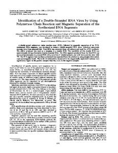
Methods Proteoglycan monomer (fraction AlAlD1) was prepared as the sodium salt from bovine nasal cartilage as described previously (Sandy, 1979). [35S]Sulphate-labelled proteoglycan was prepared by 24 h radiolabelling of post-confluent cultures of rabbit articular chondrocytes as described previously (Plaas et al., 1983); medium in which less than 10% of the monomer was present as link-stabilized aggregate was stored at -20 °C before use. Proteoglycan was determined as total glycosaminoglycan with dimethyl-Methylene Blue as described by Farndale et al. (1982), with chondroitin sulphate as standard. The percentage of proteoglycan in aggregates on Sepharose CL-2B chromatography was calculated as previously described (Plaas & Sandy, 1984). RESULTS Post-confluent cultures of immature rabbit articular chondrocytes have been shown to secrete proteoglycan monomer without active link protein (Plaas et al., 1983). In untreated medium samples this monomer was found to form aggregates with endogenous hyaluronate only after stabilization by the addition of link protein; the absence of aggregates in medium alone was presumably due to the concentration of proteoglycan being below the dissociation constant for the proteoglycan-hyaluronate interaction. To show that such monomer, secreted without link protein, was nonetheless capable of aggregating with hyaluronate alone, carrier monomer was added before the addition of hyaluronate and chromatography on Sepharose CL-2B. The percentage aggregate formed by newly synthesized [3S]sulphatelabelled monomer and carrier monomer at various hyaluronate concentrations was then determined (Fig. 1). Interestingly, a marked difference was seen in the binding behaviour of the two proteoglycan preparations. The bovine monomer showed an increase in percentage aggregate with increasing hyaluronate concentration
A. H. K. Plaas and J. D.
222 80 r
80
E 70
70
o
E
I
a)
0
'z
60
C" m
50
.._
Sandy
0,
601-
ca, C" Em w
501-
0
E40 0E u4
E 0 E
CR
0 C
~.cr
0
40)
401.
0,
E 30
30
a,
F
am
02 0 2 C.,
0~
201-
0-
io10 o 0
1
3
4
5 6 100 x Hyaluronate/proteoglycan ratio (w/w)
2
7
Fig. 1. Percentage -aggregate formed from radiolabelied and bovine monomer with various amounts of hyaluronate 1d containing 3 x 10 d.p.m. of Chondrocyte [3&S]sulphate-labelled pr3oteoglycan) was added to 6 mg of bovine AlAIDI fraction and 2.0 ml of 5 M-guanidinium chloride/0.05 M-sodium *acetate buffer, pH 6.8. The mixture was dialysed for 16 h at 4 °C against 500 ml of 0.5 M-sodium acetate buffer, pH 6.8, and hyaluronic acid was added to portions of the dialysis residue to obtain hyaluronate/proteoglycan ratios (w/w) of 0.5%, 1%, 2%, 3%, 5% and 7%. After 2 h at 22 °C samples containing about 0.4 mg of proteoglycan and 2 x 10⁵ d.p.m. of [35Slsulphate-labelledproteoglycan were applied to columns (0.5 cm x l00 cm) of Sepharose CL-2B, eluted at 2 ml/h with 0.5 M-sodium acetate buffer, pH 6.8. Fractions (0.7 ml) were analysed for proteoglycan and radioactivity content, and the percentages ofbovine (A) and radiolabelled (0) monomer in aggregate form were calculated as described in the Experimental section.
mediumf(500
similar to that observed previously (Hardingham & Muir, 1974); however, the radiolabelled monomer showed a sigmoidal bindamg curve consistent with a much lower affinity for hyahironic acid. A similar result (not shown) wasobtainedin'bindingexperimentswith thisradiolabelled monomer and a purified aggregating monomer preparation (aAIDi) from rabbit articular cartilage, showing that the di-fferential (Fig. 1) was not due simply to the different sources of proteoglycan. Although the radiolabelled monomers showed a low apparent affinity at between 1 % and 3 % hyaluronate, the majority of the molecules were capable of forming aggregates; thus with excess hyahiurnate (55% and above) about 75%O of the [35Sjsulphates-abelled proteoglycan was recovered in the aggregate form. These results suggested that proteoglycan accumulates in these chlondrocyte cultures in a precursor form with a low affinity for hyaluronate. To find conditions that might enhance -this affinity, a mixture of radiolabelled and bovine monomer was dialysed against buffers in the alkaline pH range before adjustment to pH 6.8 and addition oflimiting hyaluronate
7.0
7.5
8.0
8.5
9.0
9.5
pH
Fig. 2. Effect of treatment at alkaline pH on the percentage aggregate formed with radiolabelled and bovine monomer Chondrocyte medium (1.5 ml containing about 107 d.p.m. of [35S]sulphate-labelled proteoglycan) was added to 30 mg of bovine AlAlDI fraction and 6.0 ml of 5 M-guanidinium chloride/0.05 M-sodium acetate buffer, pH 6.8. Portions (1 ml) were dialysed for 24 h against 500 ml of 0.5 Msodium acetate buffered with 0.1 M-Tris/HCl at pH values of 6.95, 7.15, 7.45, 7.74, 8.04, 8.65 and 9.19. Each dialysis residue was adjusted to pH 6.8 with acetic acid, and hyaluronic acid was added to obtain a hyaluronate/proteoglycan ratio of 2.0% (w/w). After 2 h at 22 °C, samples containing about 0.4 mg of proteoglycan and 2 x 10⁵ d.p.m. of [35S]sulphate-labelled proteoglycan were applied to columns of Sepharose CL-2B and analysed as described in the legend to Fig. 1, and the percentages of bovine (A) and radiolabelled (c) monomer in aggregate form were calculated as described in the Experimental section.
(Fig. 2). This treatment made essentially no difference to the binding properties of the bovine monomer. However, treatment above about pH 7.7 markedly increased the affinity of the newly synthesized proteoglycan; indeed, treatment at pH 8.65 and 9.15 generated radiolabelled monomer that now exhibited a much higher affinity for hyaluronate than the carrier. The time required for the conversion from the low-affinity into the high-affinity form was determined next (Table 1); although a significant increase was seen after 1 h at pH 8.6, between 8 h and 19 h was required for maximum affinity to be acquired. The increase observed here (Table 1) was shown to depend on continuous exposure of the proteoglycan to the high pH, since samples incubated atpH 8.6 for 2 h and subsequently at pH 6.8 for 20 h did not show high affinity. Further, the high affinity acquired at pH 8.6 was stable to dialysis of samples for 24 h at 22 °C against buffer at pH 6.8. The increase in affinity was not dependent on the presence of the carrier monomer during treatment; thus, when chondrocyte medium alone was dialysed at pH 8.6 for 20 h before addition of bovine monomer and 1986
Affinity of proteoglycan for hyaluronic acid
223.
Table 1. Time-dependence of conversion from the low-affinity into the high-affinity form of newly synthesized rabbit proteoglycan
Chondrocyte medium (1.5 ml containing about 107 d.p.m. of [35S]sulphate-labelled proteoglycan) was added to 30 mg of bovine AlAlDI fraction and 6.0 ml of 5 M-guanidinium chloride/0.05 M-sodium acetate buffer, pH 6.8, and the mixture was dialysed for 24 h at 22 °C against 0.5 M-sodium acetate/0. 1 M-Tris/HCl buffer, pH 6.8. The dialysis residue was adjusted with NaOH to pH 8.6 and stirred gently at 22 'C. At various times, portions (0.5 ml) were removed and adjusted to pH 6.8 with 10% (v/v) acetic acid before addition of hyaluronic acid to obtain a hyaluronic acid/proteoglycan ratio of 2% (w/w). Samples containing about 0.4 mg of proteoglycan and 2 x 10⁵ d.p.m. of [35S]sulphate-labelled proteoglycan were applied to columns of Sepharose CL-2B and analysed as described in the legend to Fig. 1, and the percentages of bovine and radiolabelled monomer in aggregate form were calculated as described in the Experimental section. Percentage of monomer in aggregate form
Period of incubation (h)
35S-labelled proteoglycan
Bovine AIAlDI fraction
0 0.5 1.0 4.0 8.0 19.0 24.0
9.2 10.7 17.7 22.2 38.0 56.4 55.5
26.0 29.0 25.5 24.5 29.5 23.0 25.5
hyaluronic acid, the binding of the [35S]sulphate-labelled monomer was again very high (82%) relative to carrier
(23%).
DISCUSSION The binding of extracted proteoglycans to hyaluronate has been shown to depend on the maintenance of binding-region structure by multiple intramolecular disulphide bridges; loss of binding on reduction with dithiothreitol occurred with a half-life of 14 min, and on re-oxidation at pH 9 about 85% of monomers regained binding activity (Hardingham et al., 1976). We have shown here that the affinity for hyaluronate of newly synthesized monomer can be markedly enhanced by exposure to alkaline pH. Maximum affinity was obtained only after prolonged exposure to pH 8.6; however, once acquired, the high-affinity form appeared to be stable, since binding was not altered by treatment at pH 6.8. It therefore seems possible that the alkali-catalysed increase in affinity shown here (Fig. 2 and Table 1) is due to reactions altering the extent or pattern of disulphide bonding in the binding region of newly synthesized molecules; thus the non-enzymic formation of protein disulphides has in general been found to proceed more rapidly in mild alkali since the thiol groups involved have proton ionization pK values in the range of 8-10 (Wetlaufer, 1984). Alternatively, the newly synthesized monomer may have a low affinity owing to masking ofthe binding region by some other secretory product of the chondrocytes; such a masking agent might then be displaced by the exposure to high pH. The results described appear to be closely related to earlier studies (Oegema, 1980; Bayliss et al., 1983) that described a time-dependent increase in the hyaluronate affinity of proteoglycan after secretion from chondrocytes Received 4 September 1985/4 November 1985; accepted 28 November 1985
Vol. 234
in mature human cartilage explants. In these explant studies the increase in affinity also required long-term incubation, but the change occurred in the cartilage matrix, presumably near to neutral pH. Perhaps the increase in affinity brought about by alkali in the present study can also be achieved by the interaction of newly synthesized monomer with other matrix components, such as link protein in cartilage explants. This work was aided by a grant from the Orthopaedic Research and Education Foundation. A. H. K. P. is the recipient of an Arthritis Foundation Postdoctoral Fellowship Award.
REFERENCES Bayliss, M. T., Ridgway, G. D. & Ali, S. Y. (1983) Biochem. J. 215, 705-708 Bonnet, F., Dunham, D. G. & Hardingham, T. E. (1985) Biochem. J. 228, 77-85 Caterson, B. & Baker, J. (1978) Biochem. Biophys. Res. Commun. 80, 496-503 Farndale, R. W., Sayers, C. A. & Barrett, A. J. (1982) Connect. Tissue Res. 9, 247-248 Hardingham, T. E. & Muir, H. (1972) Biochim. Biophys. Acta 279, 401-405 Hardingham, T. E. & Muir, H. (1974) Biochem. J. 139,565-581 Hardingham, T. E., Ewins, R. J. F. & Muir, H. (1976) Biochem. J. 157, 127-143 Kimura, J. H., Thonar, E. J.-M. A., Hascall, V. D., Reiner, A. & Poole, A. R. (1981) J. Biol. Chem. 256, 7890-7897 Kimura, J. H., Lohmander, S. & Hascall, V. C. (1984) J. Cell. Biochem. 26, 261-278 Oegema, T. R. (1980) Nature (London) 288, 583-585 Plaas, A. H. K. & Sandy, J. D. (1984) Biochem. J. 220, 337-340 Plaas, A. H. K., Sandy, J. D. & Muir, H. (1983) Biochem. J. 214, 855-864 Sandy, J. D. (1979) Biochem. J. 177, 569-574 Wetlaufer, D. B. (1984) Methods Enzymol. 107, 301-304