The Arabidopsis DCR Encoding a Soluble BAHD Acyltransferase Is Required for Cutin Polyester Formation and Seed Hydration Properties1[C][W][OA] David Panikashvili, Jian Xin Shi, Lukas Schreiber, and Asaph Aharoni* Department of Plant Sciences, Weizmann Institute of Science, Rehovot 76100, Israel (D.P., J.X.S., A.A.); and Institute of Cellular and Molecular Botany, Department of Ecophysiology, University of Bonn, D–53115 Bonn, Germany (L.S.)
The cuticle covering every plant aerial organ is largely made of cutin that consists of fatty acids, glycerol, and aromatic monomers. Despite the huge importance of the cuticle to plant development and fitness, our knowledge regarding the assembly of the cutin polymer and its integration in the complete cuticle structure is limited. Cutin composition implies the action of acyltransferase-type enzymes that mediate polymer construction through ester bond formation. Here, we show that a member of the BAHD family of acyltransferases (DEFECTIVE IN CUTICULAR RIDGES [DCR]) is required for incorporation of the most abundant monomer into the polymeric structure of the Arabidopsis (Arabidopsis thaliana) flower cutin. DCR-deficient plants display phenotypes that are typically associated with a defective cuticle, including altered epidermal cell differentiation and postgenital organ fusion. Moreover, levels of the major cutin monomer in flowers, 9(10),16-dihydroxy-hexadecanoic acid, decreased to an almost undetectable amount in the mutants. Interestingly, dcr mutants exhibit changes in the decoration of petal conical cells and mucilage extrusion in the seed coat, both phenotypes formerly not associated with cutin polymer assembly. Excessive root branching displayed by dcr mutants and the DCR expression pattern in roots pointed to the function of DCR belowground, in shaping root architecture by influencing lateral root emergence and growth. In addition, the dcr mutants were more susceptible to salinity, osmotic, and water deprivation stress conditions. Finally, the analysis of DCR protein localization suggested that cutin polymerization, possibly the oligomerization step, is partially carried out in the cytoplasmic space. Therefore, this study extends our knowledge regarding the functionality of the cuticular layer and the formation of its major constituent the polymer cutin.
One of the most crucial adaptations of plants to the terrestrial environment 450 million years ago was the formation of their surface, the cuticle. The cuticular layer, which is covalently attached to the cell wall, plays multiple roles in the plant interaction with its surroundings, including the regulation of epidermal permeability and nonstomatal water loss (Sieber et al., 2000). It is also recognized to be vital for plant growth 1 This work was supported by Mrs. Louise Gartner (Dallas) and Mr. and Mrs. Mordechai Segal (Israel), by the Israel Science Foundation (to A.A.), by a fellowship from the Dean of the Faculty of Biochemistry, Weizmann Institute of Science (to D.P.), and by the Deutsch Forschungsgemeinschaft. A.A. is the incumbent of the Adolpho and Evelyn Blum Career Development Chair of Cancer Research. The work in the laboratory of A.A. was supported by the Y. Leon Benoziyo Institute for Molecular Medicine. * Corresponding author; e-mail
[email protected]. The author responsible for distribution of materials integral to the findings presented in this article in accordance with the policy described in the Instructions for Authors (www.plantphysiol.org) is: Asaph Aharoni (
[email protected]). [C] Some figures in this article are displayed in color online but in black and white in the print edition. [W] The online version of this article contains Web-only data. [OA] Open Access articles can be viewed online without a subscription. www.plantphysiol.org/cgi/doi/10.1104/pp.109.143388
and development, for example through mediating the prevention or promotion of postgenital organ fusion and the interaction between the pollen and the pistil (Lolle et al., 1998). The major component of the cuticle is cutin, a polyester insoluble in organic solvents, consisting of aliphatics (C16 and C18 fatty acids), aromatics (mainly ferulic and coumaric acids), and glycerol, which are likely linked by the action of different acyltransferases. Cutin insolubility could be explained either by covalent linkage to the cell wall or by cross-linking within its aliphatic domain (Pollard et al., 2008). Recently, a, v-dicarboxylic and in-chain hydroxy fatty acids have been reported as the characteristic monomers of cutin in Arabidopsis (Arabidopsis thaliana; Bonaventure et al., 2004; Franke et al., 2005). Cutin polymerization possibly involves the formation of an oligomeric building block for lipid polyesters composed of the three components mentioned above. Oligomerization putatively occurs within the epidermal cells, and the oligomers are further relocated with the aid of ATP-binding cassette (ABC) transporters to the extracellular matrix, where the polymerization itself might occur (Pollard et al., 2008). The recently identified GLYCEROL-3PHOSPHATE ACYLTRANSFERASE4 (GPAT4) and GPAT8 are likely involved in oligomer formation through CoA-activated aliphatic fatty acid attachment
Plant PhysiologyÒ, December 2009, Vol. 151, pp. 1773–1789, www.plantphysiol.org Ó 2009 American Society of Plant Biologists
1773
Panikashvili et al.
to glycerol-3-phosphate (Li et al., 2007). However, GPATs represent only one component of the more complex machinery required for cutin oligomer and polymer formation. Recently, lipase-type enzymes have been proposed to be involved in the polymerization step that occurs in the apoplastic space of the epidermal cell extracellular matrix. The BODYGUARD (BDG) gene encodes a member of the a/b-hydrolase fold protein and is polarly localized in the outer cell walls of the Arabidopsis epidermal cells. It was suggested that BDG is involved in the completion of the apoplastic polymerization process, although the mechanism of its activity remains unclear (Kurdyukov et al., 2006a). A second gene identified in Agave americana (AgaSGNH) encodes a protein belonging to the SGNH hydrolase superfamily of lipases. Similar to BDG, AgaSGNH is polarly localized in the epidermal cell outer cell wall. It is mostly expressed in the expanding parts of young leaves where cutin biosynthesis is most active. The authors suggested that AgaSGNH is involved in cutin polymer formation through a yet unknown mechanism (Reina et al., 2007). Dicarboxylic fatty acids are the major cutin monomers in leaves and stem tissues of Arabidopsis, representing nearly half of its load. In addition to dicarboxylic acids, leaves and stems of Arabidopsis contain in-chain hydroxy fatty acids, among them 9 (10),16-dihydroxy-hexadecanoic acid (up to 15% of total cutin; Nawrath, 2006). 9(10),16-Dihydroxy-hexadecanoic acid is the major cutin monomer of most angiosperms and gymnosperms (Holloway, 1982) and dominates the cutin composition of reproductive organs in many plant species, such as Vicia faba flower petals (Kolattukudy et al., 1974) and fruits of tomato (Solanum lycopersicum; Saladie´ et al., 2007), cherry (Prunus avium; Peschel et al., 2007), and gooseberry (Ribes uva-crispa; Kolattukudy, 2001). Early studies showed that at least half of secondary and all primary hydroxy groups of polyhydroxy fatty acids are esterified within the cutin polymer (Kolattukudy, 2001; Pollard et al., 2008). Thus, the existence of acyltransferases responsible for the acylation of either the primary or the secondary hydroxy groups of, for example, 9(10),16-dihydroxy-hexadecanoic acid, is anticipated. It is also possible that a second type of acyltransferase could utilize the CoA ester of the acid in order to incorporate it into the cutin polymeric structure. In this study, we show that the DEFECTIVE IN CUTICULAR RIDGES (DCR) gene encoding a putative acyltransferase of the Arabidopsis BAHD family is indispensable for the incorporation of 9(10),16dihydroxy-hexadecanoic acid into the cutin polymer of reproductive and vegetative tissues. Chemical analysis shows that this acid is the most abundant Arabidopsis flower cutin monomer, representing nearly half of the cutin load. The characterization of DCR highlighted two new functions of the cuticle in decorating petal conical cells and the release of mucilage from the 1774
seed coat epidermis cells. The dramatic phenotypes of DCR mutant lines and the susceptibility of the mutant plants to water deprivation, salt, and osmotic stresses emphasize the importance of the intact cuticle in the protection against abiotic stresses. Furthermore, localization experiments of the DCR protein suggest that the process of cutin oligomerization or polymerization might take place in the cytoplasmic space. These findings shed light on cutin oligomer/polymer formation and the cuticle function in organ development.
RESULTS Identification of DCR, a Member of the Large Arabidopsis BAHD Family of Acyltransferases
In order to identify novel genetic factors associated with cuticle assembly, we searched for genes that are tightly coexpressed with those known to be involved in Arabidopsis cutin and wax metabolism (http:// atted.jp/). A prime candidate that originated from the in silico screen was the At5g23940 gene (termed here DCR), which putatively encodes a member of the BAHD family of acyltransferases that utilize CoA thioesters and catalyze the formation of a diverse set of secondary metabolites (St-Pierre and De Luca, 2000; D’Auria, 2006; Fig. 1A; Supplemental Table S4). Phylogenetic analysis of the Arabidopsis BAHD family revealed that the DCR protein sequence is unique, being relatively distant from the dozen other members of the same clade (Supplemental Fig. S1B). An Array of Cuticle-Associated Phenotypes Displayed by the dcr Mutants
We examined three independent insertion lines (dcr-1 to -3) that showed reduced DCR transcript levels (Fig. 1, B and C; Supplemental Fig. S1A; Supplemental Table S1). In the dcr-1 line, the insertion was located in the second exon (2,588 bp downstream of ATG), in dcr-2, it was located in the second exon (2,904 bp downstream of ATG), and in dcr-3, it was located in the single intron present in the gene (800 bp downstream of ATG). The dcr mutant plants showed a clear visible phenotype, as they exhibited smaller rosettes as compared with wild-type plants (Fig. 2, A and B; Supplemental Fig. S2, G and H). Several mutant plants displayed postgenital fusions between the rosette leaves and flower buds (Fig. 2, C and G; Supplemental Fig. S2, B and C). A simple assay that detects altered cuticle permeability by the use of toluidine blue (TB; Tanaka et al., 2004) showed that the interference with DCR expression perturbed cuticle integrity in both leaves and inflorescences of all three independent mutant alleles (Fig. 2, D–F; Supplemental Fig. S2, A, D, and E). The weakest TB staining was observed in the dcr-3 line; therefore, we carried on the study with the dcr-1 and dcr-2 alleles. Plant Physiol. Vol. 151, 2009
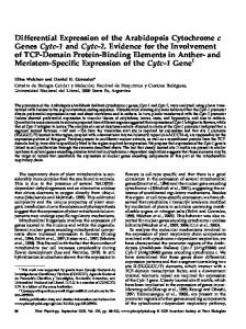
Acyltransferase Required for Cutin Formation Figure 1. Phylogenetic analysis of DCR and isolation of the dcr mutant alleles. A, Unrooted phylogenetic tree of DCR orthologs from various plant species and additional BAHD family acyltransferases. The protein sequences were analyzed using ClustalA 1.81 with a PAM350 matrix. The alignment editing was performed using GeneDoc. Multiple alignment parameters were as follows: gap opening set at 10 (default), gap extension set at 2.0, and the neighbor-joining method was used for calculating the tree. The bootstrapped tree was corrected for multiple substitutions. The scale bar of 0.1 is equal to 10% sequence divergence. The phylogenetic tree was constructed using the TreeView program. Full names and accession numbers of the proteins depicted are described in Supplemental Methods S1. B, Approximate locations of the three different insertions in the DCR locus. Exons are represented by boxes, introns by single lines, and T-DNA insertions by triangles. T-DNA positions in the genomic sequence are indicated as bp downstream of the ATG start codon. C, RTPCR analysis of the DCR transcript in inflorescence tissues of dcr-1 and wildtype (WT) plants (Nossen). Each lane represents an individual biological replicate. The UBIQUITIN-C gene served as a control for equal cDNA loading. [See online article for color version of this figure.]
In order to verify that the phenotype of the dcr mutant lines indeed originated from a lesion in the DCR gene, we first backcrossed dcr-1 and dcr-2 with their respective wild-type backgrounds. Segregation analysis of the F2 backcross progeny showed a linkage between the dcr mutant phenotype and the insertion in the DCR gene. Moreover, the homozygous dcr-1 plants were transformed with the DCR genomic sequence expressed under the control of the constitutive 35S cauliflower mosaic virus (CaMV) promoter (35S:DCR). The results indicated a functional complementation of DCR activity in the dcr-1 mutant background, as the 35S:DCR/dcr-1 plants were similar to wild-type plants and did not stain with TB (Fig. 2, H–J). The dcr mutant lines displayed abnormal flower and silique development: they had short, folded petals, shorter sepals, and semisterile siliques (Fig. 2, K and L; see Fig. 6B below; Supplemental Fig. S2F). In order to verify the origin of the semisterility, we carried out reciprocal backcrosses. The number of seeds in siliques derived from crosses in which dcr-1 pollen served as donor was fewer than the number of seeds in siliques Plant Physiol. Vol. 151, 2009
derived from crosses in which dcr-1 ovules served as acceptors (see Fig. 6A below). Thus, we concluded that the semisterility in dcr-1 primarily originates from defects in the male gametophyte. Epidermal Cell Differentiation Is Affected in Both Vegetative and Flower Organs of the dcr Mutants, Including Formation of Epicuticular Ridges on Epidermal Cells of Petals
Scanning electron microscopy (SEM) revealed that leaf epidermal cell pattern is disrupted in the dcr-1 mutant line (Fig. 3, A and B). Moreover, collapsed trichomes were often observed (Fig. 3, C and D). In addition, transmission electron microscopy (TEM) showed that the cuticle, which is typically observed as a continuous layer (Fig. 3F), was discontinuous in the dcr-1 mutant leaf epidermis (Fig. 3E). In flowers, fusions between sepals were often observed (Fig. 3, G and H). A closer examination of the sepal epidermis revealed that the adaxial epidermis is defective in the mutant plants, as abnormal cell patterns and fractures 1775
Panikashvili et al. Figure 2. Light microscopy images of different dcr mutant allele phenotypes. A, Phenotypes of 1-month-old dcr-1 versus wild-type (WT; Nossen) plants. B, Phenotypes of 1-month-old dcr-2 and dcr-3 versus wild-type (Col-0) plants. C, Five-week-old dcr-1 plant displaying postgenital fusion between rosette leaves. The site of the fusion is indicated by an arrow. D, TB staining of dcr-1 and wild-type rosettes. E and F, TB staining of dcr-1 and wild-type inflorescences. G, Fusion between flower buds in the dcr-1 inflorescence. H and I, TB staining of a dcr-1 plant complemented with the 35S:DCR construct (H) versus the wild type (I). J, TB staining of dcr-1 versus dcr-1 complemented with the 35S:DCR construct (blue staining in petals is indicated by arrows). K, Flower phenotype of dcr-2 versus the wild type. Folded petals are indicated by arrows. L, Semisterility (indicated by arrows) is obvious in the dcr-1 plant compared with the wild type.
were evident (Fig. 3, I and J). Moreover, the sepal abaxial epidermis was smoother in the mutants as compared with that of wild-type plants (Fig. 3, K and L). One of the most intriguing characteristics of the Arabidopsis petal epidermal cells is the presence of cuticular foldings. These are typically present on surfaces of both sides of the petals (Fig. 4, B, D, F, and H). Interestingly, SEM analysis revealed that in the dcr mutant petals, the cuticular foldings disappeared and the surface of both sides was smooth (Fig. 4, A, C, E, and G). These observations were confirmed using TEM examination (Fig. 4, I–L). In addition, SEM analysis of dcr-1 anther epidermal cells revealed that, compared with wild-type anthers, they are devoid of cuticular ridges, as seen in the case of dcr mutant petals (Supplemental Fig. S3). Seeds of the dcr Mutants Display Surface Alterations, Including a Defect in Mucilage Extrusion and Germination Capacity
TB test indicated that the surface of developing mutant seeds was more porous (Fig. 5, A and B). Seeds of dcr plants were wider, longer, heavier, and darker than wild-type seeds (Fig. 5, C–E). Moreover, they 1776
were often deformed (e.g. shrunken; Supplemental Table S1) and in some cases even fused to each other (Fig. 5, C–F). In addition, safranin O intensively stained cell walls of mature dcr-1 seeds, and as a result, the columella cells were less visible as compared with those in wild-type seeds (Fig. 5, G and H; Supplemental Fig. S2, I and J). This difference in staining provided additional evidence that dcr mutant seeds are altered in their surface. In mature Arabidopsis seeds, mucilage enclosed between epidermal columella cells and the outer cell wall is present in a dehydrated form. Upon contact with water, the mucilage expands and extrudes from the seed coat. Normally, this leads to visualization of the columella cells that are formed during seed coat development as cytoplasmic elevations with secondary cell wall deposited on top of them. Interestingly, in water-imbibed dcr-1 seeds, columella cells were not clearly visualized as in wild-type seeds (Fig. 5, I and J). TEM analysis of seeds fixed in a water-based fixative showed that mucilage was released from the wild-type seed coat while it was retained in the mutants (Fig. 5, K and M). In addition, we could detect a densely osmium-stained layer that resembled the cuticle coating the seed surface (Fig. 5L). When mature wild-type seeds were imbibed in ruthenium red, which stains Plant Physiol. Vol. 151, 2009
Acyltransferase Required for Cutin Formation Figure 3. Electron microscopy images of dcr mutant epidermal cells. A and B, SEM images of the leaf epidermal cell pattern in dcr-1 (A) and the wild type (WT; Nossen; B). C, A collapsed trichome in dcr-1. The site of collapse is indicated by an arrow. D, A wild-type trichome. E, TEM image of a dcr-1 leaf epidermis. A discontinuous cuticle (cut) is indicated by arrows. F, TEM image of wild-type leaf epidermis. An intact cuticle is indicated by an arrow. G, SEM image of a dcr-1 flower with fused sepals. H, Enlarged image of the sepal fusion area boxed in G. I, dcr-1 sepal adaxial epidermis. Fractures in the epidermis are indicated by arrows. J, Wild-type sepal adaxial epidermis layer. K, dcr-1 sepal abaxial epidermis layer. L, Wild-type sepal abaxial epidermis layer.
pectin, it was apparent that the dcr-1 mutant seeds are defective in mucilage extrusion (Fig. 5, N and O). As observed with safranin O staining of mutant and wildtype seeds (see above), the columella cells were not clearly visible in mutants seeds stained with ruthenium red. Even pretreatment with a cationic chelator
(0.05 M EDTA) did not facilitate mucilage release in the dcr-1 mutant seeds (Fig. 5, P and Q). The ability of the seed to take up water is a crucial quality trait, but this trait is dispensable under laboratory conditions. In order to test the ability of the mutant seeds to germinate under water-limiting conFigure 4. Electron microscopy images of dcr mutant petals. A, SEM image of dcr-1 petal abaxial epidermis layer. B, SEM image of wild-type (WT; Nossen) petal abaxial epidermis layer. C and D, Enlarged images of abaxial petal epidermal cells of dcr-1 (C) and the wild type (D). Note the cuticular foldings on the surface of wild-type cells (spaghetti-like structures) that are absent in the dcr-1 cells. E, SEM image of dcr-1 petal adaxial epidermis. F, SEM image of wild-type petal adaxial epidermis. G and H, Enlarged images of adaxial petal epidermal cells of dcr-1 (G) and the wild type (H). I and J, TEM images of dcr-1 (I) and wild-type (J) petals. The cuticular foldings in wildtype petals (J) are indicated by black arrows (absent in the mutant cells). K and L, Enlarged images of dcr-1 (K) and wild-type (L) petal abaxial epidermal cells. Note the presence of cuticular foldings in the wild type (black arrows).
Plant Physiol. Vol. 151, 2009
1777
Panikashvili et al. Figure 5. Seed surface phenotypes in the dcr mutants. A and B, TB staining of dcr-1 (A) and wild-type (WT; Nossen; B) developing seeds. C and D, Shrunken and fused seeds of dcr-1 (C) versus the wild type (D). E and F, SEM pictures of dcr-1 (E) and wild-type (F) mature seeds. G and H, Confocal microscopy pictures of dcr-1 (G) and wild-type (H) mature seeds stained with safranin O (a cell wall-specific dye). H, Visualized columella cells in wild-type seed are marked by small circles. I and J, Water imbibition of dcr-1 (I) and wild-type (J) seeds. Protruded columella cells in wild-type seeds are indicated by arrows. K to M, TEM images of dcr-1 (K and L) and wild-type (M) seed coats. L shows an enlarged image of the boxed region in K; a cuticle-like densely stained structure is indicated by black arrows. The columella cells are marked in K and M by white arrows. N and O, Mucilage release in seeds stained with ruthenium red in dcr-1 (N) and the wild type (O). P and Q, Mucilage release in 0.05 M EDTA-pretreated seeds stained with ruthenium red in dcr-1 (P) and the wild type (Q).
ditions, we tested the germination rate of dcr-1 and dcr-2 seeds on a filter paper soaked with a polymer (10% polyethylene glycol 8000; Fig. 6F). The results showed that the dcr mutant seed germination rate was to a great extent lower than that of wild-type seeds. DCR Expression Is Not Restricted to the Aerial Plant Parts and Is Epidermis Specific in Stems, Sepals, and Anther Filaments
Data from the Genevestigator database (https:// www.genevestigator.com/gv/index.jsp) indicated that DCR is strongly expressed in the inflorescences. In the flowers, the strongest expression was noted in petals. To substantiate the expression data described above and further examine the spatial and temporal expression of DCR, its full putative promoter region was fused to the GUS reporter gene (pDCR:GUS) and transformed to Arabidopsis. Expression from the putative DCR promoter was monitored extensively in two out of the 20 pDCR:GUS transgenic lines generated, starting from day 1 after germination up to senescence. At the principal growth stage 0.7 (Boyes et al., 2001), reporter expression was detected in the root cap, lateral root emerging sites, and cotyledons (Fig. 7, A–C). At the principal growth stage 1.02, reporter expression was noted in lateral roots and young emerging leaves (Fig. 7D). At principal growth stages 6.00 to 6.50, strong GUS staining was observed only in the upper stem, inflorescences, and trichomes 1778
of the mature leaves and stems (Fig. 7, E–H). Crosssections of the upper stems and inflorescences revealed epidermis-specific GUS staining at least in the stems, sepals, and anther filaments (Fig. 7, G, I, and J). Furthermore, GUS staining was also observed in pollen grains and torpedo stage seeds (Fig. 7, J and K). Cross-sections of the seeds showed that in the torpedo stage, the reporter is expressed in the inner and outer layers of the seed coat integumenta (Fig. 7L). At the bent cotyledon stage, when the inner integumenta and outer integumenta crush into one layer (Beeckman et al., 2000), the reporter expression was noted only in the inner integumenta (Fig. 7M). During embryo development, the reporter was strongly expressed at all stages examined (Fig. 7N). The DCR Protein Is Predominantly Localized in the Cytosol
Analysis of the DCR protein using prediction tools revealed no specific targeting signals or transmembrane domains, suggesting that DCR might encode a cytosolically localized protein. In order to determine its subcellular localization, the coding region was fused to the N termini of GFP and constitutively expressed in Arabidopsis. Confocal microscopy examination of leaf epidermis indicated that DCR might be localized in the cytosol or in the plasma membrane (Fig. 8, A–D). Examining protoplasts derived from the same plants provided additional evidence that DCR is Plant Physiol. Vol. 151, 2009
Acyltransferase Required for Cutin Formation
Figure 6. Sterility and altered seed characteristics in the dcr mutants. A, Defective male gametophyte in the dcr-1 mutant line. Reciprocal backcrosses show that a normal number of seeds is formed in cases of crossing dcr-1 with wild-type (WT; Nossen) pollen and a significant reduction in seed number per silique when dcr-1 pollen is used for crossing. At least three flowers were used from each genotype for reciprocal crosses. B, Average number of seeds in siliques of dcr-1 and wild-type plants. Seeds from three to five siliques from three to four plants from each genotype were collected for seed counting. C, Weight of 100 seeds of dcr-1 and the wild type. D, Seed width measurement of dcr-1 and the wild type. E, Seed length measurement of dcr-1 and the wild type. Seed pools derived from four to five plants of each genotype were used in C, D, and E. F, Percentage of nongerminating seeds of dcr mutant alleles and the corresponding wild-type ecotypes (Nossen [Nos.] and Col-0) under water-limiting conditions (10% polyethylene glycol 8000). The values indicate means of three biological replicates 6 SD. * P , 0.05.
likely a cytoplasmically localized protein, although some colocalization with the plasma membrane marker FM4-64 was observed (Fig. 8, E and F). In addition, protoplasts derived from Arabidopsis cell culture cells were cotransformed with DCR-GFP and markers for endoplasmic reticulum (ER) and Golgi localization. DCR-GFP displayed a clear cytoplasmic localization pattern that did not overlap either with the ER or with Golgi markers (Fig. 8, G–J). The easy detection of the heterologously expressed DCR protein (in Escherichia coli) in the soluble fraction provided further support that it encodes a soluble protein (Supplemental Fig. S7). The Amount of In-Chain Hydroxy Fatty Acids Is Profoundly Affected in the dcr Mutants
SEM examination of the dcr mutant vegetative organs did not reveal any difference in epicuticular wax coverage (data not shown). In order to investigate the effect of a lesion in DCR on lipid polyester metabolism, flower and leaf cutin, seed coat polyester, and root suberin were depolymerized and analyzed using gas chromatography-mass spectrometry. The results revealed that the amount of in-chain hydroxy fatty acids was profoundly affected in the dcr mutants. In flower cutin, the load of 9(10),16-dihydroxy-hexadecanoic acid was reduced to nearly undetectable levels in Plant Physiol. Vol. 151, 2009
the dcr-1 and dcr-2 plants. The amount of 9(10),16hydroxy-hexadecanedioic acid was reduced 3.2 times in dcr-1 and 2.92 times in dcr-2 flowers (Fig. 9A; Table I; Supplemental Table S2; Supplemental Fig. S4A). The amount of v-hydroxy-octadecenoic and v-hydroxyoctadecadienoic acids was also significantly reduced in flowers of the dcr plants (Table I; Supplemental Table S2). In contrast, the amount of hexadecanedioic acid in flowers was increased in both mutant alleles (3.17 times in dcr-1 and 3.06 times in dcr-2). The total cutin load was reduced nearly two times in both mutant alleles (Table I; Supplemental Table S2). Cutin composition of seed coat polyester was altered in a similar manner, as 9(10),16-dihydroxyhexadecanoic acid was reduced 13.2 and 4.83 times in dcr-1 and dcr-2 plants, respectively (Fig. 9B; Supplemental Table S2; Supplemental Fig. S4B). The amount of hexadecanedioic acid was slightly but significantly increased only in the dcr-2 plants (Fig. 9B; Supplemental Fig. S4B). No change in total seed coat polyester load between the two mutant alleles and corresponding wild-type plants was observed. Strikingly, changes similar to the ones observed in flowers and seed coats were detected in leaf cutin monomers analyzed after methanol (MeOH)/ HCl depolymerization. Again, the major monomer affected was the 9(10),16-dihydroxy-hexadecanoic acid, which was undetected in the mutants. The 1779
Panikashvili et al. Figure 7. Spatial and temporal expression of a DCR promoter-GUS reporter in Arabidopsis tissues. A, ReporterGUS expression in a 3-d-old seedling. The expression in a lateral root emergence site and the root cap region is indicated by black arrows. B and C, Close-up views of a root cap region (B) and a lateral root emergence site (C). D, Reporter-GUS expression in a 15-dold seedling. The expression in the emerging leaves and lateral roots is indicated by arrows. E and F, In the mature leaves (E) and stems (F), GUS expression is clearly observed in trichomes. G, Epidermis expression in the elongating stem. H, GUS expression in inflorescence. I, GUS expression is epidermis specific in anther filaments and sepals (indicated by arrows). J, Expression in anthers and pollen grains (indicated by an arrow). K, Whole mount view of seed-specific GUS expression. L, Torpedo stage seed GUS expression. The outer and inner integumenta layers are indicated by a bracket. M, Bent cotyledon stage seed GUS expression. Expression in the inner integumenta is indicated by an arrow. N, GUS expression in different embryonic developmental stages: i, torpedo; ii, walking stick; iii, bent cotyledon.
amount of v-OH-hexadecanoic acid was increased 1.76 times (Fig. 10; Supplemental Table S3). However, the total load of dcr-1 leaf cutin monomers was increased slightly compared with the wild type (Supplemental Table S3). Since pDCR:GUS expression was observed in roots due to the similarity in chemical composition between the cutin and suberin polymers, we analyzed depolymerized suberin from roots of the dcr mutants. The analysis showed no change in the mutant suberin composition (Supplemental Fig. S5). These results suggested that DCR is required for the incorporation of the in-chain hydroxylated fatty acids to the cutin polymer and that its most likely substrate is 9(10),16-dihydroxyhexadecanoic acid, the most abundant Arabidopsis flower cutin monomer detected by our analysis. 1780
Enhanced Water Loss and Increased Sensitivity to Salinity, Osmotic Stresses, and Drought Conditions Displayed by the dcr Mutants
To assay how the DCR deficiency in the mutant plants could affect the rate of water loss, we monitored changes to the fresh weight of detached rosettes. Roots and emerging inflorescence stems of 4-week-old seedlings were detached from the rosettes, and the percentage of water loss was examined during 6 h. The results showed that fresh weight loss from the rosette tissues was increased in the dcr-1 and dcr-2 mutants compared with the corresponding wild-type rosettes (Fig. 11A). While the difference in the water loss rate of dcr-1 was obvious already after 40 min, in the dcr-2 line, the difference from the wild type was significant only after 4 h. Plant Physiol. Vol. 151, 2009
Acyltransferase Required for Cutin Formation
Figure 8. Subcellular localization of the DCR protein. A to D, Confocal microscopy images through Arabidopsis leaf epidermal cells and protoplasts expressing 35S:DCR-GFP. The images were acquired through a GFP filter (A), chlorophyll filter (B), transmission filter (C), and a merge between GFP, chlorophyll, and transmission filters (D). E and F, A confocal series showing DCR-GFP localization in a protoplast derived from a transgenic plant expressing DCR-GFP. The images with GFP were acquired through a GFP filter (E) and with the plasma membrane marker FM4-64 through a chlorophyll filter (F). G to J, A confocal series showing DCR-GFP localization in Arabidopsis cell culture protoplasts transformed with DCR-GFP (G and I) and with ER (H) and Golgi (J) markers. Golgi stacks are indicated by white arrows.
To assess whether DCR deficiency affects the response to environmental stresses, we subjected seedlings of dcr-1 to salinity (200 mM NaCl) and osmotic (400 mM mannitol) stress conditions. We observed that dcr-1 seedlings are more susceptible to salinity and osmotic stress as compared with the wild type (Fig. 11B). Six days following the application of salt stress, 100% of the dcr-1 seedlings were entirely bleached, whereas only 40% of the wild type were either entirely or partially bleached. Eight days following the application of osmotic stress, 23% of the dcr-1 seedlings were bleached. After 15 d, the amount of bleached dcr-1 seedlings reached 75%. None of the wild-type seedlings were bleached after 8 d of osmotic stress, and only 10% of them were bleached after 15 d of osmotic stress. We also subjected the dcr-1 and dcr-2 mutants and their corresponding wild-type plants (ecotypes Nossen and Columbia [Col-0], respectively) to water deprivation conditions. The results showed that the dcr-1 and dcr-2 plants were more susceptible to water deprivation than the corresponding wild-type plants (Fig. 11C). In addition to the conditional stress phenotypes described above, we noted that the dcr mutant plants displayed increased levels of chlorosis and death upon maturation as compared with wild-type plants (Fig. 11D). Altered Root Architecture in DCR-Deficient Plants
While growing mutant and wild-type seedlings on vertical agar plates, we observed that the dcr-1 plants display excessive lateral roots and hairs as compared Plant Physiol. Vol. 151, 2009
with the wild-type seedlings (Fig. 11, E and F). Moreover, observing mutant seedlings 9 d after germination on 0.25% diphenylboric acid (staining flavonoids) with the epifluorescence microscope revealed more lateral root initials in the dcr-1 seedlings as compared with the wild-type seedlings (Supplemental Fig. S6).
DISCUSSION DCR Is Required for Oligomer/Polymer Formation of Cutin in Reproductive and Vegetative Organs
Regardless of cutin being one of the most central plant biopolymers, the current knowledge on its biosynthesis is very limited. Here, we show that DCR, a member of the BAHD family of acyltransferases, is required for cutin oligomer/polymer formation in Arabidopsis. Cutin polymer production likely requires the action of different acyltransferases in order to link its aliphatic, aromatic (ferulic and coumaric acids), and glycerol monomers to each other. While GPAT function has been attributed to the aliphatic acylation of glycerol-3-phosphate (Li et al., 2007), chemical analysis of the dcr mutant alleles suggested that the major substrate of DCR activity is 9(10),16-dihydroxy-hexadecanoic acid. The first indication that in-chain hydroxylated fatty acids represent the major cutin monomers of reproductive organs was provided by Kolattukudy and coworkers (1974), who found that 9(10),16-dihydroxyhexadecanoic acid represents about 80% of the V. faba 1781
Panikashvili et al.
Figure 9. The lipid polyester profiles of dcr-1 and the wild type (WT; Nossen) in flowers and seed coats after BF3 depolymerization. A, The flower cutin polyester profile. Differential monomers are indicated by arrows. B, The seed coat polyester profile. Differential monomers are indicated by arrows. The values indicate means of four biological replicates 6 SD. * P , 0.05. For the full names of identified monomers, see Supplemental Table S5. DW, Dry weight.
flower petal cutin. Moreover, 9(10),16-dihydroxyhexadecanoic acid represents between 50% and 83% of the fruit cutin monomer load in tomato (Saladie´ et al., 2007), Malabar papaiarnarum, gooseberry (Kolattukudy, 2001), and cherry (Peschel et al., 2007). In this study, we found that this particular acid dominates the Arabidopsis flower cutin, representing nearly half of its load (51.53% in the case of the Col-0 ecotype and 55.37% in the case of the Nossen ecotype), and its levels were dramatically decreased in the flower cutin of the dcr lines. Other groups detected dicarboxylic acids as the main components of Arabidopsis flower cutin using a MeOH/HCl depolymerization method (Kurdyukov et al., 2006b; Kannangara et al., 2007), and this difference might originate from the dissimilar depolymerization methods used. The depolymerization method with BF3 used here allows better identification of this particular acid (Nawrath, 2006). Seed 1782
coat polyester and leaf cutin analysis corroborated the findings in the flower, since once more, 9(10),16dihydroxy-hexadecanoic acid was the main cutin constituent decreased in the mutants. Despite the fact that 9(10),16-dihydroxy-hexadecanoic acid presence in the seed coat polyester and leaf cutin is only up to 7%, as was found by us and others (Molina et al., 2006, 2008; Lu¨ et al., 2009), the seed coat and leaf surface in dcr mutants was profoundly affected. The prominent expression of DCR in flower organs combined with the pronounced developmental phenotypes of the dcr mutant alleles complement the chemical information described above regarding the role of DCR in the incorporation of 9(10),16-dihydroxy-hexadecanoic acid and possibly additional monomers into the cutin polymer. Already 35 years ago, it was shown that ATP and CoA together with the presence of hydroxylated fatty Plant Physiol. Vol. 151, 2009
Acyltransferase Required for Cutin Formation
Table I. List of flower cutin monomers identified after BF3 depolymerization with their respective concentrations in dcr-1 mutants and wild-type plants Data presented here are in mg mg21 dry weight and represent averages of four replicates. Differences with P , 0.05 were regarded as significant. Identified Cutin Monomers
Aromatics trans-Coumaric acid trans-Ferulic acid cis-Sinapic acid trans-Sinapic acid Subtotal Saturated fatty acids C24-FA (tetracosanoic acid) Long-chain alcohols C20-1-ol (docosanol) Dicarboxylic fatty acids C16:2-DA [hexadecadiene-(1,16)-dioic acid] C16:1-DA [hexadecene-(1,16)-dioic acid] C16-DA [hexadecane-(1,16)-dioic acid] C18:2-DA [octadecadiene-(1,18)-dioic acid] C18:1-DA [octadecene-(1,18)-dioic acid] C18-DA [octadecane-(1,18)-dioic acid] Subtotal Mid-chain hydroxylated fatty acids C16-9/10-HDA [9/10-hydroxy-hexadecane-(1,16)-dioic acid] C16-9/10,16-DHFA (9/10,16-dihydroxy-hexadecanoic acid) Subtotal Terminal-hydroxylated fatty acids C16:1-v-HFA (16-hydroxy-hexadecenoic acid) C16-v-HFA (16-hydroxy-hexadecanoic acid) C18:3-v-HFA (18-hydroxy-octadecatrienoic acid) C18:2-v-HFA (18-hydroxy-octadecadienoic acid) C18:1-v-HFA (18-hydroxy-octadecenoic acid) Subtotal 2-Hydroxylated fatty acids C16-2-HFA (2-hydroxy-hexadecanoic acid) C22:1-2-HFA (2-hydroxy-docosenoic acid) C22-2-HFA (2-hydroxy-docosanoic acid) C24:1-2-HFA (2-hydroxy-tetracosenoic acid) C24-2-HFA (2-hydroxy-tetracosanoic acid) C26:1-2-HFA (2-hydroxy-hexacosenoic acid) C26-2-HFA (2-hydroxy-hexacosanoic acid) Subtotal Total
acids are the primary prerequisites for a successful cutin polymer biosynthesis (Croteau and Kolattukudy, 1974). The same authors showed that hydroxyl groups of aliphatic acids act as donors for the transesterification reaction during cutin polymer formation, whereas the free carboxyl groups are not necessary for this process. This implies the existence of putative cutin transacylase, which utilizes dihydroxy or polyhydroxy fatty acyl CoA in order to acylate free hydroxyl groups of the growing cutin polymer. In addition, it has been shown recently that a high number of hydroxy fatty acids, especially those having hydroxyl groups in terminal and midchain positions like 9 (10),16-dihydroxy-hexadecanoic acid, are the most effective in creating an extended two-dimensional cutin monolayer (Benitez et al., 2008). Plant Physiol. Vol. 151, 2009
dcr-1
Wild Type (Nossen) Mean
Mean
SD
0.0377 0.1002 0.0317 0.2600 0.4297
0.0118 0.0079 0.0059 0.0048 0.0304
0.1053 0.1075 0.0183 0.3026 0.5336
0.0148 0.0087 0.0028 0.0077 0.0339
0.0670
0.0147
0.0922
0.0024
0.2059
0.0027
0.1819
0.0185
0.0718 0.0757 2.4604 0.8523 0.9994 0.2870 4.7466
0.0013 0.0262 0.0763 0.0324 0.0407 0.0051 0.1819
0.0837 0.0659 0.7769 1.2207 0.9981 0.2454 3.3906
0.0071 0.0050 0.0274 0.1288 0.0459 0.0375 0.2518
0.2453 0.0542 0.2994
0.0080 0.0409 0.0488
0.7840 9.7399 10.5240
0.1134 1.2405 1.3538
0.1683 0.8362 0.3059 0.1207 0.0485 1.4796
0.0527 0.0258 0.0714 0.0033 0.0016 0.1547
0.1637 0.5442 0.5671 0.3954 0.1549 1.8254
0.0216 0.0336 0.1445 0.0446 0.0224 0.2666
0.0452 0.0436 0.1725 0.2637 0.3934 0.0685 0.1473 1.1343 8.3624
0.0006 0.0147 0.0062 0.0131 0.0040 0.0031 0.0038 0.0455 0.4787
0.0396 0.1162 0.1514 0.2563 0.2825 0.0664 0.1296 1.0421 17.5898
0.0008 0.0066 0.0021 0.0042 0.0862 0.0035 0.0027 0.1062 2.0332
SD
Change versus the Wild Type
Down
Down
Up
Down Down Down
Up Down Down
Up Down Up
Up Down
With respect to the actual DCR enzyme activity, three possible reactions could be proposed. In the first, DCR might utilize aromatic CoAs or aliphatic CoAs in order to acylate the primary or secondary hydroxy groups of 9(10),16-dihydroxy-hexadecanoic acid. The second possibility is that DCR might use the CoA of 9 (10),16-dihydroxy-hexadecanoic acid in order to acylate either aromatics or other hydroxy fatty acids. In the third option, the first two reactions could occur when the cutin dimers or trimers serve as acceptors for the acylation reaction. In the course of this study, we generated a recombinant DCR enzyme by heterologous expression in E. coli (Supplemental Fig. S7) and examined the first possibility using various CoAs (feruloyl-CoA, coumaroyl-CoA, palmitoyl-CoA, and stearoyl-CoA) as donors and 9(10),16-dihydroxy1783
Panikashvili et al. Figure 10. The leaf cutin profile of dcr-1 and the wild type (Nossen) after MeOH/HCl depolymerization. Differential monomers are indicated by arrows. The values indicate means of four biological replicates 6 SD. * P , 0.05. For the full names of identified monomers, see Supplemental Table S5. n.d., Not detected. White bars, Ecotype Nossen; black bars, dcr-1.
hexadecanoic acid, 16-hydroxy-hexadecanoic acids, long-chain alcohols (C18 and C20), and glycerol as acceptors for enzyme assays. While we could not detect any new product in all substrate combinations used so far, the two other options of DCR enzyme activity should be examined in a future study once the appropriate substrate, namely the 9(10),16-dihydroxy-hexadecanoyl CoA, has been purified or synthesized chemically. The Importance of DCR in the Arabidopsis Seed Coat
Seed coat polyester analysis in the dcr mutants revealed a significant reduction in the levels of several cutin monomers, although fairly mild as compared with those displayed in flowers. In Arabidopsis, the seed coat consists of three layers of inner integumenta and two layers of an outer integumenta that differ greatly in their morphological characteristics (Haughn and Chaudhury, 2005). At the embryo maturity stage, the inner integumenta crushes into a single-cell-thickwalled layer (Beeckman et al., 2000). Recently, it has been shown using promoter-reporter gene fusions that the suberin-associated gene GPAT5 is mostly expressed in the outer integumenta whereas the cutin-associated gene ATT1 is detected in the inner integumenta (Molina et al., 2008). Based on these observations, the authors suggested the existence of cutin in the inner integumenta and suberin in the outer integumenta. Interestingly, while DCR is ubiquitously expressed in all integumenta layers in young seeds, it is specifically expressed in the inner integumenta of the mature seeds. During seed coat formation, the epidermal cells produce large quantities of mucilage that upon contact with water hydrates, and this leads to the rupture of the outer seed coat surface (Penfield et al., 2001). To date, several genes associated with mucilage release upon hydration have been reported; all encoded enzymes that are either involved in mucilage production or transcription factors regulating seed coat cell dif1784
ferentiation (Western et al., 2004). In this study, we provide evidence that DCR is required for the proper release of mucilage upon hydration. Further support to the conclusion that a fully functional DCR protein is vital for seed hydration properties was provided by germination assays showing that the germination rate under water-limiting conditions of seeds derived from both dcr mutant alleles was lower as compared with wild-type seeds. Prior to its maturation, between 1 d after pollination and up to 12 d after pollination, the embryo is protected by the seed coat, which is not receptive to hydrophilic substances. Later, when the embryo is grown to maturity, the seed coat becomes receptive to water, as proper hydration properties at maturity are vital for seed germination. Surprisingly, the lesion in DCR affects seed coat surface properties both at early stages of seed development and later in the dry seed. Although for a long time it was suggested that the outer surface of the seed coat is composed of a primary cell wall (Western et al., 2000), it is likely that apart from the primary cell wall, a lipidic cutin-like structure covers the cell wall in order to protect it. This is supported by the dense, osmium-stained layer (resembling a cuticular layer) observed in the developing seed coat (Watanabe et al., 2004; Fig. 5L). The completeness of this structure could be examined by the TB test (Tanaka et al., 2004), and developing dcr seeds are in fact intensely stained with the TB stain as compared with wild-type seeds. DCR is not the only gene found to be associated with Arabidopsis seed coat surface formation and required for its intactness. The developing seed coats of the acr4-1 and desperado-2 (dso-2) mutants were also positively stained with TB. The ACR4 receptor-like protein kinase was suggested to be required for leaf and seed coat surface formation (Watanabe et al., 2004). Seeds of the dso-2 mutant lines were suggested to be impaired in the integrity of the seed surface (Panikashvili et al., Plant Physiol. Vol. 151, 2009
Acyltransferase Required for Cutin Formation Figure 11. Water loss, root architecture, and tolerance to various stress conditions of the dcr mutants. A, Rate of water loss of dcr-1, dcr-2, and wild-type (WT) plants. Four rosettes per genotype were weighed during the time intervals depicted. The results are derived from three independent experiments and depicted with SE of the mean for each time point. The values indicate means of three biological replicates 6 SE. * P , 0.05. ec. Nos., Ecotype Nossen; ec. Col., ecotype Col-0. B, Salt and mannitol stress experiments. Six-day-old dcr-1 and wild-type (Nossen) seedlings grown on MS agar plates were transferred to MS plates supplemented with 200 mM NaCl or 400 mM mannitol for an additional 2 weeks of growth. The results were documented 6 d after application of the salt stress and 15 d of osmotic stress. C, Water deprivation tolerance experiment. Three-weekold dcr-1, dcr-2, and wild-type plants were exposed to 10 d of water deprivation. Subsequently, seedlings were rewatered once and their appearance was documented after 1 week. D, Increased chlorosis and death in rosette leaves of 8-week-old dcr-1 plants grown in normal laboratory conditions as compared with wild-type plants (damaged leaves marked by arrows). E and F, Increased lateral roots (E) and hair formation (F) in dcr-1 plants compared with the wild type (Nossen).
2007). DSO/ABCG11 is an ABC transporter that is required for cutin and wax transport to plant organs (Bird et al., 2007; Luo et al., 2007; Panikashvili et al., 2007), and it might be active with DCR and ACR4 in the same genetic pathway. Contribution of DCR and Cutin to the Decoration of Conical Petal Cell Surface
Cuticular folds are found on the outer surface of a large number of plant species, frequently on flower organs such as petals (Jetter, 2006; Koch et al., 2009). The importance of these structures is not clear, but they might play a role in the pollination process, either by increasing the cell surface to facilitate emission of scent molecules or by influencing the reflection from Plant Physiol. Vol. 151, 2009
petal cells for pollinator attraction (Jetter, 2006). In addition, these structures could provide favorable structure to pollinators to walk on, or they could serve as a tactile cue to find the nectar. Regarding their composition, they might be formed as a result of the cuticle or cell wall thickening (Koch et al., 2009). The absence of these structures from the dcr mutant petal epidermal surfaces and the fact that the use of organic solvents did not remove them from the petal surface suggest that they are made of cutin that is tightly linked to the epidermal cell wall. Trichome Development in the dcr Mutants
The Arabidopsis trichomes or epidermal hairs are considered as “simple” and nonglandular, although 1785
Panikashvili et al.
little direct evidence exists that they are indeed incapable of low-level secretion (Wagner et al., 2004). Upon maturation, the Arabidopsis trichomes are surrounded by a ring of eight to 12 rectangular basement or support cells (Hu¨lskamp et al., 1994). Cuticle-related Arabidopsis mutants and transgenic plants frequently display defects in trichome development, as in the case of SHINE1/WIN1 overexpressors (Aharoni et al., 2004), bdg (Kurdyukov et al., 2006a), and dso/abcg11 (Bird et al., 2007; Luo et al., 2007; Panikashvili et al., 2007). The lesion in DCR affected the support cells, as trichomes were collapsed into the epidermal cell layer. Cuticle-associated genes are also often expressed in trichome cells, as we observed here for DCR expression in leaves and stems. A general characteristic, common to almost all secretory trichomes, is complete cutinization of the side walls of stalk cells (Fahn, 1988). The presence of these cells in trichomes ensures that the flow of substances or their precursors into the trichome takes place exclusively through the symplast and that the flow of substances back into the plant through the apoplast is prevented. It was also reported that lipid polyesters are found in boundaries between the plant and its secretory organs (e.g. trichome support cells; Kolattukudy, 2001). Adding to the importance of DCR in trichome development and function was the fact that trichomes were the only tissue in which its expression was maintained throughout all of the developmental stages we examined. DCR Might Play a Role in the Metabolism of a Root Cutin-Like Polymer But Not of Suberin
As anticipated for a gene associated with cutin formation DCR expression was epidermis specific in vegetative and reproductive organs. Nevertheless, it is also expressed in the root cap, lateral root primordia, and developing lateral root. Its root expression might point to involvement in the biosynthesis of suberin. However, the expression pattern observed in this study for DCR does not match the profile of a typical suberin biosynthetic gene such as DAISY/KCS2 (Franke et al., 2009). In accordance, we did not find any significant change in the root suberin polyester composition of the dcr mutants as compared with wild-type roots. This is not the first report on root expression of cuticular genes, as transcripts of KCS1 (Todd et al., 1999), BDG (Kurdyukov et al., 2006a), SHINE3 (Aharoni et al., 2004), DSO (Panikashvili et al., 2007), and HOTHEAD (Kurdyukov et al., 2006b) were detected in roots. Interestingly, expression of both SHINE3 and DSO, which were previously associated with the regulation of the cutin biosynthetic pathway (Aharoni et al., 2004; Kannangara et al., 2007) and transport (Bird et al., 2007; Luo et al., 2007; Panikashvili et al., 2007), was specifically detected in emerging lateral root primordia and the lateral root cap. The detection of excessive root branching and root hair formation in the dcr-1 line supports the involvement of DCR in the process of lateral root emergence. As in dcr-1, root 1786
branching was also affected in the bdg (Kurdyukov et al., 2006a) and the dso (Panikashvili et al., 2007) mutants. It is possible that the formation of a robust, cutin-like protective barrier is required for lateral root primordia and tip infiltration between cells and through the soil. Thus, DCR might be involved in the synthesis of a cutin-like polymer in the young, emerging roots. DCR Is Also Required in the Vegetative Organ Surface, and Its Deficiency Results in Reduced Ability to Cope with Environmental Stresses
The cuticle covering both reproductive and vegetative organ surfaces serves as a primary barrier against various environmental cues, including water deprivation, salt, and osmotic stress conditions. This function of the surface is mediated through the control of both transpirational and nontranspirational water loss (Shepherd and Wynne Griffiths, 2006). While this study was mostly focused on the effects of DCR deficiency in reproductive organ surfaces (i.e. flowers and seeds), DCR gene expression in young emerging leaves and the elongating part of stems suggested that it is also involved in cutin polymer formation in vegetative organs. Indeed, chemical analysis of cutin in leaves could not detect 9(10),16-dihydroxyhexadecanoic acid in the dcr mutants. The increase in the levels of two other cutin monomers, hexadecanedioic and v-OH-hexadecanoic acids, in leaves and flowers of the dcr mutants implies that the same compensatory mechanism was activated in both tissues. We suggest that DCR is involved in the incorporation of in-chain hydroxy fatty acids into the cutin polymer during early stages of leaf expansion, when the formation of the cutin polymeric matrix is especially active (Reina et al., 2007). Indeed, analysis of pDCR:GUS plants showed that DCR is especially active in young expanding leaves and expressed only in trichomes upon leaf maturation. The reduced ability of dcr mutants to withhold water, demonstrated by a water loss experiment from detached rosettes and the susceptibility to water deprivation and osmotic stresses, may possibly be attributed to impaired cuticular integrity. While we did not examine stomatal index in the dcr mutants, cuticular mutants are often perturbed in stomata development (Aharoni et al., 2004); therefore, the dcr mutant stress phenotypes could also be due to excessive transpirational water loss. To summarize, here we show that DCR, a member of the BAHD family of acyltransferases, is required for the incorporation of the most abundant monomer into the polymeric structure of flower organ cutin. Its activity in constructing the plant lipid polyester is required for epidermal cell differentiation and patterning, trichome development, and the prevention of undesired postgenital fusions in aboveground organs. It is also essential underground, in shaping the root through lateral root formation. The nature of the lipid Plant Physiol. Vol. 151, 2009
Acyltransferase Required for Cutin Formation
polyester associated with DCR activity in roots is unclear; nevertheless, it does not appear to be a suberin. In seeds, DCR provides specific characteristics to the surface, most probably by the construction of a lipidic layer that influences dehydration properties, including the release of mucilage. It is apparent that cuticle-associated proteins, such as DCR, do not only play a role in plant development but are also active in stress response programs mediated by the cuticle (e.g. salinity, drought, and osmotic stress). The characterization of DCR augments the current knowledge regarding plant surface polymer formation and highlights the range of physiological roles played by the cutin polyester (as part of the cuticle) in both vegetative and reproductive organs. It further emphasizes the complexity of constructing such a barrier and the consequences to plant development and fitness when the assembly of this polymer is perturbed.
MATERIALS AND METHODS Plant Material and Mutant Lines Arabidopsis (Arabidopsis thaliana) plants were grown in the climate room at 20°C, 70% relative humidity, and 16-h/8-h light/dark cycle (Col-0, Wassilewskija, and Nossen ecotypes). Salk (SALK_128228, termed dcr-2) and Wisconsin (WiscDsLox245B03, termed dcr-3) T-DNA insertion lines were obtained from the Arabidopsis Biological Resource Center. The RIKEN Ds transposon line 12-1765-1, termed dcr-1, was received from the RIKEN Arabidopsis Ac/Ds transposon-tagged lines resource. The plant growth stage was defined as described elsewhere (Boyes et al., 2001).
Construction of Phylogenetic Trees and Sequence Alignments The protein sequences were analyzed using ClustalA 1.81 with the PAM350 matrix (Thompson et al., 1997). The alignment editing was performed using GeneDoc (Nicholas et al., 1997). The multiple alignment parameters were as follows: gap opening set at 10 (default), gap extension set at 2.0, and the neighbor-joining method was used for calculating the tree. The bootstrapped tree was corrected for multiple substitutions. The scale bar of 0.1 is equal to 10% sequence divergence. The phylogenetic trees were constructed using the TreeView program (Page, 1996).
Staining, Electron Microscopy, and Mucilage Release Experiments The method for examination of cuticular integrity was performed as described by Tanaka et al. (2004), and the safranin O staining of cell walls and confocal microscopy of seed coats were as reported by Truernit and Haseloff (2008). For SEM, flowers were collected and fixed with glutaraldehyde using standard protocols and dried using critical point drying. Stems were sputter coated without prior fixation directly after 24 h of air drying, while mature seeds were sputter coated directly after harvest. Samples were mounted on aluminum stubs and sputter coated with gold. SEM was performed using an XL30 ESEM FEG microscope (FEI) at 5 to 10 kV. For TEM, leaves from 1-month-old plants, flowers from 7-week-old plants, and mature seeds from 60-d-old plants were collected and processed using a standard protocol (Chuartzman et al., 2008). The Epon-embedded samples were sectioned (70 nm) using an ultramicrotome (Leica) and observed with a Technai T12 transmission electron microscope (FEI). The mucilage release experiments were performed as described previously (Rautengarten et al., 2008).
Stress Experiments For the detached rosette air-drying experiments, the rosettes were detached from 1-month-old mutant plants and weighed every 10 to 30 min up to
Plant Physiol. Vol. 151, 2009
6 h. For the water deprivation experiment, seeds were sown in soil, watering was stopped at 3 weeks after germination for 10 d, the plants were subsequently rewatered, and the recovery was followed for 1 additional week. For the salt and mannitol stress assays, 6-d-old seedlings were transferred to Murashige and Skoog (MS) plates supplemented with 200 mM NaCl or 400 mM mannitol for an additional 2 weeks of growth, and plant survival was monitored.
Generation of DNA Constructs For the generation of the DCR (At5g23940) overexpression construct, a genomic sequence (3,062 bp) from the start codon to the stop codon was amplified from wild-type (Col-0) plant genomic DNA with the following oligonucleotides (sense, 5#-BamHI-GCGGATCCATGAAGATAAAGATTATGAG-3#; antisense, 5#-NcoI-CGCCATGGTCAAACAAACCCATTGCCAT-3#) and cloned into the pFLAP100 vector under the control of the 35S CaMV promoter. The cassette including the 35S promoter, the DCR genomic sequence, and the NOS terminator was digested with PacI and AscI and cloned into the binary vector pBIN(+) (van Engelen et al., 1995). The complete DCR 5# upstream region (2,930 bp; termed pDCR) was amplified from wildtype (Col-0) plant genomic DNA using the following oligonucleotides (sense, 5#-BamHI-GGATCCGTTGTCTTGCGATATGCTTG-3#; antisense, 5#-NcoI-CCATGGGGTGAAAGAGATTTTAACTG-3#), excised with the appropriate restriction enzymes, and subcloned into the pMAX vector containing the GUS coding sequence and the NOS terminator. The pDCR fragment together with the GUS gene and the NOS terminator was excised with PacI and AscI and cloned into the binary vector pBIN(+) (van Engelen et al., 1995). For examination of DCR protein subcellular localization, the DCR cDNA was amplified without the stop codon from wild-type (Col-0) plant inflorescence using the following oligonucleotides (sense, 5#-BamHI-GCGGATCCATGAAGATAAAGATTATGAG-3#; antisense, 5#-NotI-CGCGGCCGCAACAAACCCATTGCCATTTCC-3#) and cloned into pENTER 1A (Invitrogen). The LR Clonase (Invitrogen) was subsequently used to recombine the DCR cDNA into the pMDC85 binary vector upstream of the GFP coding sequence and under the control of the 35S CaMV promoter, creating a construct bearing DCR coding sequence translationally fused to the N termini of GFP (Curtis and Grossniklaus, 2003). For plant transformation, inflorescences were dipped into Agrobacterium tumefaciens strain GV3101 carrying the various constructs as described (Clough and Bent, 1998).
Reverse Transcription-PCR Analysis For semiquantitative reverse transcription (RT)-PCR analysis, a fixed amount of DNase-treated total RNA was reverse transcribed using the AMV Reverse Transcriptase (EurX). The semiquantitative RT-PCR was performed using the GreenTaq DNA polymerase (Promega) with the following conditions: initial 94°C denaturation step for 2 min, followed by 45 s at 94°C, 45 s at 55°C, 1 min at 72°C, and 5 min of final elongation. The oligonucleotide sequences were as follows: for DCR (At5g23940), sense, 5#-GACCACTCCTTGCTGTCCAG-3#, and antisense, 5#-GAAAATAGTTATGTCCTCCG-3#. UBIQUITIN-C (At5g25760) was used as an endogenous control using the following oligonucleotides: sense, 5#-AGCGCGACTGTTTAAAGAATACA-3#, and antisense, 5#-TTGTGCCATTGAATTGAACCC-3#. The following numbers of amplification cycles were used: 28 for UBIQUITIN-C and 29 for DCR.
DCR Promoter Analysis and Subcellular Localization Assays Out of 25 independent transgenic lines, two with uniform GUS expression were chosen for detailed staining and histological analysis as described before (Baum and Rost, 1996). For the DCR subcellular localization assays, either leaves or protoplasts (prepared as described [Yoo et al., 2007]) derived from transgenic Arabidopsis plants expressing DCR-GFP were used. The plasmid DNA for protoplast transformation was purified using the Plasmid Maxi Kit (Geneaid). The protoplasts were prepared from Arabidopsis cell culture and transformed as described (http://genetics.mgh.harvard.edu/sheenweb/ protocols_reg.html). The ER (CD3-960) and Golgi (CD3-968) markers and confocal microscopy conditions were described elsewhere (Nelson et al., 2007). Fluorescence was observed by an Olympus CLSM500 microscope with argon laser at 488 nm for excitation and images for GFP, plasma membrane marker FM4-64, and chlorophyll signals and 580 nm for mCherry. The signals
1787
Panikashvili et al.
were collected through 505 to 525 nm for GFP, 620 to 750 nm for chlorophyll and FM4-64, and 605 to 630 nm for mCherry. The presence of the GFP and mCherry signals was verified using the laser photobleach approach with subsequent follow-up of the recovery.
coumaroyl-CoAs, Patrick Dussault for 9(10),16-dihydroxy-hexadecanoic acid, Sergey Malitsky for help with gas chromatography-mass spectrometry, and Guy Gafni for technical assistance. Received June 22, 2009; accepted October 7, 2009; published October 14, 2009.
Lipid Polyester Analysis For flower cutin analysis, soluble lipids were extracted from inflorescences by dipping in 10 mL of a methanol:chloroform (1:1, v/v) mixture for 14 d (solvent was changed daily). The inflorescence material was dried, weighed (about 10–20 mg), and used for analysis. For leaf cutin analysis, the leaves were photographed using light microscopy, delipidated as described above, and used for analysis. Leaf areas were determined using ImageJ software (National Institutes of Health). The seed coats were delipidated, and the roots were treated before suberin depolymerization as described (Ho¨fer et al., 2008). The flowers, seed coats, and root lipid polyesters were depolymerized with BF3 and analyzed further as described (Franke et al., 2005), except for the leaf cutin, which was depolymerized with MeOH/HCl containing 7% (v/v) methylacetate (Lu¨ et al., 2009) before analysis as described (Franke et al., 2005).
Supplemental Data The following materials are available in the online version of this article. Supplemental Figure S1. Identification of the Arabidopsis DCR gene and its mutant alleles. Supplemental Figure S2. Phenotypes of the dcr mutants. Supplemental Figure S3. SEM pictures of dcr-1 and wild-type (Nossen) anthers. Supplemental Figure S4. The lipid polyester profile in flower and seed coat tissues of the dcr-2 mutants and wild-type plants. Supplemental Figure S5. Root suberin polyester profile of the dcr-1 mutant allele versus the wild type (Nossen) and the dcr-2 mutant allele versus the wild type (Col-0). Supplemental Figure S6. Excessive root branching in dcr-1 mutant plants visualized using diphenylboric acid staining. Supplemental Figure S7. Purification of the DCR recombinant protein expressed in E. coli cells. Supplemental Table S1. A summary of DCR insertion lines. Supplemental Table S2. List of flower cutin monomers identified after BF3 depolymerization with their respective concentrations in dcr-2 mutants and wild-type plants. Supplemental Table S3. List of leaf cutin monomers identified after MeOH/HCl depolymerization with their respective concentrations in dcr-1 mutants and wild-type plants. Supplemental Table S4. A list of putative AtDCR homologs from various plant species. Supplemental Table S5. A full list of cutin and suberin monomers obtained from various tissues of dcr mutant and wild-type plants. Supplemental Methods S1.
Note Added in Proof During the review of this article, Marks et al. (Marks MD, Wenger JP, Gilding E, Jilk R, Dixon RA [2009] Transcriptome analysis of Arabidopsis wild-type and gl3-sst sim trichomes identifies four additional genes required for trichome development. Mol Plant 2: 803–822) described the trichomerelated phenotypes related to the gene described in our article.
ACKNOWLEDGMENTS We thank Eyal Shimoni for assistance with TEM, Eugenia Klein for help with SEM, Vladimir Kiss for assistance with confocal microscopy, Wilfried Schwab for hosting D.P. in his laboratory and for providing feruloyl- and
1788
LITERATURE CITED Aharoni A, Dixit S, Jetter R, Thoenes E, van Arkel G, Pereira A (2004) The SHINE clade of AP2 domain transcription factors activates wax biosynthesis, alters cuticle properties, and confers drought tolerance when overexpressed in Arabidopsis. Plant Cell 16: 2463–2480 Baum SF, Rost TL (1996) Root apical organization in Arabidopsis thaliana. 1. Root cap and protoderm. Protoplasma 192: 178–188 Beeckman T, De Rycke R, Viane R, Inze´ D (2000) Histological study of seed coat development in Arabidopsis thaliana. J Plant Res 113: 139–148 Benitez JJ, Heredia-Guerrero JA, Serrano FM, Heredia A (2008) The role of hydroxyl groups in the self-assembly of long chain alkylhydroxyl carboxylic acids on mica. J Phys Chem C 112: 16968–16972 Bird D, Beisson F, Brigham A, Shin J, Greer S, Jetter R, Kunst L, Wu X, Yephremov A, Samuels L (2007) Characterization of Arabidopsis ABCG11/WBC11, an ATP binding cassette (ABC) transporter that is required for cuticular lipid secretion. Plant J 52: 485–498 Bonaventure G, Beisson F, Ohlrogge J, Pollard M (2004) Analysis of the aliphatic monomer composition of polyesters associated with Arabidopsis epidermis: occurrence of octadeca-cis-6,cis-9-diene-1,18-dioate as the major component. Plant J 40: 920–930 Boyes DC, Zayed AM, Ascenzi R, McCaskill AJ, Hoffman NE, Davis KR, Gorlach J (2001) Growth stage-based phenotypic analysis of Arabidopsis: a model for high throughput functional genomics in plants. Plant Cell 13: 1499–1510 Chuartzman SG, Nevo R, Shimoni E, Charuvi D, Kiss V, Ohad I, Brumfeld V, Reich Z (2008) Thylakoid membrane remodeling during state transitions in Arabidopsis. Plant Cell 20: 1029–1039 Clough SJ, Bent AF (1998) Floral dip: a simplified method for Agrobacterium-mediated transformation of Arabidopsis thaliana. Plant J 16: 735–743 Croteau R, Kolattukudy PE (1974) Biosynthesis of hydroxyfatty acid polymers: enzymatic synthesis of cutin from monomer acids by cellfree preparations from the epidermis of Vicia faba leaves. Biochemistry 13: 3193–3202 Curtis MD, Grossniklaus U (2003) A Gateway cloning vector set for highthroughput functional analysis of genes in planta. Plant Physiol 133: 462–469 D’Auria JC (2006) Acyltransferases in plants: a good time to be BAHD. Curr Opin Plant Biol 9: 331–340 Fahn A (1988) Secretory tissues in vascular plants. New Phytol 108: 229–257 Franke R, Briesen I, Wojciechowski T, Faust A, Yephremov A, Nawrath C, Schreiber L (2005) Apoplastic polyesters in Arabidopsis surface tissues: a typical suberin and a particular cutin. Phytochemistry 66: 2643–2658 Franke R, Hofer R, Briesen I, Emsermann M, Efremova N, Yephremov A, Schreiber L (2009) The DAISY gene from Arabidopsis encodes a fatty acid elongase condensing enzyme involved in the biosynthesis of aliphatic suberin in roots and the chalaza-micropyle region of seeds. Plant J 57: 80–95 Haughn G, Chaudhury A (2005) Genetic analysis of seed coat development in Arabidopsis. Trends Plant Sci 10: 472–477 Ho¨fer R, Briesen I, Beck M, Pinot F, Schreiber L, Franke R (2008) The Arabidopsis cytochrome P450 CYP86A1 encodes a fatty acid omegahydroxylase involved in suberin monomer biosynthesis. J Exp Bot 59: 2347–2360 Holloway PJ (1982) The chemical constitution of plant cutins. In D Cutler, K Alvin, C Price, eds, The Plant Cuticle. Academic Press, London, pp 45–85 Hu¨lskamp M, Misr´a S, Ju¨rgens G (1994) Genetic dissection of trichome cell development in Arabidopsis. Cell 76: 555–566 Jetter R (2006) Examination of processes involved in the emission of scent volatiles from the flowers. In N Dudareva, E Pichersky, eds, Biology of Floral Scent. Taylor and Francis Group, Boca Raton, FL, pp 125–146 Kannangara R, Branigan C, Liu Y, Penfield T, Rao V, Mouille G, Hofte H, Pauly M, Riechmann JL, Broun P (2007) The transcription factor WIN1/ SHN1 regulates cutin biosynthesis in Arabidopsis thaliana. Plant Cell 19: 1278–1294
Plant Physiol. Vol. 151, 2009
Acyltransferase Required for Cutin Formation
Koch K, Bhushan B, Barthlott W (2009) Multifunctional surface structures of plants and their occurrence in various environments: an inspiration for biomimetics. Prog Mater Sci 54: 137–178 Kolattukudy PE (2001) Polyesters in higher plants. Adv Biochem Eng Biotechnol 71: 1–49 Kolattukudy PE, Croteau R, Brown L (1974) Structure and biosynthesis of cuticular lipids: hydroxylation of palmitic acid and decarboxylation of C(28), C(30), and C(32) acids in Vicia faba flowers. Plant Physiol 54: 670–677 Kurdyukov S, Faust A, Nawrath C, Bar S, Voisin D, Efremova N, Franke R, Schreiber L, Saedler H, Metraux JP, et al (2006a) The epidermisspecific extracellular BODYGUARD controls cuticle development and morphogenesis in Arabidopsis. Plant Cell 18: 321–339 Kurdyukov S, Faust A, Trenkamp S, Bar S, Franke R, Efremova N, Tietjen K, Schreiber L, Saedler H, Yephremov A (2006b) Genetic and biochemical evidence for involvement of HOTHEAD in the biosynthesis of long-chain alpha-,omega-dicarboxylic fatty acids and formation of extracellular matrix. Planta 224: 315–329 Li Y, Beisson F, Koo AJ, Molina I, Pollard M, Ohlrogge J (2007) Identification of acyltransferases required for cutin biosynthesis and production of cutin with suberin-like monomers. Proc Natl Acad Sci USA 104: 18339–18344 Lolle SJ, Hsu W, Pruitt RE (1998) Genetic analysis of organ fusion in Arabidopsis thaliana. Genetics 149: 607–619 Lu¨ S, Song T, Kosma DK, Parsons EP, Rowland O, Jenks MA (2009) Arabidopsis CER8 encodes LONG-CHAIN ACYL-COA SYNTHETASE 1 (LACS1) that has overlapping functions with LACS2 in plant wax and cutin synthesis. Plant J 59: 553–564 Luo B, Xue XY, Hu WL, Wang LJ, Chen XY (2007) An ABC transporter gene of Arabidopsis thaliana, AtWBC11, is involved in cuticle development and prevention of organ fusion. Plant Cell Physiol 48: 1790–1802 Molina I, Bonaventure G, Ohlrogge J, Pollard M (2006) The lipid polyester composition of Arabidopsis thaliana and Brassica napus seeds. Phytochemistry 67: 2597–2610 Molina I, Ohlrogge JB, Pollard M (2008) Deposition and localization of lipid polyester in developing seeds of Brassica napus and Arabidopsis thaliana. Plant J 53: 437–449 Nawrath C (2006) Unraveling the complex network of cuticular structure and function. Curr Opin Plant Biol 9: 281–287 Nelson BK, Cai X, Nebenfuhr A (2007) A multicolored set of in vivo organelle markers for co-localization studies in Arabidopsis and other plants. Plant J 51: 1126–1136 Nicholas KB, Nicholas HB Jr, Deerfield DW (1997) GeneDoc: analysis and visualization of genetic variation. EMBNET News 4: 1–4 Page RDM (1996) TREEVIEW: an application to display phylogenetic trees on personal computers. Comput Appl Biosci 12: 357–358 Panikashvili D, Savaldi-Goldstein S, Mandel T, Yifhar T, Franke RB, Hofer R, Schreiber L, Chory J, Aharoni A (2007) The Arabidopsis DESPERADO/AtWBC11 transporter is required for cutin and wax secretion. Plant Physiol 145: 1345–1360 Penfield S, Meissner RC, Shoue DA, Carpita NC, Bevan MW (2001) MYB61 is required for mucilage deposition and extrusion in the Arabidopsis seed coat. Plant Cell 13: 2777–2791 Peschel S, Franke R, Schreiber L, Knoche M (2007) Composition of the cuticle of developing sweet cherry fruit. Phytochemistry 68: 1017–1025
Plant Physiol. Vol. 151, 2009
Pollard M, Beisson F, Li Y, Orhlogge J (2008) Building lipid barriers: biosynthesis of cutin and suberin. Trends Plant Sci 13: 236–246 Rautengarten C, Usadel B, Neumetzler L, Hartmann J, Bu¨ssis D, Altmann T (2008) A subtilisin-like serine protease essential for mucilage release from Arabidopsis seed coats. Plant J 54: 466–480 Reina JJ, Guerrero C, Heredia A (2007) Isolation, characterization, and localization of AgaSGNH cDNA: a new SGNH-motif plant hydrolase specific to Agave americana L. leaf epidermis. J Exp Bot 58: 2717–2731 Saladie´ M, Matas AJ, Isaacson T, Jenks MA, Goodwin SM, Niklas KJ, Xiaolin R, Labavitch JM, Shackel KA, Fernie AR, et al (2007) A reevaluation of the key factors that influence tomato fruit softening and integrity. Plant Physiol 144: 1012–1028 Shepherd T, Wynne Griffiths D (2006) The effects of stress on plant cuticular waxes. New Phytol 171: 469–499 Sieber P, Schorderet M, Ryser U, Buchala A, Kolattukudy P, Metraux JP, Nawrath C (2000) Transgenic Arabidopsis plants expressing a fungal cutinase show alterations in the structure and properties of the cuticle and post-genital organ fusions. Plant Cell 12: 721–738 St-Pierre B, De Luca V (2000) BAHD superfamily of acyltransferases involved in secondary metabolism. In JT Romeo, R Ibrahim, L Varin, V De Luca, eds, Evolution of Metabolic Pathways. Pergamon, Amsterdam, pp 285–316 Tanaka T, Tanaka H, Machida C, Watanabe M, Machida Y (2004) A new method for rapid visualization of defects in leaf cuticle reveals five intrinsic patterns of surface defects in Arabidopsis. Plant J 37: 139–146 Thompson JD, Gibson TJ, Plewniak F, Jeanmougin F, Higgins DG (1997) The CLUSTAL-X Windows interface: flexible strategies for multiple sequence alignment aided by quality analysis tools. Nucleic Acids Res 25: 4876–4882 Todd J, Post-Beittenmiller D, Jaworski JG (1999) KCS1 encodes a fatty acid elongase 3-ketoacyl-CoA synthase affecting wax biosynthesis in Arabidopsis thaliana. Plant J 17: 119–130 Truernit E, Haseloff J (2008) Arabidopsis thaliana outer ovule integument morphogenesis: ectopic expression of KNAT1 reveals a compensation mechanism. BMC Plant Biol 8: 35 van Engelen FA, Molthoff JW, Conner AJ, Nap JP, Pereira A, Stiekema WJ (1995) pBINPLUS: an improved plant transformation vector based on pBIN19. Transgenic Res 4: 288–290 Wagner GJ, Wang E, Shepherd RW (2004) New approaches for studying and exploiting an old protuberance, the plant trichome. Ann Bot (Lond) 93: 3–11 Watanabe M, Tanaka H, Watanabe D, Machida C, Machida Y (2004) The ACR4 receptor-like kinase is required for surface formation of epidermis-related tissues in Arabidopsis thaliana. Plant J 39: 298–308 Western TL, Skinner DJ, Haughn GW (2000) Differentiation of mucilage secretory cells of the Arabidopsis seed coat. Plant Physiol 122: 345–356 Western TL, Young DS, Dean GH, Tan WL, Samuels AL, Haughn GW (2004) MUCILAGE-MODIFIED4 encodes a putative pectin biosynthetic enzyme developmentally regulated by APETALA2, TRANSPARENT TESTA GLABRA1, and GLABRA2 in the Arabidopsis seed coat. Plant Physiol 134: 296–306 Yoo SD, Cho YH, Sheen J (2007) Arabidopsis mesophyll protoplasts: a versatile cell system for transient gene expression analysis. Nat Protoc 2: 1565–1572
1789