Rhizoferrin is a novel carboxylate-type siderophore which has recently been isoIated from Rhizopus microsporus and other fungi of the Mucorales.
Journal of Industrial Microbiology, (1995) 14, 105-112 9 1995 Society for Industrial Microbiology 0169-4146/95/$09.00
The carboxylate type siderophore rhizoferrin and its analogs produced by directed fermentation H. Drechsel t, M. Tschierske 2, A. Thieken 2, G. Jung 1, H.
Z~ihner 2 and G. W i n k e l m a n n 2
llnstitute of Organic Chemistry, Auf der Morgenstelle 18 and 2[nstitute of Biology, Microbiology and Biotechnology, Auf der MorgenstelIe 1, University of Tiibingen, Germany (Received 29 March 1994; accepted 20 July 1994)
Key words: Iron chelators; Rhizoferrin; Rhizoferrin analogs; Carboxylate siderophores; Directed fermentation
SUMMARY Rhizoferrin is a novel carboxylate-typesiderophore which has recently been isoIated from Rhizopus microsporus and other fungi of the Mucorales (Zygomycetes).The present investigationshows that a variety of rhizoferrin analogs can be produced by directed fermentation. Thus both the diaminobutane backbone and the citric acid side chains of rhizoferrinhave been substitutedby diamine and citric acid analogs added to the culture medium. The new Iigands as well as their iron complexeshave been characterizedby physicochemicalmethods. Conditionsof precursor incorporationand implicationsfor the biosynthesis of the new siderophoresare discussed.
INTRODUCTION Iron is an essential element for almost every living organism. Based on its redox activity and its ability to form coordination compounds with a variety of different ligands, it is present in a large number of vital enzymes. However, its bioavailability is limited by its low solubility in the normal and physiological pH range. As an evolutionary response siderophores have evolved. Siderophores are low molecular weight iron chelators produced by bacteria and fungi under iron-limiting conditions [41]. They are excreted into the growth medium, where they form chelate complexes with iron(III), solubilizing the ferric hydroxides. The ferric complexes are taken up into the microbial cells by specific receptors and energy-dependent membrane transport systems. Siderophores have traditionally been divided into two main groups, based on the chemical nature of their coordination sites. The first group, which includes enterobactin [25,29], regarded until recently as the strongest iron chelator [27], coordinates iron with catecholate hydroxy groups. The second group are hydroxamate ligands, possessing N-hydroxylated amide bonds. Examples are the bacterial ferrioxamines [4,23,35] and the fungal ferrichromes [2,8,42]. In cases where donating groups other than aromatic hydroxy functions or hydroxamates are involved, they have been treated as mixed types of siderophores. N, O and S atoms in heterocycles, c~-hydroxycarboxylic acids, c~-keto-carboxylic acids and carboxylate groups can also participate in the coordination of iron.
Correspondence to: G. Winkelmann, Mikrobiologie und Biotechnologie, Universit~t Tabingen, Auf der Morgenstelle 1, 72076 Ttibingen, Germany.
A new type of siderophores has been discovered recently, which contain only hydroxy and carboxy functions as donor groups to iron(III). With a steadily increasing number of representatives, this so called 'carboxylate type' class of siderophores is being established as a third class of siderophores besides catecholate and hydroxamate siderophores. The first siderophore of this class, rhizobactin, containing three carboxyl and one hydroxyl function, was isolated from a strain of Rhizobiurn meliloti [30]. Another example is staphyloferrin A, containing two citryl residues linked by amide bonds to D-ornithine [14,22]. Staphyloferrin B, likewise produced by numerous staphylococci [13], is not a homolog of staphyloferrin A, but is instead unsymmetrically built with only citric acid in common with staphyloferrin A [10]. In fungi, previously known to produce only hydroxamate type siderophores, a new carboxylate siderophore, rhizoferrin, has been detected [12]. Rhizoferrin functions as the main siderophore of the Zygomycetes. Its production, isolation and structure will be described in this review. Moreover, a directed fermentation has been developed to produce a considerable number of analogs. The constituent building blocks of rhizoferrin, 1,4-diaminobutane and citric acid, could thus be modified in a wide range. PRODUCTION AND ISOLATION OF RHIZOFERRIN
Rhizopus microsporus var. rhizopodiformis has been reported to be the causative agent of mucormycosis in dialysis patients after therapy with the iron chelator desferrioxamin B [6,36]. This is due to the ability of the fungus to utilize the exogenous siderophore ferrioxamine B as an iron supply [5]. This observation led us to examine the fungus' specific siderophore. As a result, a novel carboxylate type siderophore, rhizoferrin, was isolated [12].
The carboxylate type siderophore rhizoferrin H Drechselet al
106
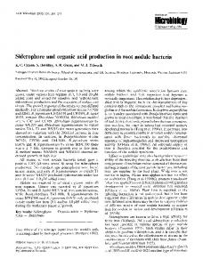
Although rhizoferrin (Fig. 1) represents a fairly simple molecule, it may have potential application in biotechnology due to its appreciable metal-binding properties and the ability to be easily degraded by various microorganisms. Rhizopus microsporus var. rhizopodiformis was grown on a glucose/asparagine/salts medium under iron-limiting conditions [40]. Since neither hydroxamate nor catecholate siderophores could be detected, the organic acid fraction was analyzed. The culture filtrate was first passed through an Amberlite, XAD-2 (ICN, Costa Mesa, CA, USA) column. Then the acidic components were purified by a two step ion exchange procedure with Dowex 50WX8 and Dowex 2X8 and subsequent gel permeation chromatography with Biogel P2 (BioRad, Richmond, CA, USA). Fractions positive on chrome azurole S plates were collected and lyophilized. Final purification was achieved on reversed phase HPLC with a gradient of water/acetonitrile yielding a pure compound. By this procedure, rhizoferrin could later also be obtained from strains of Mucor mucedo and Phycomyces nitens (Mucoraceae), Chaetostylum fresenii and Cokeromyces recurvatus (Thamnidiaceae), Cunninghamella elegans and Mycotypha africana (Choanephoraceae), Mortierella vinacea (Mortierellaceae) and Basidiobolus microsporus (Basidioboloceae, Entomophthorales) [33]. STRUCTURE OF RHIZOFERRIN Rhizoferrin consists of two molecules of citric acid linked to 1,4-diaminobutane through two amide bonds (Fig. 1). The hydrolytic fragments could be determined by derivatization and gas chromatography with mass spectrometric identification. The connectivity of the components was determined by nuclear magnetic resonance spectroscopy. Details of the structure elucidation are described in [12]. Although rhizoferrin appears constitutionally symmetric, it contains two chiral centers in the citric acid residues. The configuration of the quaternary carbon atoms could be determined by CD (circular dichroism) spectroscopy of aqueous solutions of the iron free ligand and comparison with reference compounds of known chirality [11]. It was found that rhizoferrin is naturally produced with R,R-configuration. The implications for biosynthesis will be discussed below. The iron complex of rhizoferrin showed a metal to ligand charge transfer band at 335 nm which was found for all members of the rhizoferrin family and is thus characteristic for purely carboxylate-type siderophores. The pH stability of the iron complex was estimated by pH-dependent UV spec-
0
_....~CH2
N
~.C /
H
/
CH 2
\
...... COO-
~
_CH2
/
CH2
"q
CH2
I"',.
H~C//
O
/ o.~ /
c
C ,,CH~ N...----C.H: __CH 2 N / ~ ~.."" H ~C'X~ "~'CH I I "~-' ' ~ /~o. o//C__CI.la
CH2
~
./eC 0 0
COOFig. 2. Structural formula of imido-rhizoferrin.
troscopy. The charge transfer band at 335 nm appeared at pH 4, reached maximum intensity at pH 5.5 and remained unchanged to a pH of 9. This agrees well with the spectrophotometric titrations of rhizoferrin and its iron complex, performed by Rochel [28]. The iron complex of rhizoferrin showed a positive Cotton effect at 340 nm. This is in good agreement with the positive absorption of ferric complexes of the ferrichromes which by comparison with known crystal structures have been assigned a A-configuration (the donor atoms are arranged in the shape of a left-handed propeller) around the iron center [37]. The CD spectra of rhizoferrin are thus indicative for a A-configuration around the metal center [11]. Acidic solutions of rhizoferrin gradually decompose with formation of two dehydration products, the structures of which have been determined [11]. The first dehydration product, named imidorhizoferrin, is formed by cyclization of one citryl residue to a succinimidyl derivative (Fig. 2). The chirality of the quaternary carbon atom is retained. The second dehydration product is cyclized on both citryl residues and was therefore named bisimido-rhizoferrin (Fig. 3). Both compounds can be easily detected by capillary electrophoresis in coated capillaries (Fig. 4). In citric acid buffer, 20 raM, pH 2.5, rhizoferrin has a slight net negative charge. This is in agreement with titration experiments of the ligand [28]. Referenced against the neutral marker mesityl oxide, rhizoferrin showed a shorter retention time. Imido-rhizoferrin has fewer negative charges, since it has lost one carboxy group by cyclization to the succinimidyl derivative. Its peak is found between rhizoferrin and the neutral marker. Bisimido-rhizoferrin has one less free carboxy group and is found between imido-rhizoferrin and the neutral marker. The relative amounts of the imido forms increase upon standing in citric acid buffer at pH 2.5. A similar behavior has been observed with all analogs of rhizoferrin described below. DIRECTED FERMENTATION OF RHIZOFERRIN ANALOGS In directed fermentation, building blocks or analogs of constituents of a targeted biomolecule are added to the growth
\
HO,,
/
COO-
CH2
C ,.o
-o
o
o II
/
C-..CH, c ..CH, \^/c.~./c~ / C ~ 1N / ~C"" . O" ~ )" --CH. CH, \ /XO. CH,__C%~
COO
COO-
o//C
CH=
OOC
Fig. 1. Structural formula of rhizoferrin.
Fig. 3. Structural formula of bisimido-rhizoferrin.
The carboxylate type siderophore rhizoferrin H Drechsel et al
@
I1
A
described below. Starting with a low rhizoferrin-concentration (about 50 mg L-'), the yield of rhizoferrin was increased to 800 mg L -1 in flask cultures. This was achieved by variation of the type of inoculum and the medium composition. The production of rhizoferrin and its derivatives, like other siderophores, occurred simultaneously with biomass formation in the trophophase. As expected, the biosynthesis of the rhizoferfins was strongly regulated by the iron content of the medium. The addition of 10/xM iron(III) decreased production to 1015%, although growth was not affected.
Diamine analogs of rhizoferrin
B
C
Fig. 4. Capillary electrophoretic separation of rhizoferrin (I), imidorhizoferrin (H) and bisimido-rhizoferrin (liD. (A) Separation of a purified rhizofen'in sample which still contains a small amount of imidorhizoferrin. (B) Separation of rhizoferrin after incubation in citric acid buffer (pH 2.5 at 25 ~ 1 day), resulting in an increase of imidorhizoferrin. ((2) Separation of a sample of imido-rhizoferrin after incubation for 1 (lay in citric acid buffer (pH 2.5, 25 ~ 1 day) showing residual amounts of rhizoferrin and bisimido-rhizoferrin. Fused silica capillary, 72 cm x 50/xm, coated with /x-Coat (Applied Biosystems, Weiterstadt, Germany), 15 kV, polarity negative at inlet side, buffer 20 mM citric acid, pH 2.5, detection by UV absorption at 200 nm.
medium of the producing strain. Provided the precursors are taken up into the cell and linked into the biosynthetic pathways, this will result in altered and often increased levels of production of the biomolecule. By this means, isotopically labelled precursors or constitutionally similar constituents can be incorporated into biomolecules and analogs of the naturally occurring biomolecule can be obtained by isolation of the fermentation products. In the case of rhizoferrin, two components can be modified. First, the chain length of the diamine can be varied and branched dJiamines and diamines with additional functional groups may be introduced. Second, citric acid may be replaced by similar molecules with fewer or different functional groups, affecting strongly the complexing properties of the resulting siderophore analogs. In both cases, a number of analogs of rhizoferrin have been obtained. The femmntative production of rhizoferrin was carried out with several producer strains (see above). From these, Cunninghamella elegans and Mucor rouxii were chosen for an optimization of the fermentation process. Finally Cunninghamella elegans was used for production of the analogs
Chain elongation as well as chain shortening was achieved by feeding appropriate precursors to the producing strain. Addtion of 1,5-diaminopentane (cadaverine) resulted in the production of an analog with a bridge of five methylene units, named homorhizoferrin (Fig. 5). 1,3-diaminopropane yielded an analog with three methylene units, named norrhizoferrin (Fig. 6). Product formation and yield of both analogs were approximately the same (see Table 1). The structures were confirmed by gas chromatographic detection of 1,5-diaminopentane and of 1,3-diaminopropane in the hydrolyzates of homorhizoferrin and norrhizoferrin, respectively. The calculated masses of the new compounds were confirmed by pneumatically assisted elecu'ospray mass spectrometry and by fast atom bombardment mass spectrometry. The proton and 13C NMR signals of both compounds have been completely assigned [9]. The iron-free ligands of homorhizoferrin and norrhizoferrin have been studied by circular dichroism spectroscopy. The chirality of the citric acid residues manifests itself in the overall chirality of the desferri ligands. At 205 nm, a negative Cotton effect demonstrates the chirality of the asymmetric centers of citric acid in both analogs to be the same as in rhizoferrin, earlier found to be R,R [11]. The CD spectra of the iron complexes at different pH showed a minimum at 310 nm and a maximum at 370 nm, indicating a A-configuration around the metal center. The shape of the CD-bands of ferri-homorhizoferrin was approximately the same as for ferri-rhizoferrin, whereas the Cotton effect of ferri-norrhizoferrrin showed only about half that intensity. This correlates with the observation of likewise reduced intensity of the charge transfer band of ferri-norrhizoferrin measured by UV/Vis spectroscopy. These findings indicate a strained geometry of the coordination sites around the iron center in the case of norrhizoferrin, whereas the complexation of homorhizoferrin is apparently not influenced by its elongated diamine bridge. This will be further evaluated by spectrophotometric titration of the ligands and calculation of the complex formation constants of the ferric complexes, as has been done in the case of rhizoferrin [28]. The variation in chain length has been continued by the substitution of the middle methylene group of eadaverine by an oxygen atom. Feeding of the precursor bis-(2-aminoethyl)ether to cultures of Cunninghamella elegans, resulted in the production of an analog named oxahomorhizoferrin (Fig. 7). Product formation was approximately one third of the diaminoalkanes described above. Yield of the new derivative showed a decrease at higher concentration of precursor, indi-
107
The carboxylate type sider0ph0re rhiz0ferrin H Drechselet al
108
OH