Original Article
The Effects of First Premolar Extractions on Third Molar Angulations Mustafa Yig˘it Saysela; Go¨kce Deniz Meralb; ˙Ilken Kocaderelic; Ferda Tas¸ard Abstract: The purpose of this study was to determine the relationship between the inclinations of second and third molars during a two- to 2.5-year period in patients treated orthodontically both with and without premolar extractions. Records of 37 first premolar extraction patients and 33 nonextraction patients were examined. The pretreatment and posttreatment panoramic radiographs were analyzed. The angles were measured between the long axis of the third molar and the occlusal plane and between the long axis of the third molar and the long axis of the second molar. Changes in third molar angulations from pretreatment to posttreatment for two groups were compared by Mann–Whitney U-test. Statistical analysis revealed that mandibular third molars showed an improvement in angulation relative to the occlusal plane in the first premolar extraction group. (Angle Orthod 2005;75:719–722.) Key Words: Premolar extraction; Nonextraction; Orthodontic treatment; Third molar angulation
INTRODUCTION
Similarly the lack of compensatory periosteal apposition at the posterior outline of the maxillary tuberosity could prevent eruption of the maxillary third molar.4 The eruption space for the mandibular third molars is also affected by the direction of tooth eruption during the functional phase of eruption. If the posterior teeth erupt more anteriorly, the retromolar space will increase.8,9 The impact of third molar eruption on mandibular incisor crowding has been the subject of many studies.1–6 Causes for third molar impaction and predictions of third molar eruption have also been studied extensively.12–16 Richardson17 investigated cephalometric methods for the prediction of third molar impaction, but the results of the study were inconclusive. The purpose of this study was to investigate the changes in maxillary and mandibular third molar angulation relative to the occlusal plane and changes relative to second molar long axis in a group treated with four first premolar extractions and to compare these changes with a nonextraction group.
The development of third molars and their influence on the dental arches has long been of concern to the dental profession.1 Mandibular third molar impaction is a major problem in modern dentistry.2 The developmental path of third molars in human beings is very irregular and the formation, calcification timing, and the position and course of eruption of these teeth show great variability. Frequently, third molars are impacted or congenitally missing.3 In modern populations, the impaction rate is higher for third molars than for any other tooth.4–7 One explanation could be that the retromolar space frequently is inadequate. If the remodeling resorption at the anterior aspect of the mandibular ramus is limited, the eruption of the mandibular third molars might be blocked.8–11 Associate Professor, Department of Oral Surgery, Faculty of Dentistry, Hacettepe University, Ankara, Turkey. b Research Assistant, Department of Oral Surgery, Faculty of Dentistry, Hacettepe University, Ankara, Turkey. c Professor and Head, Department of Orthodontics, Faculty of Dentistry, Hacettepe University, Ankara, Turkey. d Professor and Head, Department of Oral Surgery, Faculty of Dentistry, Hacettepe University, Ankara, Turkey. b Corresponding author: Go¨kce Deniz Meral, DDS, PhD, Turan Gunes Bulvarı, Korman Sitesi, 3, block no:23 C ¸ ankaya, Ankara 06550, Turkey (e-mail:
[email protected]). a
MATERIALS AND METHODS Pretreatment and posttreatment panoramic radiographs of 70 patients orthodontically treated at the Department of Orthodontics at Hacettepe University were selected. The inclusion criteria for selecting the patients were an Angle Class I skeletal and dental relationship, full eruption of second premolars and upper/lower bilateral unerupted third molars seen on panoramic radiograph. Thirty-three of the patients (12 boys; 21 girls) were treated without extractions
Accepted: July 2004. Submitted: May 2004. Q 2005 by The EH Angle Education and Research Foundation, Inc. 719
Angle Orthodontist, Vol 75, No 5, 2005
720
SAYSEL, MERAL, KOCADERELI, TAS¸AR
TABLE 1. Mean Deviations of Age and Orthodontic Treatment Periods for Extraction and Nonextraction Groups Mean age (y) 6 SD
n Extraction mandible Extraction maxilla Non–extraction mandible Non–extraction maxilla
37 33 32 30
13.17 13.03 12.02 12.04
6 6 6 6
Mean treatment time (y) 6 SD
1.50 1.77 1.62 1.58
2.50 2.37 2.13 1.95
6 6 6 6
1.06 1.10 1.00 0.73
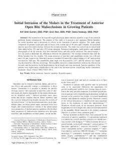
within one month before the start of orthodontic treatment. All the posttreatment panoramic radiographs were taken on the day the active orthodontic appliances were removed or within one week of debonding. All the radiographs were taken on the same panoramic unit (Planmeca-Proline 2002 CC, Helsinki, Finland). Radiographs were evaluated using a standardized technique of tracing the images of the molar teeth on matte acetate paper. The occlusal line was constructed through the cusp tips of the first molar and the second premolar. All second premolars were fully erupted at the beginning of the treatment period. The anterior angles formed by the long axis of the third molar and the occlusal plane plus the angle between the long axes of the second and third molar were measured (Figure 1). For each measurement, pretreatment values were subtracted from posttreatment values to obtain the change that occurred during treatment. The changes in third molar angulations relative to occlusal plane and relative to the second molar from pretreatment to posttreatment for each group were compared with Mann–Whitney U-test and Wilcoxon test (P , .05). FIGURE 1. (1) The anterior angle between the long axis of the maxillary third molar and occlusal plane. (2) The angle between maxillary second and third molar. (3) The angle between mandibular second and third molar. (4) The anterior angle between the long axis of the mandibular third molar and occlusal plane.
(nonextraction group) and 37 of the patients (13 boys; 24 girls) were treated with the extraction of mandibular and maxillary first premolars (extraction group). All the patients in the extraction and nonextraction groups were treated with fixed appliances using the edgewise technique by the same clinician (Dr Kocadereli). The second molars were not included in fixed appliances. The mean ages of the patients and the treatment time are shown in Table 1. All pretreatment panoramic radiographs were taken
RESULTS The method error was assessed by retracing 30 randomly selecting panoramic radiographs after 15 days. Method error coefficients for all measurements were calculated and were within acceptable limits (range 0.98–0.99).18 Mandibular third molars Table 2 shows the descriptive statistics for changes in mandibular third molar angulations related to the occlusal plane and relative to the second molar. There was a statistically significant difference between the extraction and nonextraction groups in the median third molar angulation relative to the occlusal plane (P
TABLE 2. Median Values of Changes in Mandibular Third Molar Angulation (P , .05*). The Values in Parentheses are Minimal and Maximal Ranges. ns: Not significant Pretreatment (Median Values) Mandibula
Extraction
Nonextraction
Posttreatment (Median Values)
P
Third molar–occlusal plane 130 (96–142) 126.5 (112–145) ns Second molar–third molar 28 (3–56) 30.5 (8–50) ns
Angle Orthodontist, Vol 75, No 5, 2005
Extraction
Nonextraction
P
125 (8–150) 23 (2–55)
130 (109–147) 28 (5–43)
ns ns
Difference (Median Values) Extraction Nonextraction 11 (1–134) 9 (1–36)
5.5 (1–21) 3.5 (1–37)
P * *
721
THIRD MOLAR ANGULATION
TABLE 3. Mean Values for the Changes in Angulations of Maxillar Third Molars (P , .05*). The Values in Parentheses are Minimal and Maximal Ranges. ns: not significant Pretreatment (Median Values) Maxilla Third molar–occlusal plane Second molar–third molar
Extraction
Nonextraction
P
68 (16–104) 17 (2–52)
70 (116–39) 18 (2–39)
ns ns
, .05). There also was a statistically significant difference between the extraction and nonextraction groups in the median angle formed by the long axis of third molar angulation relative to the second molar (P , .017; a Bonferroni correction was made in Mann–Whitney test). Maxillary third molars Table 3 lists the descriptive statistics for changes in the maxillary third molar angulations. Comparing the change in maxillary third molar angulations resulting from orthodontic treatment showed no significant difference between the extraction and nonextraction and control groups with Mann–Whitney U-test (P . .05). In the extraction group, at the end of the orthodontic treatment, the mandibular third molars showed more uprighting than did the maxillary third molars. DISCUSSION The mandibular third molar is by far the most frequently impacted tooth after the maxillary third molar.19 The prevalence of mandibular third molar impaction is variable in different populations, ranging from 9.5% to 39%.17 Skull materials indicate that third molar impaction was relatively infrequent in primitive populations.4,10,12,13 This has been attributed to mesial drift of the posterior teeth because of excessive interproximal attrition, thereby increasing the retromolar space. Similarly, third molar impaction is rarely observed after second molar extraction.14,15,20,21 Richardson and Richardson20 reported that normalsized lower third molars make adequate replacements for second molars in the majority of their cases, and lower third molars can upright and erupt for a wide variety of mesioangular positions by extraction of second molars. Orton-Gibbs et al22 showed that all 178 mandibular third molars were uprighted after the extraction of second molars. They also reported that more mandibular third molars than maxillary third molars erupting into an excellent position. Moffitt21 investigated the evaluation of the effect of extracting maxillary second molars on the eruption and function of third molars in 56 patients. Their study indicates that the maxillary third molars usually erupt into acceptable positions within the arch and with positive occlusal contacts.
Posttreatment (Median Values) Extraction
Nonextraction
74 (42–112) 65 (44–115)16 14 (1–45) (1–50)
Difference (Median Values)
P
Extraction Nonextraction
P
ns ns
15 (1–51) 8 (1–23)
ns ns
9.5 (1–25) 8.5 (1–30)
These studies suggest that mesial movement of the molars and a concomitant increase in eruption space are likely to reduce the frequency of third molar impaction. In our study, the changes in angulation of mandibular third molars in the extraction group were significantly more upright (P , .05). All the patients in the present study were dentally and skeletally Class I. Therefore, there was no need to protract mandibular molars or retract maxillary molars to obtain a Class I molar relationship. We can explain the uprighting of the mandibular third molars by the increase in the retromolar area by growth. Mesial movement of the molars during closure of the extraction site could have a larger effect on third molar impaction in the mandible than in the maxilla. The average age at third molar eruption ranges from 17 to 21 years, but the roots are not fully formed until 18 to 25 years of age. Accordingly, third molar impaction could have been diagnosed in studies examining subjects more than 20 years old.4 In our study, because all patients were less than 17 years old at the end of the treatment period, we could not determine the final clinical eruption or impaction of the third molars. We just determined the uprighting of third molars. The results of this study suggest that factors other than extractions could influence the inclination and subsequent eruption of third molars. This study did not reveal any basis to predict the eruption of third molars because third molar angulations improved whether or not teeth were extracted. Also, even with this improvement in angulations, third molars may still become impacted. If the patients had been Class II dentally, and mandibular molar protraction had been used to correct the molar relationship, an even more favorable change in mandibular third molar angulations may have occurred. Conversely, if maxillary molar distalization had been used to correct a molar relationship, an even more unfavorable change in maxillary third molar angulations may have occurred. It is possible that the type of mechanics used and anchorage considerations have more of an effect on third molar angulations than the actual extraction of first premolars. Although many impacted teeth may remain symptom free throughout life, they are a potential source of trouble and their early removal is generally recomAngle Orthodontist, Vol 75, No 5, 2005
722
SAYSEL, MERAL, KOCADERELI, TAS¸AR
mended. The orthodontist is constantly aware of the developing third molar and its possible effect on the dentition during and after orthodontic treatment. The effect of orthodontic treatment on developing third molars should also be considered and measures to relieve developing impactions included in the treatment plan. Begg23 claimed that there was insufficient forward movement of the dentition of modern man because of lack of attrition resulting in lack of space for the third molar. Faubion24 has shown that the prevalence of third molar impaction is reduced but not eliminated in patients treated by extraction of premolars. Richardson25 found that extraction of a molar almost eliminated the occurrence of third molar impaction. The initial angulations of the third molars may also influence their subsequent eruption. Richardson26 found that third molars with a small degree of angulation erupted earlier than those with steeper angulations. Richardson,26 Bjo¨rk et al,27 and Svendsen and Bjo¨rk,28 all stated that mandibular growth is a contributing factor in mandibular third molar eruption, but its exact role is uncertain. CONCLUSIONS This study’s data support the concept that orthodontic treatment involving premolar extractions improves mandibular third molar angulations. However, an improvement in angulation does not necessarily mean that third molars will erupt in good position. Third molar angulation and eruption can be influenced by factors other than first premolar extractions. Consequently, it may be prudent for orthodontists to advise patients that premolar extractions will not ensure that the third molars will erupt and have sufficient space to achieve good alignment. REFERENCES 1. Staggers JA, Germane N, Fortson WM. A comparison of the effects of first premolar extractions on third molar angulation. Angle Orthod. 1992;2:135–138. 2. Richardson ME. The etiology and prediction of mandibular third molar impaction. Angle Orthod. 1977;47:165–172. 3. Forsberg CM. Tooth size, spacing, and crowding in relation to eruption or impaction of third molars. Am J Orthod Dentofacial Orthop. 1988;94:57–62. 4. Kim TW, A˚rtun J, Behbehani F, Artese F. Prevalence of third molar impaction in orthodontic patients treated nonextraction and with extraction of 4 premolars. Am J Orthod Dentofacial Orthop. 2003;123:138–145. 5. Dachi SF, Howell FV. A survey of 3874 routine full-mouth radiographs II. A study of impacted teeth. Oral Surg Oral Med Oral Pathol. 1961;14:1165–1169.
Angle Orthodontist, Vol 75, No 5, 2005
6. Bishara SE, Andreasen G. Third molars: a review. Am J Orthod. 1983;83:131–137. 7. Grover PS, Lorton L. The incidence of unerupted permanent teeth and related clinical cases. Oral Surg Oral Med Oral Pathol. 1985;59:420–425. 8. Bjo¨rk A, Jensen E, Palling M. Mandibular growth and third molar impaction. Acta Odontol Scand. 1956;14:231–271. 9. Bjo¨rk A. Variations in the growth pattern of the human mandible: longitudinal radiographic study by the implant method. J Dent Res. 1963;42:400–411. 10. Alling CC, Alling RD. Indications for Management of Impacted Teeth. Impacted Teeth. Philadelphia, Pa: WB Saunders Co; 1993:46–49. 11. Silling G. Development and eruption of the mandibular third molar and its response to orthodontic therapy. Angle Orthod. 1973;43:271–278. 12. Begg PR. Stone Age man’s dentition. Am J Orthod. 1954; 40:298–312,373–383,517–531. 13. Murph TR. Reduction of the dental arch by approximal attrition. Br Dent J. 1964;116:483–488. 14. Cavanaugh JJ. Third molar changes following second molar extractions. Angle Orthod. 1985;55:70–76. 15. Gooris CGM, A˚rtun J, Joondeph DR. Eruption of mandibular third molar after second molar extractions: a radiographic study. Am J Orthod Dentofacial Orthop. 1990;98:161–167. 16. Dierkes DD. An investigation of the mandibular third molars in orthodontic cases. Angle Orthod. 1975;45:207–212. 17. Richardson M. Changes in lower third molar position in the young adult. Am J Orthod Dentofacial Orthop. 1992;102: 320–327. 18. Winner BJ. Statistical Principles in Experimental Design. 2nd ed. New York, NY: McGraw-Hill; 1971:79–84. 19. Hattab F, Rawashdeh M, Fahmi M. Impaction status of third molars in Jordanian students. Oral Surg Oral Med Oral Pathol. 1995;79:24–29. 20. Richardson ME, Richardson A. Lower third molar development subsequent to second molar extraction. Am J Orthod Dentofacial Orthop. 1993;104:566–574. 21. Moffitt AH. Eruption and function of maxillary third molars after extraction of second molars. Angle Orthod. 1998;68: 147–152. 22. Orton-Gibbs S, Crow V, Orton HS. Eruption of third permanent molars after the extraction of second permanent molars. Part 1: assessment of third molar position and size. Am J Orthod Dentofacial Orthop. 2001;119:226–238. 23. Begg PR. Begg Orthodontic Theory and Technique. 2nd ed. Philadelphia, Pa: WB Saunders Co; 1971:21–22. 24. Faubion BH. Effect of extraction of premolars on eruption of mandibular third molars. J Am Dent Assoc. 1968;76:316– 320. 25. Richardson ME. The early developmental position of the lower third molar relative to certain jaw dimension. Angle Orthod. 1970;40:226–230. 26. Richardson ME. Some aspects of lower third molar eruption. Angle Orthod. 1974;44:141–145. 27. Bjo¨rk A, Jensen E, Palling M. Mandibular growth and third molar impaction. Acta Odontol Scand. 1956;14:231–272. 28. Svendsen H, Bjo¨rk A. Third molar impaction: a consequence of late M3 mineralization and early physical maturity. Eur J Orthod. 1988;10:1–12.