tumor which were radiolabeled overnight with tri- ... identification in association with nuclei ofandrogen- sensitive cells. Surprisingly ... Address reprint requests to William C. Beckman, Jr., ...... Claflin AJ, Pollack A, Malinin T, Block NL, Irvin GL:.
American Journal of Pathology, Vol. 128, No. 3, September 1987
Copyright © American Association of Pathologists
The Epithelial Origin of a Stromal Cell Population in Adenocarcinoma of the Rat Prostate BECKMAN, Jr., PhD, L. CAMPS, Jr., MD, ROBERT M. WEISSMAN, MD, STEVEN L. KAUFMAN, MD, PhD, STEPHEN J. SANOFSKY, MD, ROBERT L. REDDICK, MD, and GENE P. SIEGAL, MD, PhD
From the Divisions of General and Urologic Surgery, Department of Surgery, Divisions of Oncologic and Surgical Pathology, Department ofPathology, and the Lineberger Cancer Research Center, School of Medicine, University of North Carolina at
Dunning R3327-H rat prostate adenocarcinoma cells, when grown in syngeneic (Copenhagen) rats or nude mice, produce tumors with prominent hypercellular stroma. The authors have previously demonstrated the of anomalous steroid-sensitive cells in both presence the epithelium and stromal compartments of this model system. In order to better understand the histoof these cells, the authors studied samples ofthe genesis tumor which were radiolabeled overnight with tritiated dihydrotestosterone (3H-DHT). Frozen sections of the tissues were thaw-mounted onto autoradioemulsion-coated slides to permit silver grain graphic identification in association with nuclei of androgensensitive cells. Surprisingly, numerous silver grains were found to be associated with nuclei of large cells within the stroma. Therefore, these cells were termed "epithelioid" pending confirmation of their origin. To further define these cells and their relationship to the surrounding matrix, autoradiograms have now been examined immunohistochemically with antibodies directed against the basement membrane glycoprotein, laminin, as well as antibodies specific for intermediate filaments. Following identification of cytoskeletal acinar basement membranes, epithelioid cells were identifiable both in the stroma and in the acinar epithe-
lial cell layer. Histochemical staining with acid phosa marker for prostatic epithelium, was perphatase, formed and shown to be present in acinar epithelial cells as well as in epithelioid cells. Additionally, fluorescence-activated cell sorting was employed to characterize the DNA content of cell types within the H tumor. Epithelioid cells were found to be in highest concentration in an aneuploid peak with a ploidy of immunoapproximately 6N. The autoradiographic, histochemical, cytometric, and ultramicroscopic studies suggest that 1) epithelioid cells are epithelial derived stromal cells; 2) these epithelioid cells arise by division of aneuploid neoplastic precursor pathologic cells of approximately 3N ploidy, which are found within the prostatic epithelium; and 3) the resulting 6N cells degrade the basement membrane locally, invade the stroma, and populate it. Here, they can be fibroblasts by their size, acid phosdistinguished from and activity, hormone receptor content. Thus, phatase the term "epithelioid" is inappropriate; and these cells should be regarded simply as large neoplastic epithelial (LNE) cells. The presence ofthis cell type suggests that this tumor subline represents a useful naturally occurring model for the study of the initial stages of neoplastic transformation. (Am J Pathol 1987, 128:555-565)
THE DUNNING R3327-H (H) rat prostatic adenocarcinoma is a neoplastic cell subline which originated from one of the original rats to which the R3327 tumor was passed.' The hormone sensitivity of this tumor was initially described by Voight and Dunning,2 who observed that the tumor grew more rapidly in intact males than in castrated males. The original tumor was a well-differentiated papillary adenocarcinoma that bore considerable histologic resemblance to the normal dorsal prostate.l These neoplasms occasionally lose their characteristic well-differentiated morphology and begin to proliferate rapidly. Thus, numerous sublines of this
Supported by NIH Grants R25 17973 and CA35915, ACS institutional Grant IN-15-Y, and UNC Biomedical Research Support Grant USPHS 5-S01-FR-05406. At the time this work was performed, G.P.S. was a Junior Faculty Clinical Fellow of the American Cancer Society (JFCF #739) and a UNC Junior Faculty Development awardee. R.L.R. was a Junior Faculty Clinical Fellow of the American Cancer Society (JFCF #732). Accepted for publication May 8, 1987. Dr. Weissman's present address is Department of Sur-
WILLIAM C.
JOSEPH
Chapel Hill, Chapel Hill, North Carolina
gery (Urology), Mason Clinic, Seattle, Washington. Address reprint requests to William C. Beckman, Jr., PhD, Department of Surgery, Clinical Sciences Bldg.
(229H), School of Medicine, University of North Carolina, Chapel Hill, NC 27514. 555
556
BECKMAN ET AL
tumor have arisen.3-7 Investigators at the Johns Hopkins University School of Medicine who extensively characterized the original R3327 tumor used the suffix "H" to identify it as a hormonally sensitive subline.6'8 Thus, studies describing the original R3327 adenocarcinoma may refer to it as R3327 or R3327-H.8'9 The present study uses the latter designation. The H tumor contains androgen9-'2 and estrogen receptors.9,10 Following orchiectomy'0 or estrogen priming,13 progestogen receptors can be demonstrated as well, suggesting the functionality of the estrogen receptor.13 Orchiectomy of male rat'4 or athymic mouse15 hosts results in a transient growth suppression lasting approximately 60 days, followed by a return to the former growth rate. The resulting tumor is insensitive to further alterations in circulating androgen concentration and is designated the HI
(hormone-insensitive) tumor.16-17 The stroma ofthis tumor model is characterized by enhanced cellularity, compared with the stroma of the rat dorsal prostate from which it was derived. ' We have previously shown that nuclei of a prominent subpopulation of these cells have a high concentration of receptors for dihydrotestosterone (DHT) and estradiol (E2).18 Since the histogenesis of these cells was uncertain, we suggested the name "epithelioid cells" to distinguish them from normal fibroblasts
which lack substantial hormone receptors.18 As we now show, these cells can be identified both within the boundary of the acinar basement membrane and in the stroma. Thus, the term "epithelioid" is no longer appropriate. This cell type represents a population of large neoplastic epithelial (LNE) cells. We have further clarified the localization of these large cells possessing enhanced capacity to bind androgens. Electron-microscopic and immunohistochemical studies have been performed to demonstrate the relationship between these cells and the extracellular matrix and have provided further evidence of their invasive capacity. Flow cytometric analysis was performed, and the uniqueness of the DNA ploidy exploited for separation and study of this population.
Materials and Methods Tumor Inoculation and Incubation Ten male nude (bg/bg) mice (NIH) were implanted subcutaneously under acepromazine/ketamine anesthesia with 3 cu mm pieces of Dunning R3327-H tumor in the inguinal region. After 6 months the tumors had grown to approximately 1 cu cm in volume. Mice were castrated 24 hours before surgical excision of the tumors to allow for clearance ofendog-
AJP ·
September 1987
enous serum androgens. Tissues were placed in 4 C RPMI 1640 culture medium containing 100 U/ml penicillin and 100 ug/ml streptomycin and minced with razor blades into small pieces 1-2 cu mm. Tissues were incubated overnight as described below in closed 50-ml tubes in a shaking water bath (37 C). Tissue viability was maintained in RPMI 1640 culture medium with antibiotics, 2 mM HEPES buffer, and 1 nM 3H-DHT (210 Ci/mM, New England Nuclear, Cambridge, Mass) to permit maximal hormone receptor binding.18 Competition studies were performed by the addition of 100 nM unlabeled DHT to the incubation medium. After incubation, the tissues were washed for 4 hours with hourly changes of 10 ml RPMI 1640 medium containing 3.5% (wt/vol) bovine serum albumin (Fraction V, Sigma Chemical Co., St.
Louis, Mo).19
Autoradiography After incubation, tissues were prepared for autoradiography by the method of Beckman et al.'8 Briefly, tissues were blotted, mounted on brass cryostat stubs, frozen in isopentane at -70 C, and stored in liquid nitrogen until processing for autoradiography. Sections (4,u) were cut and thaw-mounted onto emulsion-coated slides (Kodak NTB-3). Slides were developed weekly and examined for the presence of cells with nuclear androgen receptor complexes, as evidenced by a concentration of silver grains over cell nuclei. When sufficient silver grains had accumulated over nuclei to permit identification of steroid-sensitive ells, the remaining slides in the box were removed in a darkroom, and then developed in Kodak D-19 developer (45 seconds at 15 C) followed by Kodak Fixer (5 minutes at 15 C).20 Immunohistochemistry Autoradiograms which were prepared for combined autoradiography and immunohistochemistry were immunostained following the method of Keefer et al.2' Prior to photographic development, slides were placed in a 4% paraformaldehyde solution (4 C, pH 7.0) for 60 seconds. Subsequent to photographic development, slides were washed for 5 minutes in running tap water and then placed in 0.05 M Tris buffer (pH 7.6) for 5 minutes. Slides were treated for an additional 5 minutes in 1% normal sheep serum (NSS) to block nonspecific binding sites prior to application of the antibody bridge. A double peroxidase-antiperoxidase method22 was used for immunohistochemical demonstration of la-
Vol. 128
*
No. 3
minin and keratin proteins. Rabbit anti-laminin antiserum (Bethesda Research Laboratories, Bethesda, Md) was used at a concentration of 1:100 in Tris buffer supplemented with 1% NSS. Rabbit anti-keratin antiserum (Lipshaw, Detroit, Mich) was used undiluted as supplied. This latter polyclonal antibody recognizes a large family of both high and intermediate weight keratins. Both antisera were applied overnight at room temperature (RT). Each set of slides immunostained included one control slide for which normal rabbit serum was substituted for the primary antiserum to control for nonspecific deposition of reaction product. Intracellular vimentin filaments were demonstrated immunohistochemically with a monoclonal antibody kit (Hybritech, Inc., San Diego, Calif) following the published protocol. Acid Phosphatase Cytochemistry Cells containing acid phosphatase enzyme were demonstrated utilizing Barka's modification of Burstone's acid phosphatase method as outlined by Brinn and Pickett.23 This procedure yields a red reaction product wherever the acid phosphatase enzyme is located.
Fluorescence-Activated Cell Sorting H tumors were dissociated enzymatically in a solution of 0.25% collagenase with constant stirring for 60 minutes at 37 C. Free cells in the supernatant were removed every 10 minutes and diluted in 3.5% BSA to stop the enzymatic reaction. Fresh enzyme solution was added to replace the volume of supernatant removed. Viable tumor cells were maintained in the same culture medium as described above for organ culture of solid tumor pieces. Hoechst dye #33342 was added to achieve a final concentration in the media of 1 ,uM. This dye concentration was 20% of that recommended by Shapiro.24 Preliminary studies indicated that the low concentration of Hoechst dye in the solution broadened the peaks of the histograms (C V = 7.5% versus 4% for 5 ,uM Hoechst), but superior cell viability was obtained over the long period of time (3 hours) required for the sort. Cells were incubated for 45 minutes at 37 C before sorting. Flow cytometry was performed using a Coulter Epics V system equipped with an argon laser and the MDADS (Coulter Electronics, Hialeah, Fla) data acquisition system. Fluorescence of Hoechst dye bound to DNA was activated by a 363-nm argon laser excitation beam at 20 mW. Emission was recorded through a 418-nm short-pass interference filter. For-
EPITHELIAL ORIGIN OF STROMAL CELLS
557
ward angle light scatter and DNA content parameters were used for sorting cells. The three peaks of fluorescence identified by DNA fluorescence intensity were set at Channels 52, 109, and 166. These peaks were sorted into two populations. Cells with a mean channel position of 52 were sorted together by collecting all cells falling between Channels 30 and 71 into a single collection vial. Cells in the two peaks between Channels 71 and 180 were collected together. Subsequently, the distinct peaks of cells with mean values at Channels 109 and 166 were separated by a second sorting pass. Cells in each of the three populations resulting from this two-step sorting were stained with the Giemsa stain and examined microscopically.
Transmission Electron Microscopy H tumor pieces 1-2 mm in diameter were fixed in a mixture of 0.1 M phosphate buffer, 2% paraformaldehyde, and 1.25% glutaraldehyde with 0.01% picric acid (Ito-Karnovsky fixative).25 After overnight fixation, tissues were dehydrated through a graded ethanol series and embedded in Epon resin. Thin (900 nm) sections were cut and mounted on 100mesh copper grids for ultrastructural study utilizing a Zeiss 109 electron microscope.
Morphometric Analysis Measurement of nuclear areas was performed on the Zeiss Videoplan Image Analysis System with digitizing tablet. Photomicrographs (1000X) were placed on the tablet, and a stylus was used to circumscribe the nuclear boundaries of epithelial and LNE cells (recognized by the large numbers of silver grains present over the nucleus). Silver grains were recorded by the point counting function. This made it possible to record both the number ofsilver grains within each nucleus and the nuclear area within which they were contained. The standard measurement programs which are contained within the Zeiss Videoplan statistical program package were used.
Results Histologic comparison oftissues from the H tumor (Figure 1) and the normal rat dorsal prostate (Figure 2) revealed both to be characterized by variable
numbers of glandular acini consisting ofcytologically bland epithelial cells and myoepithelial-like basal cells. The stroma from the normal prostate was loose with a supporting fibrovascular network and infiltrating macrophage population. The H tumor acinar epithelium consists of one to two cell layers. The adlu-
558
BECKMAN ET AL
AJP · September 1987
..
.. li:·
:/.!=', ·i.i ***y
.'
, '.-
,,
*
2
t
.iiiift.
i.
*..rn .* 3l *
^, : .
·t'
*
*
-'..,' r--313
*.
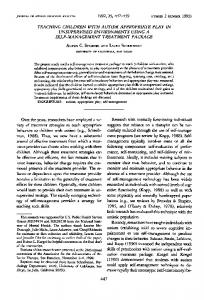
- tne uunning n/R37-n tumor ollowing incubation witn raaloactve oanyorotestosterone. iiver grains iniacate presence OT Frgure 1-Hstologic section (4i) Ot larger quantities of radioactive steroid in nuclei of "epithelioid" cells. (Methyl green-pyronin [MGP] stain, X750) Figure 2-Histologic section (4p ) of normal rat dorsal prostate.stained histochemically for acid phosphatase (dark areas at apical portion of epithelial cells). Few cells are present in the stroma, which is largely free of acid phosphatase reaction product with the exception of lysosomal activity in occasional macrophages. (X750) Figure 3-A well-developed
'S
basement membrane surrounds the basal cell layer of acini. A light immunohistochemical reaction reveals the presence of laminin present within the epithelial basement membrane which surrounds the acinus. Intraacinar "epithelioid" cells (arrows) were identified by the presence of increased numbers of nuclear-associated silver grains. Extraacinar cells as well as large intraacinar cells contain increased numbers of nuclear silver grains, resulting from receptor bound 3H-DHT. (MGP, X 350) Figure 4-A large cell at the margin of the acinus appears to be exiting from the basement membrane, which exists as a filmy "halo" around the cell (/arge arrow). Other cells with increased numbers of nuclear silver grains appear in the stroma. (MGP, X350)
Vol. 128
·
EPITHELIAL ORIGIN OF STROMAL CELLS
No. 3
layer is composed of small epithelial cells, which, by morphometric analysis, have a mean nuclear area of 36.8 ± 4.9 sq p and smaller basal cells having a mean nuclear area of 17 ± 4.3 sq p (Table 1) which usually form a continuous layer surrounding the acinus. Additionally, the stromal compartment of the H tumor contained many cells with a large nuclear area (93.0 ± 4.3 sq ,; Figure 1) not found in the normal prostate stroma. These cells were identifiable by large numbers ofsilver grains concentrated within minal
the boundaries of the nuclear membrane. Localization ofsilver grains in cells of H tumors grown in nude mice and incubated in vitro with 3H-DHT indicated the presence of androgen associated with nuclei of large cells in the stroma which we have previously termed "epithelioid" cells.'8 Autoradiograms ofthe H tumor were evaluated to determine the numbers of silver grains present over nuclei of basal, epithelial and "epithelioid" cell types. Although "epithelioid" cell nuclei were roughly three times as large as epithelial cell nuclei, their mean silver grain content was greater than ten times that of the epithelial cells (Table 1). Thus, it is unlikely that the increased DHT-associated radioactivity can be explained by the difference in nuclear area. Further examination of tumor tissue sections which had been immunohistochemically stained with antibodies to laminin revealed similar large cells Table 1
Mean Cell Sizes and Silver Grain Content of Different Cell
Types Within the H Tumor NUCLEAR AREA ± S.E.M. )
SILVER GRAINS / NUCLEUS ( MEAN + S.E.M.
(MEAN
0 +1 o
+1
-25
-20
(n
z
E1
s#
,
. st
i I
9 A
,
Is
,
*. ;.
a I
4,-,
8
.1
4.0 4 5-Electron of normal acinus H of the tumor. Cells appear relatively innocuous. A large "light cell" is present in the center of the field, which is Figure micrograph poorly attached to the adjacent epithelial cells. "Light cells" are believed to be precursors of epithelioid cells. (X1500) Figure 6-Higher magnification of the in cell" 5. Desmosomes are absent and a fluid filled area exists in the vicinity of thecell. (X3400) "light Figure Figure 7-Normal basal cells have well-formed basal laminae and normal ultrastructure. (X7000) Figure 8-"Light cells" which have reached the basal lamina reveal increased distortion of the ultrastructure, including vacuolization within the RER and distortion of the nuclear membrane. Continuities frequently exist between the RER and the nucleolemma (X3400) S,
I..
.
Vol. 128
o
EPITHELIAL ORIGIN OF STROMAL CELLS
No. 3
561
S.:A-
:
.
i "
--
fe^
..
~~
~
., ,
6
I..
~
~
~
.1
Figure 9-Light cells which migrate into the stroma from the acinar epithelium degrade basement membrane and stroma in their path (large arrow) leaving behind collagen fragments, reduplicated basal lamina, and cytoplasmic debris. The prominent, irregularly shaped nucleus is a key morphologic feature of this cell type. Continuity of the epithelial basal lamina with this cell is still evident in the region of the small arrow, enlarged in the inset. (X2100; inset, X3400)
tate cells (Figure 2) and in the H tumor (Figure 10). Within the H tumor, the amount of acid phosphatase
reaction product produced in large "epithelioid" cells was so great that, for illustrative purposes, the reaction had to be stopped prior to the appearance of product in the acinar epithelial cells (Figure 10). Long-term incubations revealed product in acinar epithelial cells as well. A polyclonal antibody to keratin stained periacinar cells intensely (Figure 11). Longer incubation times revealed the presence of weak keratin immunoreactivity in acinar epithelial cells and epithelioid cells as well (not illustrated). A monoclonal antibody to vimentin filaments revealed intense immunoreactivity in occasional "epithelioid" cells and lower levels of immunoreactivity in fibroblasts and other stromal "epithelioid" cells (Figure 12). The acinar epithelium did not stain with vimentin. Fluorescence activated cell sorting following enzymatic digestion separated the H tumor into three populations of cells on the basis of DNA content (Fig-
ure 13). Microscopic examination of cells within each of these peaks following separation revealed that the first peak consisted primarily of fibroblasts and normal diploid epithelial cells. Cells in the second peak consisted of a mixture of dividing cells and other cells which did not appear to be undergoing mitosis. Occasional cells in this peak were characterized by larger nuclei, which appeared to be morphologically irregular. Cells in the third population consisted mainly of very large cells with highly enlarged nuclei, although some smaller cells (