Microbiology (2007), 153, 1499–1507
DOI 10.1099/mic.0.2006/004838-0
The Escherichia coli yhjA gene, encoding a predicted cytochrome c peroxidase, is regulated by FNR and OxyR Jonathan D. Partridge, Robert K. Poole and Jeffrey Green Correspondence Jeffrey Green
[email protected]
Received 1 December 2006 Revised
19 January 2007
Accepted 22 January 2007
Department of Molecular Biology and Biotechnology, The University of Sheffield, Sheffield S10 2TN, UK
The Escherichia coli FNR protein is an oxygen-responsive global transcription factor, and OxyR is a key regulator of the peroxide stress response. Here both FNR and OxyR are shown to regulate expression of the E. coli yhjA gene. The yhjA gene encodes a predicted cytochrome c peroxidase, a bacterial haem-containing protein involved in the peroxide stress response through its ability to convert hydrogen peroxide to water. It is shown that the yhjA gene of E. coli possesses a class II FNR site and an OxyR site upstream of the yhjA transcript start. Expression of yhjA was found to be dependent on this unusual combination of FNR and OxyR under conditions of oxygen starvation. Phenotypic analysis of the yhjA mutant revealed increased sensitivity to exogenous hydrogen peroxide and organic peroxides during growth under anaerobic conditions, consistent with the observed regulation and predicted function of the yhjA gene product.
INTRODUCTION The Escherichia coli FNR protein is an oxygen-responsive global transcription factor (Constantinidou et al., 2006; Covert et al., 2004; Gonzalez et al., 2003; Kang et al., 2005; Partridge et al., 2006; Salmon et al., 2003). Under anaerobic conditions FNR contains a [4Fe–4S] cluster that promotes dimerization of the protein and enhances site-specific DNA binding (Lazazzera et al., 1996). In the presence of oxygen, the iron–sulphur clusters disassemble, leading to the formation of FNR monomers and subsequent inhibition of DNA binding, thereby switching off FNR-activated genes (Crack et al., 2004, 2006; Kiley & Beinert, 2003; Lazazzera et al., 1996; Sutton et al., 2004a, b). Upon binding of the active protein to DNA, FNR activates transcription by recruiting RNA polymerase, or can alternatively repress transcription by inhibiting the formation of productive promoter–RNA polymerase interactions (reviewed by Browning et al., 2002). Transcription activation is facilitated by the formation of specific protein–protein contacts between the activating regions (AR1, AR2 and AR3) of FNR and regions of RNA polymerase (Bell & Busby, 1994; Blake et al., 2002; Lamberg et al., 2002). The DNA sequence recognized by FNR consists of an inverted repeat with a consensus TTGATnnnnATCAA (Eiglmeier et al., 1989). There are two basic types of FNR-dependent promoter. Class I promoters possess FNR sites located close to 261.5, 271.5, 282.5 or 292.5 (Wing et al., 1995) that permit the formation of only one protein–protein contact, between Abbreviations: CCP, cytochrome c peroxidase; RACE, random amplification of cDNA ends.
2006/004838 G 2007 SGM
Printed in Great Britain
AR1 of FNR and the C-terminal domain of the RNA polymerase a-subunit. However, the more common architecture is that of the class II promoters, which have an FNR site centred at, or close to, 241.5 relative to the transcript start (Browning et al., 2002; Guest et al., 1996; Wing et al., 2000). This arrangement allows the formation of multiple protein–protein contacts between RNA polymerase and all three activating regions of FNR (Bell & Busby 1994; Blake et al., 2002; Li et al., 1998; Williams et al., 1997). The FNR protein primarily coordinates gene regulation in remodelling central metabolism in response to changes in oxygen availability in many bacteria (Guest et al., 1996; Korner et al., 2003), but it also exerts effects over other systems, and new members of the FNR regulon are still being uncovered (Covert et al., 2004; Kang et al., 2005). For example, FNR has been implicated in the peroxide stress response as an anaerobic activator of cytochrome c peroxidase (CCP) in a number of bacteria including Neisseria gonorrhoeae, Paracoccus denitrificans and Pseudomonas stutzeri (Turner et al., 2003; van Spanning et al., 1997; Vollack et al., 1999). Furthermore, the FNR homologue, Anr, activates CCP expression in Pseudomonas aeruginosa (Zimmermann et al., 1991). The primary role of CCP is thought to be in peroxide stress resistance, because of its ability to catalyse the conversion of hydrogen peroxide to water (Minard & McAlister-Henn, 2001; Seib et al., 2004, reviewed by Atack & Kelly, 2006). However, because typical bacterial CCP proteins are periplasmic dihaem-containing proteins that use a monohaem cytochrome c as an electron donor they could 1499
J. D. Partridge, R. K. Poole and J. Green
potentially allow the use of hydrogen peroxide as a terminal electron acceptor. Here, a search for new members of the E. coli FNR regulon identified the yhjA gene as a likely candidate. The expression of this gene, which is predicted to encode a CCP, is shown to be driven from an FNR-dependent class II promoter that is also dependent on OxyR. The phenotype of the yhjA mutant indicates a role for YhjA in the peroxide stress response of E. coli.
METHODS Bacterial strains and growth conditions. All the bacterial strains and plasmids used in this work are listed in Table 1. For b-galactosi-
dase assays, L broth (Sambrook & Russell, 2001) containing appropriate antibiotics was inoculated (1 : 50) from overnight cultures. Cultures were incubated under aerobic (250 ml conical flasks containing 10 ml medium shaken at 250 r.p.m.), micro-aerobic (250 ml conical flasks containing 150 ml medium shaken at 100 r.p.m.), or anaerobic (bottles filled to the neck, without shaking) conditions at 37 uC until an OD600 of 0.4–0.6 was reached, at which point b-galactosidase activities were measured (Miller, 1972). Oxygen transfer rates were measured as described by Wainwright et al. (2005). Nucleic acid methods. DNA was isolated and manipulated by
conventional methods (Sambrook & Russell, 2001). Promoter regions of interest were amplified from the chromosome of MC1000 by PCR using the following oligonucleotides engineered to contain EcoRI/BamHI restriction sites: yjhB, JDP1 and JDP2; yfgF, JDP3 and 4; yhjA, JDP5 and 6; yecI, FyecI and RyecI; smf, Fsmf and Rsmf (Table 1). The sequence of each fragment was verified by DNA sequencing after ligation into EcoRI/BamHI-digested pRS415 (Simons et al., 1987). Selected constructs were then transferred to lRZ5 (Simons et al., 1987) and the resulting (promoter–lacZ) fusions were introduced in single copy into the l attachment site of isogenic E. coli strains. Lysogeny status was verified using the method of Powell et al. (1994). Further transfer of the promoter fusions into different genetic backgrounds was achieved using bacteriophage P1vir-mediated transduction (Sambrook & Russell, 2001). For 59 random amplification of cDNA ends (RACE)-PCR transcript mapping, RNA was isolated from anaerobic cultures of E. coli MC1000 using Qiagen RNeasy mini kits according to the manufacturer’s instructions. The yhjA transcript start was identified using 2 mg E. coli RNA per RACE reaction according to the manufacturer’s instructions (Roche) with oligonucleotides JDP7 and JDP8 (Table 1). Site-directed mutagenesis of the consensus FNR site (TTGAT-N4ATCAA to ATCAT-N4-ATGAT) and putative OxyR-binding site (ATAGgcacaggCTATcttattgATAGtTtatAttcAT to TATCgcacaggGATActtattgTATCtAtatTttcTA) was achieved using PCR and appropriate synthetic oligonucleotides, with all mutations confirmed by DNA sequencing. Construction of mutants. The yhjA gene of W3110 was disrupted
by linear transformation based on the method of Yu et al. (2000). Oligonucleotides containing 39-end sequences complementary to the first or last 20 bp of the chloramphenicol resistance cassette of plasmid pACYC184 (Martinez et al., 1988) and 59-end sequences flanking yhjA were constructed. Linear DNA carrying the resistance cassette and flanking regions was generated by PCR. E. coli strain W3110 containing the plasmid pTP223 (TetR) (Poteete & Fenton, 1984), which carries the l red recombinase genes under the control of an IPTG-inducible promoter, were grown overnight at 37 uC and diluted (1 : 100) in L broth containing tetracycline (25 mg ml21) and 1500
IPTG (2 mM) and grown to an OD600 of ~0.3. Electrocompetent cells were prepared and transformed with approximately 5 mg of PCR product then recovered in 1 ml L broth for 1 h before plating on selective medium (CmR). The resulting colonies were immediately cured of pTP223 and mutations were screened by PCR and DNA sequencing. Further transfer of the mutations into clean genetic backgrounds was achieved using bacteriophage P1virmediated transduction (Sambrook & Russell, 2001). The oxyR-deficient mutant for use in promoter assays was generated through P1vir-mediated transduction (Sambrook & Russell, 2001) of the mutation from GSO47 (Zheng et al., 1999) to MC1000, to generate JRG5393. Overexpression
and
purification
of
OxyR
and
FNR*.
Overexpression of OxyR from MV247 was as described by Haagmans & van der Woude (2000) with modifications (Correnti et al., 2002). Solubilization was aided by the addition of 0.1 % Tween 20 to the bacterial pellet and incubation at room temperature for 30 min prior to sonication. Protein was purified from the cell-free extract using a HiTrap heparin column (Amersham) as described previously (Storz et al., 1990) with modifications according to Kullik et al. (1995). The final preparations contained ~60 % OxyR as judged by Coomassie-stained SDS-polyacrylamide gels. The FNR protein FNR-D154A (designated FNR*) retains DNAbinding activity under aerobic conditions (Ziegelhoffer & Kiley, 1995) and was isolated as described by Meng et al. (1998). Gel retardation assays. Radiolabelled promoter fragments (~50 ng) were incubated with 6 mM FNR* and/or 0.06 mM OxyR in binding buffer (Correnti et al., 2002) for 30 min before separating the protein–DNA complexes from DNA on 6 % TBE-buffered polyacrylamide gels. After electrophoresis the gels were transferred to filter paper (3MM, Whatman) and dried for autoradiography. Disc diffusion assays. Soft-top agar (3 ml, 0.65 % agar) was mixed with an aliquot of a culture (200 ml, OD600 ~0.3) of the
strain under investigation grown under anaerobic conditions. The mix was poured evenly onto L agar plates and, once set, a sterile antibiotic filter disc (6 mm) was placed in the centre of the plate. The indicated reagents were added to the centre of the disc and the plates were incubated under anaerobic conditions at 37 uC for 16 h before measuring the zone of growth inhibition around the disc. Killing curve studies. Bacterial cultures were grown to OD600
~0.3 under anaerobic conditions and then challenged with a stress reagent. Aliquots were removed at t=0, t=20, t=40 and t=60 min and serially diluted in L broth before being plated onto L agar plates incubated under aerobic conditions. The numbers of viable bacteria were calculated from the numbers of c.f.u. after 16 h growth at 30 uC under aerobic conditions.
RESULTS Identification of the yhjA gene as a member of the FNR regulon The E. coli FNR protein recognizes DNA sequences related to the FNR consensus (TTGATnnnnATCAA) (Eiglmeier et al., 1989). The key features of this inverted repeat are the A–T base pairs at positions +7 and 27 (underlined), which allow discrimination between FNR sites and sites recognized by related proteins, and the G–C base pairs at +5 and 25 (bold), which form a core interaction with the DNA recognition helices of this family of transcription factors Microbiology 153
The regulation of Escherichia coli yhjA
Table 1. Bacterial strains, plasmids and oligonucleotides Relevant genotype or properties E. coli strains GS047 JRG1728 JRG3462 JRG5066 JRG5067 JRG5354 JRG5393 JRG5609 JRG5641 JRG5694 MC1000 MV247 W3110 Plasmids pACYC184 pGS1633 pGS1634 pGS1740 pGS1740a pGS1828 pGS1829 pGS2018
pMV107 pRS415 pTP223 Oligonucleotides JDP1 JDP2 JDP3 JDP4 JDP5 JDP6 JDP7 JDP8 FyecI RyecI Fsmf Rsmf
Source or reference
MC4100 oxyR-Kan MC1000 D(tyrR-fnr-rac-trg)17 zdd-230-Tn9 CAG627 pGS771 MC1000/lRZ5(PyhjA–lacZ) from pGS1634 JRG1728/lRZ5(PyhjA–lacZ) from pGS1634 W3110 DyhjA MC1000 oxyR-Kan JRG5393/lRZ5(PyhjA–lacZ) from pGS1634 MC1000/lRZ5(PyhjA0–lacZ) from pGS2018 MC1000/lRZ5(PyhjA9–lacZ) from pGS1829 D(lacIPOZYA)X74 galU galK D(araABC-leu) XL-1 Blue pMV107 F2, l2
Zheng et al. (1999) Lab collection Meng et al. (1998) This work This work
Source of cat cassette for linear transformation PyfgF–lacZ fusion in pRS415 (192 bp promoter fragment, 2108 to +84 relative to the transcript start), ApR PyhjA–lacZ fusion in pRS415 (187 bp promoter fragment, 2127 to +60 relative to the transcript start), ApR PyecI–lacZ fusion in pRS415 (399 bp promoter fragment), ApR Psmf–lacZ fusion in pRS415 (383 bp promoter fragment), ApR PyjhB–lacZ fusion in pRS415 (450 bp promoter fragment), ApR PyhjA–lacZ fusion in pRS415 with the FNR-binding site altered from TTGAT-N4-ATCAA to ATCAT-N4-ATGAT, ApR PyhjA : : lacZ fusion in pRS415 with the OxyR-binding site altered from ATAGgcacaggCTATcttattgATAGtTtatAttcAT to TATCgcacaggGATActtattgTATCtAtatTttcTA, ApR pZE24 with a 929 bp fragment encoding OxyR lacYZA-based promoter-fusion vector, ApR lam, bet and exo genes of phage l, under the control of the lac promoter, TetR
Martinez et al. (1988) This work
This work This work This work This work Lab collection Haagmans & van der Woude (2000) Lab collection
TTTTGAATCAACTGTAGGTGGGTATC TTTTGGATCCGTGTTATTCTAATATAGCCC TTTTGAATTCTGTAATAATGATGAATTAACATG TTTTGGATCCGCAGAATCCCCCACATCC TTTGAATTCCCGCAATCATGATATGCC TTTGGATCCCGGTGGTGAGTTATTGTG CGTAATCCATCAACTGTTTCG GCCTAAAACCTTATTATTTTCAC TTTTGAATCTTAATTATCACTGGGCCCATTAATTAACAG TTTTGGATCCAAGCATTCCAGCGGATCCCATACCTAATAT TTTTGAATCTCTAAAAGCTCTGAATTCARTTAACACTAGC TTTTGGATCCTTTCTGTATCGAGGATCCTTATCTCCCTGC
(Schultz et al., 1991; Spiro et al., 1990). The E. coli K-12 genome sequence was searched for the presence of consensus FNR sites (TTGATnnnnATCAA) using the search pattern tool in the Colibri database (http:// genolist.Pasteur.fr/Colibri). This analysis yielded a total of 21 sites. Eliminating those located within coding regions, and those associated with prophages, left 12 consensus FNR sites, which were all centred within 345 bp of a start codon. http://mic.sgmjournals.org
This work This This This This
work work work work
This work
Haagmans & van der Woude (2000) Simons et al. (1987) Poteete & Fenton (1984)
This This This This This This This This This This This This
work work work work work work work work work work work work
Of the 12 transcriptional units associated with consensus FNR sites, seven (cydA, ndh, narK, narG, nirB, ydhY and yfiD) are known to be FNR-regulated (Green et al., 1998; Guest et al., 1996; Kang et al., 2005). The regulation of the remaining five genes (yecI, yfgF, yhjA, yjhB and smf) had not been investigated. To test whether the FNR sites upstream of yecI, yjhB, yfgF, yhjA and smf contributed to regulation of these genes, the corresponding promoter regions (e.g. 1501
J. D. Partridge, R. K. Poole and J. Green
Table 2. Effects of oxygen availability and FNR on the regulation of yecI, yfgF, yjhB, yhjA and smf Cultures of MC1000 (lac) and JRG1728 (lac fnr) transformed with plasmids pGS1828 (PyjhB–lacZ), pGS1633 (PyfgF–lacZ), pGS1634 (PyhjA–lacZ), pGS1740 (PyecI–lacZ), pGS1740a (Psmf–lacZ), were grown under aerobic and anaerobic conditions at 37 uC in L broth to OD600 0.4–0.6. Promoter activities were determined by measuring b-galactosidase activities. Values are means±SD from three independent cultures assayed in triplicate. Promoter
Aerobic MC1000 JRG1728 (fnr)
PyecI PyfgF PyjhB PyhjA Psmf
3375±215 4350±430 2950±73 99±5 280±18
3094±245 3527±300 2805±146 75±4 250±27
Anaerobic MC1000
JRG1728 (fnr)
2295±105 56831±1220 2963±194 733±14 180±9
2025±115 4360±252 2885±186 63±5 175±18
fusions. Promoter activity assays in MC1000 (lac) revealed a ~32-fold induction under anaerobic compared to aerobic conditions (Table 3). This anaerobic activation was lowered to only ~3-fold in the fnr mutant JRG1728 (fnr lac) (Table 3). Therefore, evidence for direct regulation by FNR was sought using gel retardation assays. To simplify the experiments an FNR protein (FNR-D154A, referred to as FNR*) that retains some DNA-binding activity in the presence of air was used (Ziegelhoffer & Kiley, 1995). The assays showed that FNR* was able to bind at the native yhjA promoter region, but not at the same promoter fragment with mutations in the predicted FNR site (Fig. 1, lanes 2 and 3). Furthermore, mutation of the FNR site essentially abolished FNR-mediated regulation (Table 3). To investigate the architecture of the yhjA promoter, 59 RACE-PCR was used to identify the transcript start. This placed the centre of the FNR site at 241.5 (Fig. 2). Thus, it was concluded that database searching had identified the yhjA gene as a member of the FNR regulon with an FNRdependent class II promoter.
PyecI–lacZ) were fused to lacZ in the reporter plasmid pRS415 and transferred into MC1000 (lac) and JRG1728 (fnr lac). Expression of yecI, yjhB and smf was not affected by the presence or absence of FNR under anaerobic conditions (Table 2), although expression of both yecI and smf was apparently lower in the absence of oxygen. However, expression of yfgF, and yhjA was enhanced under anaerobic conditions in an FNR-dependent manner (Table 2). Here the results of experiments to further characterize the regulation and function of yhjA are presented. Expression of yhjA is driven from a class II FNR-dependent promoter Preliminary in vivo transcriptional analysis using a plasmidbased yhjA–lacZ fusion showed FNR-dependent regulation of yhjA. To further characterize the mechanism of regulation, the activity of the yhjA promoter region fused to lacZ was investigated using single-copy chromosomal
Activation of yhjA is co-dependent on the OxyR system Further inspection of the yhjA promoter region identified a potential binding site for the oxidized (disulphide) form of the transcriptional regulator OxyR that matched the consensus (ATAGnTnnnAnCTATnnnnnnnATAGnTnnnAnCTAT) in 16 (shown in bold) of 20 bases (Toledano et al., 1994). The 39 end of the potential OxyR site is located only 8 bp upstream of the 59 end of the FNR site (Fig. 2). The OxyR protein acts as a transcriptional regulator initiating, directly or indirectly, the synthesis of more than 40 gene products to counter the effects of peroxide stress, although transcription of some of these genes is also altered during exposure to heat shock or redox-cycling agents (Greenberg & Demple, 1989; Storz & Zheng, 2000; Walkup & Kogoma, 1989; Zheng et al., 2001).
Table 3. Regulation of yhjA Isogenic E. coli MC1000 derivatives harbouring chromosomal promoter–lacZ fusions (either yhjA–lacZ, or yhjA–lacZ with a mutated FNR site, from pGS1829, or yhjA–lacZ with a mutated OxyR site, from pGS2018) were grown under aerobic, micro-aerobic and anaerobic conditions at 37 uC in L broth to OD600 0.4–0.6. Promoter activities were determined by measuring b-galactosidase activities. Values are means±SD from three independent cultures assayed in triplicate. ND, not done. b-Galactosidase activity (Miller units)
Strain
MC1000 (yhjA–lacZ) JRG1728 fnr (yhjA–lacZ) JRG5393 oxyR (yhjA–lacZ) MC1000 (yhjA–lacZ FNR site inactivated) MC1000 (yhjA–lacZ OxyR site inactivated)
1502
Aerobic
Micro-aerobic
Anaerobic
0.8±0.2 0.7±0.1 2.0±0.2 0.8±0.1 2.1±0.2
13.3±3.0 3.5±0.2 5.9±0.7
31.0±2 2.0±0.1 4.0±0.3 1.1±0.1 5.6±0.2
ND ND
Microbiology 153
The regulation of Escherichia coli yhjA
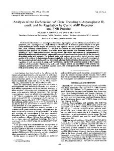
To investigate whether OxyR contributes to the regulation of yhjA, the PyhjA–lacZ fusion was transferred to an oxyR mutant. The anaerobic induction of yhjA expression observed previously was significantly lowered (Table 3). Furthermore, mutation of the OxyR site essentially abolished OxyR-mediated regulation (Table 3). These data suggest that OxyR is required for expression of yhjA. Therefore, evidence for direct regulation of yhjA expression was sought using gel retardation assays. These showed that, like FNR, OxyR was able to bind at the unaltered yhjA promoter region, but not at the same promoter fragment carrying mutations in the predicted OxyR site (Fig. 1, lanes 4 and 5). Gel retardation assays in which the yhjA promoter was incubated with increasing amounts of OxyR revealed an apparent Kd of ~10 nM for OxyR binding (not shown). Moreover, in gel retardation assays in which both FNR* and OxyR were present, the migration of the labelled yhjA promoter was slower than in the presence of either FNR* or OxyR alone, suggesting that both proteins occupy the yhjA promoter simultaneously (Fig. 1, lane 6). Thus, the regulation of yhjA is interesting because transcription appears to be dependent on two transcription factors that respond to seemingly incompatible signals, i.e. lack of oxygen and peroxide stress. Nevertheless, during micro-aerobic growth oxidative stress might be encountered
origin yhjA_FNR/OxyR yhjA_OxyR yhjA_FNR
yhjA
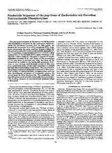
whilst some FNR activity is retained. However, yhjA expression was only partially activated under micro-aerobic (150 ml culture volumes shaken at 100 r.p.m., equivalent to an oxygen transfer rate of 12 mmol min21 l21) compared to anaerobic conditions (Table 3), indicating that these particular micro-aerobic conditions are not optimal for yhjA expression. A yhjA mutant exhibits increased sensitivity to exogenous peroxides The gene encoding the predicted CCP of E. coli, yhjA, was expected to contribute to the peroxide stress response. Therefore the sensitivity of a yhjA mutant to hydrogen peroxide and organic peroxides during anaerobic growth was tested. Initial disc diffusion assays indicated that the mutant was more sensitive than the parent to hydrogen peroxide, t-butyl hydroperoxide and cumene hydroperoxide (not shown). This enhanced sensitivity was confirmed in killing assays using cultures grown under anaerobic conditions to OD600 ~0.3 before exposure to the peroxide stress reagents for a total of 20, 40 and 60 min (Fig. 3). Complementation of the yhjA peroxide-sensitive phenotype by supplying a wild-type copy of yhjA in trans was not possible, because all attempts to create plasmids that expressed the YhjA protein in an active form were unsuccessful (not shown). Therefore, to ensure that the observed phenotype was caused by disruption of yhjA and not by point mutations located elsewhere in the genome, the lesion was transferred by P1 transduction to a clean genetic background, and the observations reported above were confirmed with the new mutant, although this approach does not exclude the possibility of polar effects on neighbouring genes. Exposure of the yhjA mutant to nitrosative stress reagents (sodium nitroprusside, S-nitrosoglutathione, 3-[2-hydroxy1-(1-methylethyl)-2-nitrosohydrazino]-1-propanamine, 3-[2-hydroxy-1-methyl-2-nitrosohydrazino]-N-methyl1-propanamine) and the antimicrobial compound sodium hypochlorite in both disc diffusion and killing curve assays
1 A
2 3 4 5 6 A d hjA d yhjA e e t t yhj y a a ctiv ctiv ina ina e e t t i i s Rs yR FN Ox
yhj
Fig. 1. Interaction between FNR* and OxyR at the yhjA promoter. Radiolabelled unaltered yhjA promoter (lanes 1, 2, 4 and 6) and corresponding promoter fragments with mutations in the predicted FNR-binding site (lane 3) or the predicted OxyRbinding site (lane 5) were incubated with FNR* (6 mM), OxyR (60 nM), or FNR* and OxyR for 30 min at 25 6C before separation of protein–DNA complexes by electrophoresis. Lane 1, no protein; lanes 2 and 3, FNR*; lanes 4 and 5, OxyR; lane 6, FNR* and OxyR. The positions of free DNA (yhjA), yhjA–protein complexes and the origin of migration (origin) are indicated. http://mic.sgmjournals.org
Fig. 2. Nucleotide sequence of the yhjA promoter region. The transcript start (arrow), putative ”10 box (italicized), FNR site (boxed), and OxyR site (underlined) are indicated. Matches to the FNR and OxyR consensus sequences are highlighted in bold. 1503
J. D. Partridge, R. K. Poole and J. Green
100 10 1
Survival (%)
0.1 100
(a)
10 1 0.1 100
(b)
10 1 0.1
(c) 20 40 Time (min)
60
Fig. 3. Sensitivity of the yhjA mutant to peroxide stress in liquid culture. Cultures of E. coli W3110 (#) and the isogenic yhjA mutant ($) were grown to OD600 ~0.3 before being treated with the following reagents: (a) hydrogen peroxide, 2 mM; (b) t-butyl hydroperoxide, 0.4 mM; (c) cumene hydroperoxide, 0.2 mM. At t=0, 20, 40 and 60 min the number of c.f.u. in the treated cultures was determined. The data presented are the means of triplicate assays from three independent cultures. Error bars (most within the sizes of the symbols) show the standard deviations.
indicated no increased sensitivity compared to the parent strain, suggesting that yhjA has a specific role in the peroxide stress response (data not shown).
DISCUSSION The work presented here indicates that the yhjA gene of E. coli, which is predicted to encode a cytochrome c peroxidase (CCP), plays a role in the peroxide stress response under anaerobic conditions. Inspection of the E. coli genome sequence indicates that the yhjA gene is a single transcriptional unit. The YhjA protein (465 amino acids) is 46 % identical (61 % similar over 291 amino acids) to the CCP from Pseudomonas aeruginosa (323 amino acids) (Samyn et al., 1995). However, the YhjA protein is somewhat atypical. Generally bacterial CCP proteins possess two haem cofactors, the first (high-potential) site accepts electrons from the donating cytochrome c before transferring them to the second (low-potential) site, often referred to as the peroxidatic site, where hydrogen peroxide is reduced to 1504
water (reviewed by Atack & Kelly, 2006). Most physiological and biochemical studies of bacterial CCP proteins have been of these typical dihaem proteins, including those of Methylococcus capsulatus (Zahn et al., 1997), Neisseria gonorrhoeae (Turner et al., 2003), Nitrosomonas europaea (Arciero & Hooper, 1994), Paracoccus denitrificans (Gilmour et al., 1993), Paracoccus pantotrophus (Goodhew et al., 1990), Pseudomonas aeruginosa (Ellfolk & Soininen, 1970; Fu¨lo¨p et al., 2001), Pseudomonas nautica (Alves et al., 1999) and Rhodobacter capsulatus (Hanlon et al., 1992). However, the YhjA amino acid sequence reveals the presence of a third haem-binding motif. The additional haembinding site is located in an N-terminal extension of ~80 amino acid residues, which is absent from the dihaem CCP proteins. The exact role of this extended domain and the third haem is unclear, because the trihaem CCP proteins are poorly characterized. However, it has been suggested that the trihaem CCP proteins may have evolved by fusion of a monohaem cytochrome c gene to a dihaem ccp gene, consequently bypassing the need for a separate donor system (Atack & Kelly, 2006). Another distinction between CCP proteins is their cellular location, being either soluble periplasmic proteins or anchored to the membrane as lipoproteins. Cellular location is controlled by the presence of N-terminal amino acid sequences that are recognized by the Sec secretion pathway (Braun & Wu, 1993) and either type I or type II signal peptidases, the former generating a soluble periplasmic protein, whilst the latter leads to anchoring as a lipoprotein. Thus, the CCP of N. gonorrhoeae possesses a signal peptidase II recognition sequence and is a lipoprotein (Turner et al., 2003). Similarly, YhjA also possesses a potential target for signal peptidase II cleavage (VAIC), suggesting it is likely to be a lipoprotein, perhaps accounting for the lack of success in attempts to create expression plasmids that yielded functional protein. The peroxide-sensitive phenotype of the yhjA mutant reported here is consistent with the assignment of YhjA as a CCP. Whilst bacterial CCP proteins are known to detoxify hydrogen peroxide, there is evidence that they also work with organic peroxides. For example, a Bacteroides fragilis ccp mutant exhibits enhanced sensitivity to organic peroxides (t-butyl hydroperoxide and cumene hydroperoxide) compared with hydrogen peroxide (Herren et al., 2003). Likewise, the E. coli yhjA mutant was sensitive to organic peroxides and hydrogen peroxide, and the differences in sensitivity to these compounds might reflect the relative contribution of YhjA to their detoxification. Thus, the bioinformatic and phenotypic analyses are consistent with the assignment of the E. coli YhjA protein as a trihaem CCP. The regulation of yhjA expression is interesting, because transcription is dependent on two transcription factors, FNR and OxyR, that respond to two seemingly incompatible signals, i.e. oxygen starvation and peroxide stress. It is often under conditions of high aeration that bacteria are exposed to hydrogen peroxide (Seaver & Imlay, 2004), leading to Microbiology 153
The regulation of Escherichia coli yhjA
activation of OxyR. However, these same conditions inactivate FNR (Becker et al., 1996). It is possible that OxyR might be activated by nitrosative stress, as suggested by Kim et al. (2002), but analysis of the yhjA mutant phenotype did not support this idea. This raises the possibility that both oxidized and reduced OxyR could be functional at the yhjA promoter, because both forms of OxyR are able to bind at some promoters. Oxidized OxyR recognizes ATAGnT elements in four adjacent major grooves on one face of the DNA helix (Toledano et al., 1994). In contrast, reduced OxyR contacts ATAGnT elements in two pairs of major grooves separated by one turn of the DNA helix (Toledano et al., 1994). The DNA sequence of the oxyRS promoter region is such that both these patterns of binding are possible, and thus OxyR regulates its own expression, when reduced and oxidized, simply by repositioning itself on the DNA. At OxyRactivated promoters, such as ahpC, dps, gorA and katG, the ATAGnT elements are arranged such that only oxidized OxyR binds with significant affinity (Toledano et al., 1994). The yhjA OxyR site resembles those of OxyR-activated promoters, lacking an additional ATAGnT element. Moreover, the fourth OxyR contact point in yhjA does not obviously resemble both ATAGnT and AnCTAT elements, which is necessary to allow redox-state-responsive repositioning of OxyR at oxyRS (Toledano et al., 1994). Furthermore, because the yhjA OxyR and FNR sites are so close, any additional downstream OxyR contacts will overlap the FNR site and are likely to prevent FNR binding. Thus, the architecture of the yhjA promoter, the phenotype of the yhjA mutant and the predicted function of the YhjA protein suggest that transcription from the yhjA promoter is likely to be restricted to specific environmental conditions in which the bacteria are exposed to peroxides, but oxygen is limited such that some FNR activity is retained. The presence of a consensus FNR site in the yhjA promoter is consistent with this idea, because a consensus site is likely to be occupied in preference to other sites when the amount of active FNR is limited. The ability of FNR, located in a typical class II position, to only weakly activate expression of yhjA in the absence of OxyR suggests that the role of OxyR might be to counteract the effects of an as-yet-unidentified regulator that inhibits the action of FNR. The extensive mutagenesis of the promoter to inactivate the OxyR site is likely to have disrupted other regulatory elements in this region (294 to 257), but this was not sufficient to enhance FNR-mediated yhjA expression (Table 3), and suggests that any such regulatory element is located upstream (2127 to 294) of the OxyR site.
binding to the katG promoter, transcript profiling suggests that FNR activates katG expression (Constantinidou et al., 2006). If the katG FNR site(s) are functional, then although the katG promoter architecture is different to that of yhjA, where the FNR and OxyR sites are very close together, the seemingly incompatible combination of FNR and OxyR might be used to control expression of at least two genes associated with the peroxide stress response in E. coli.
The regulatory combination of OxyR and FNR might not be restricted to the yhjA promoter. The katG gene encodes hydroperoxidase I, which acts as a dual-function catalase and peroxidase with a proven role in the peroxide stress response. Expression of katG is activated by OxyR binding at a site centred at 252 relative to the transcript start. In addition, there are two credible FNR sites located at 296.5 and 2112.5, and although there is no direct evidence of FNR
Transcription activation by FNR: evidence for a functional Activating Region 2. J Bacteriol 184, 5855–5861.
http://mic.sgmjournals.org
In conclusion, it has been shown that the YhjA protein is likely to be a CCP that contributes to the survival of E. coli upon exposure to peroxide stress, and that expression of the yhjA gene is driven from an FNR-dependent class II promoter that also requires OxyR for activity. This unusual use of FNR and OxyR to activate expression suggests that yhjA plays a specific role in countering oxidative stress in E. coli, possibly in particular micro-aerobic environments. Further extensive in vitro analysis is required to determine the exact mechanism by which two regulators that respond to such disparate signals combine to activate yhjA expression.
ACKNOWLEDGEMENTS The authors would like to thank: R. E. Roberts for technical assistance; G. Storz (NIH, Bethesda, MD, USA), S. C. Andrews (University of Reading, UK) and M. van der Woude (Immunology and Infection Unit, University of York, UK) for bacterial strains and phage lysates; and the Biotechnology and Biological Sciences Research Council (UK) for supporting this work.
REFERENCES Alves, T., Besson, S., Duarte, L. C., Pettigrew, G. W., Girio, F. M. F., Devreese, B., Vandenberghe, I., Van Beeumen, J., Fauque, G. & Moura, I. (1999). A cytochrome c peroxidase from Pseudomonas
nautica 617 active at high ionic strength: expression, purification and characterization. Biochim Biophys Acta 1434, 248–259. Arciero, D. & Hooper, A. (1994). A di-heme cytochrome c peroxidase
from Nitrosomonas europaea catalytically active in both the oxidized and half-reduced states. J Biol Chem 269, 11878–11886. Atack, J. M. & Kelly, D. J. (2006). Structure, mechanism and
physiological roles of bacterial cytochrome c peroxidases. Adv Microb Physiol 52, 73–106. Becker, S., Holighaus, G., Gabrielczyk, T. & Unden, G. (1996). O2 as
the regulatory signal for FNR-dependent gene expression in Escherichia coli. J Bacteriol 178, 4515–4521. Bell, A. & Busby, S. J. W. (1994). Location and orientation of an
activating region in the Escherichia coli transcription factor FNR. Mol Microbiol 11, 383–390. Blake, T., Barnard, A., Busby, S. J. W. & Green, J. (2002).
Braun, V. & Wu, H. C. (1993). Lipoproteins: structure, function, biosynthesis and model for protein export. In New Comprehensive Biochemistry, vol. 27, Bacterial Cell Wall, pp. 319–341. Edited by J.-M. Ghuysen & R. Hachenbecks. Amsterdam: Elsevier. Browning, D. F., Lee, D., Green, J. & Busby, S. J. W. (2002). Secrets
of bacterial transcription taught by the Escherichia coli FNR protein. In Signals, Switches, Regulons and Cascades. pp. 127–142. Edited by 1505
J. D. Partridge, R. K. Poole and J. Green D. Hodgson & C. Thomas. Cambridge, UK: Cambridge University Press.
coli K-12 regulates a large number of genes of unknown function. J Bacteriol 187, 1135–1160.
Constantinidou, C., Hobman, J. L., Griffiths, L., Patel, M. D., Penn, C. W., Cole, J. A. & Overton, T. W. (2006). A reassessment of the FNR
Kiley, P. J. & Beinert, H. (2003). The role of Fe-S proteins in sensing
regulon and transcriptomic analysis of the effects of nitrate, nitrite, NarXL, and NarQP as Escherichia coli K-12 adapts from aerobic to anaerobic growth. J Biol Chem 281, 4802–4815.
Kim, S. O., Merchant, K., Nudelman, R., Bayer, W. F., Keng, T., DeAngelo, J., Hausladen, A. & Stamler, J. S. (2002). OxyR: a
Correnti, J., Munster, V., Chan, T. & van der Woude, M. (2002).
Korner, H., Sofia, H. J. & Zumft, W. G. (2003). Phylogeny of the
Dam-dependent phase variation of Ag43 in Escherichia coli is altered in a seqA mutant. Mol Microbiol 44, 521–532. Covert, M. W., Knight, E. M., Reed, J. L., Herrgard, M. J. & Palsson, B. O. (2004). Integrating high-throughput and computational data
elucidates bacterial networks. Nature 429, 92–96. Crack, J., Green, J. & Thomson, A. J. (2004). Mechanism of oxygen
sensing by the bacterial transcription factor fumarate-nitrate reduction (FNR). J Biol Chem 279, 9278–9286. Crack, J., Green, J., Le Brun, N. E. & Thomson, A. J. (2006).
Detection of sulphide release from the oxygen-sensing [4Fe-4S] cluster of FNR. J Biol Chem 281, 18909–18913.
and regulation in bacteria. Curr Opin Microbiol 6, 181–185.
molecular code for redox-mediated signalling. Cell 109, 383–396. bacterial superfamily of Crp-Fnr transcription regulators: exploiting the metabolic spectrum by controlling alternative gene programs. FEMS Microbiol Rev 27, 559–592. Kullik, I., Toledano, M. B., Tartaglia, L. A. & Storz, G. (1995).
Mutational analysis of the redox-sensitive transcriptional regulator OxyR: regions important for oxidation and transcriptional activation. J Bacteriol 177, 1275–1284. Lamberg, K. E., Luther, C., Weber, K. D. & Kiley, P. J. (2002).
Characterization of activating Region 3 from Escherichia coli FNR. J Mol Biol 315, 275–283.
Eiglmeier, K., Honore, N., Iuchi, S., Lin, E. C. C. & Cole, S. T. (1989).
Lazazzera, B. A., Beinert, H., Khoroshilova, N., Kennedy, M. C. & Kiley, P. J. (1996). DNA binding and dimerization of the Fe-S
Molecular genetic analysis of FNR-dependent promoters. Mol Microbiol 3, 869–878.
containing FNR protein from Escherichia coli are regulated by oxygen. J Biol Chem 271, 2762–2768.
Ellfolk, N. & Soininen, R. (1970). Pseudomonas cytochrome c peroxidase. I. Purification procedure. Acta Chem Scand 24, 2126–2136.
Li, B., Wing, H., Lee, D., Wu, H. & Busby, S. (1998). Transcription
Fu¨lo¨p, V., Watmough, J. & Ferguson, S. J. (2001). Structure and
enzymology of two bacterial diheme enzymes: cytochrome cd1 nitrite reductase and cytochrome c peroxidase. Adv Inorg Chem 51, 163–204.
activation by Escherichia coli FNR protein: similarities to and differences from the CRP paradigm. Nucleic Acids Res 26, 2075–2081. Martinez, E., Bartolome, B. & Delacruz, F. (1988). pACYC184-
Gilmour, R., Goodhew, C. F., Pettigrew, G. W., Prazeres, S., Moura, I. & Moura, J. J. (1993). Spectroscopic characterization of cytochrome c
derived cloning vectors containing the multiple cloning site and lacZ alpha reporter gene of pUC8/9 and pUC18/19 plasmids. Gene 15, 159–162.
peroxidase from Paracoccus denitrificans. Biochem J 294, 745–752.
Meng, W., Green, J. & Guest, J. R. (1998). FNR-dependent repression
Gonzalez, R., Tao, H., Purvis, J. E., York, S. W., Shanmugam, K. T. & Ingram, L. O. (2003). Gene array-based identification of changes that
contribute to ethanol tolerance in ethanologenic Escherichia coli: comparison of KO11 (parent) to LYO1 (resistant mutant). Biotechnol Prog 19, 612–623. Goodhew, C. F., Wilson, I. B., Hunter, D. J. & Pettigrew, G. W. (1990). The cellular location and specificity of bacterial cytochrome c
peroxidases. Biochem J 271, 707–712.
of ndh gene expression requires two upstream FNR-binding sites. Microbiology 143, 1521–1532. Miller, J. H. (1972). Experiments in Molecular Genetics. Cold Spring
Harbor, NY: Cold Spring Harbor Laboratory. Minard, K. I. & McAlister-Henn, L. (2001). Antioxidant function of
cytosolic sources of NADPH in yeast. Free Radic Biol Med 31, 832–843. Partridge, J. D., Scott, C., Tang, Y., Poole, R. K. & Green, J. (2006).
Green, J., Baldwin, M. & Richardson, J. (1998). Downregulation of
Escherichia coli transcriptome dynamics during the transition from anaerobic to aerobic conditions. J Biol Chem 281, 27806–27815.
Escherichia coli yfiD expression by FNR occupying a site at 293.5 involves the AR1-containing face of FNR. Mol Microbiol 29, 1113–1123.
Poteete, A. R. & Fenton, A. C. (1984). l red-dependent growth and recombination of phage P22. Virology 134, 161–167.
Greenberg, J. T. & Demple, B. (1989). A global response induced in
Powell, B. S., Court, D. L., Nakamura, Y., Rivas, M. P. & Turnbough, C. L., Jr (1994). Rapid confirmation of single-copy lambda prophage
Escherichia coli by redox-cycling agents overlaps with that induced by peroxide stress. J Bacteriol 171, 3933–3939. Guest, J. R., Green, J., Irvine, A. S. & Spiro, S. (1996). The FNR
integration by PCR. Nucleic Acids Res 22, 5765–5766. Salmon, K., Hung, S. P., Mekjian, K., Baldi, P., Hatfield, G. W. & Gunsalus, R. P. (2003). Global gene expression profiling in
modulon and FNR-regulated gene expression. In Regulation and Gene Expression in Escherichia coli, pp. 317–342. Edited by E. C. C. Lin & A. S. Lynch. Austin, TX: R. G. Landes.
Escherichia coli K12: the effects of oxygen availability and FNR. J Biol Chem 278, 29837–29855.
Haagmans, W. & van der Woude, M. (2000). Phase variation of Ag43
Sambrook, J. W. & Russell, D. W. (2001). Molecular Cloning: a
in Escherichia coli: Dam-dependent methylation abrogates OxyR binding and OxyR-mediated repression of transcription. Mol Microbiol 35, 877–887.
Laboratory Manual, 3rd edn. Cold Spring Harbor, NY: Cold Spring Harbor Laboratory.
Hanlon, S. P., Holt, R. A. & McEwan, A. G. (1992). The 44 kDa c-type
Samyn, B., Van Craenenbroeck, K., De Smet, L., Vandenberghe, I., Pettigrew, G. & Van Beeumen, J. (1995). A reinvestigation of the
cytochrome induced in Rhodobacter capsulatus during growth with dimethylsulphoxide as an electron acceptor is cytochrome c peroxidase. FEMS Microbiol Lett 97, 283–288.
covalent structure of Pseudomonas aeruginosa cytochrome c peroxidase. FEBS Lett 377, 145–149.
Herren, C. D., Rocha, E. R. & Smith, C. J. (2003). Genetic analysis of
of a CAP–DNA complex: the DNA is bent by 90u. Science 253, 1001–1007.
an important oxidative stress locus in the anaerobe Bacteroides fragilis. Gene 316, 167–175. Kang, Y., Weber, K. D., Qiu, Y., Kiley, P. J. & Blattner, F. R. (2005).
Genome-wide expression analysis indicates that FNR of Escherichia 1506
Schultz, S. C., Shields, G. C. & Steitz, T. A. (1991). Crystal-structure
Seaver, L. C. & Imlay, J. A. (2004). Are respiratory enzymes the
primary sources of intracellular hydrogen peroxide? J Biol Chem 279, 48742–48750. Microbiology 153
The regulation of Escherichia coli yhjA
Seib, K. L., Tseng, H. J., McEwan, A. G., Apicella, M. A. & Jennings, M. P. (2004). Defenses against oxidative stress in Neisseria
Wainwright, L. M., Elvers, K. T., Park, S. F. & Poole, R. K. (2005). A
gonorrhoeae and Neisseria meningitidis: distinctive systems for different lifestyles. J Infect Dis 190, 136–147.
truncated haemoglobin implicated in oxygen metabolism by the microaerophilic food-borne pathogen Campylobacter jejuni. Microbiology 151, 4079–4091.
Simons, R. W., Houman, F. & Kleckner, N. (1987). Improved single
Walkup, L. K. B. & Kogoma, T. (1989). Escherichia coli proteins
and multicopy lac-based cloning vectors for protein and operon fusions. Gene 53, 85–96.
inducible by oxidative stress mediated by the superoxide radical. J Bacteriol 171, 1476–1484.
Spiro, S., Gaston, K. L., Bell, A. I., Roberts, R. E., Busby, S. J. & Guest, J. R. (1990). Interconversion of the DNA binding specificities
Williams, S. M., Savery, N. J., Busby, S. J. W. & Wing, H. J. (1997).
of two related transcription regulators, CRP and FNR. Mol Microbiol 4, 1831–1838. Storz, G. & Zheng, M. (2000). Oxidative stress. In Bacterial Stress
Responses, pp. 47–59. Edited by G. Storz & R. Hengge-Aronis. Washington, DC: American Society for Microbiology. Storz, G., Tartaglia, L. A. & Ames, B. N. (1990). Transcriptional
regulator of oxidative stress-inducible genes: direct activation by oxidation. Science 248, 189–194. Sutton, V. R., Mettert, E. L., Beinert, H. & Kiley, P. J. (2004a). Kinetic
analysis of the oxidative conversion of the [4Fe–4S]2+ cluster of FNR to a [2Fe–2S]2+ cluster. J Bacteriol 186, 8018–8025.
Sutton, V. R., Stubna, A., Patschkowski, T., Mu˝nck, E., Beinert, H. & Kiley, P. J. (2004b). Superoxide destroys the [2Fe–2S]2+ cluster of
Transcription activation at Class I FNR-dependent promoters: identification of the activating surface of FNR and the corresponding contact site in the C-terminal domain of the RNA polymerase a subunit. Nucleic Acids Res 25, 1112–1118. Wing, H. J., Williams, S. M. & Busby, S. J. W. (1995). Spacing
requirements for transcription activation by Escherichia coli FNR protein. J Bacteriol 177, 6704–6710. Wing, H. J., Green, J., Guest, J. R. & Busby, S. J. W. (2000). Role of
activating region 1 of Escherichia coli FNR protein in transcription activation at class II promoters. J Biol Chem 275, 29061–29065. Yu, D., Ellis, H. M., Lee, E., Jenkins, N. A., Copeland, N. G. & Court, D. L. (2000). An efficient recombination system for chromosome
engineering in Escherichia coli. Proc Natl Acad Sci U S A 97, 5978–5983.
FNR from Escherichia coli. Biochemistry 43, 791–798.
Zahn, J. A., Arciero, D. M., Hooper, A. B., Coats, J. R. & DiSpirito, A. A. (1997). Cytochrome c peroxidase from Methylococcus capsulatus
Toledano, M. B., Kullik, I., Trinh, F., Baird, P. T., Schneider, D. T. & Storz, G. (1994). Redox-dependent shift of OxyR–DNA contacts
Zheng, M., Doan, B., Schneider, T. D. & Storz, G. (1999). OxyR and
along an extended DNA-binding site: a mechanism for differential promoter selection. Cell 78, 897–909.
Bath. Arch Microbiol 168, 362–372. SoxRS regulation of fur. J Bacteriol 181, 4639–4643.
Turner, S., Reid, E., Smith, H. & Cole, J. (2003). A novel cytochrome
Zheng, M., Wang, X., Templeton, L. J., Smulski, D. R., LaRossa, R. A. & Storz, G. (2001). DNA microarray-mediated transcriptional
c peroxidase from Neisseria gonorrhoeae: a lipoprotein from a Gramnegative bacterium. Biochem J 373, 865–873.
profiling of the Escherichia coli response to hydrogen peroxide. J Bacteriol 183, 4562–4570.
van Spanning, R. J., De Boer, A. P., Reijnders, W. N., Westerhoff, H. V., Stouthamer, A. H. & Van Der Oost, J. (1997). FnrP and NNR of
Ziegelhoffer, E. C. & Kiley, P. J. (1995). In vitro analysis of a
Paracoccus denitrificans are both members of the FNR family of transcriptional activators but have distinct roles in respiratory adaptation in response to oxygen limitation. Mol Microbiol 23, 893–907. Vollack, K.-U., Hartig, E., Korner, H. & Zumft, W. G. (1999). Multiple
transcription factors of the FNR family in denitrifying Pseudomonas stutzeri: characterization of four fnr-like genes, regulatory responses and cognate metabolic processes. Mol Microbiol 31, 1681–1694.
http://mic.sgmjournals.org
constitutively active mutant form of the Escherichia coli global transcription factor FNR. J Mol Biol 245, 351–361. Zimmermann, A., Reimmann, C., Galimand, M. & Haas, D. (1991).
Anaerobic growth and cyanide synthesis of Pseudomonas aeruginosa depend on anr, a regulatory gene homologous with FNR of Escherichia coli. Mol Microbiol 5, 1483–1490. Edited by: S. J. W. Busby
1507