The Journal of Immunology
The Fas/Fas Ligand Pathway Is Important for Optimal Tumor Regression in a Mouse Model of CTL Adoptive Immunotherapy of Experimental CMS4 Lung Metastases Sheila A. Caldwell, Mary H. Ryan, Elwood McDuffie, and Scott I. Abrams1 The mechanisms of CTL-mediated tumor regression in vivo remain to be fully understood. If CTL do mediate tumor regression in vivo by direct cytotoxicity, this may occur via two major effector mechanisms involving the secretion of perforin/granzymes and/or engagement of Fas by Fas ligand (FasL) expressed by the activated CTL. Although the perforin pathway has been considered the dominant player, it is unclear whether Fas-mediated cytotoxicity is additionally required for optimal tumor rejection. Previously, we produced H-2Ld-restricted CTL reactive against the CMS4 sarcoma, which expresses a naturally occurring rejection Ag recognized by these CTL and harbors a cytokine (IFN-␥ plus TNF)-inducible, Fas-responsive phenotype. The adoptive transfer of these CTL to syngeneic BALB/c mice with minimal (day 3 established) or extensive (day 10 established) experimental pulmonary metastases resulted in strong antitumor responses. Here we investigated whether a FasL-dependent CTL effector mechanism was important for optimal tumor regression in this adoptive immunotherapy model. The approach taken was to compare the therapeutic efficacy of wild-type to FasL-deficient (gld) CTL clones by adoptive transfer. In comparison with wild-type CTL, gld-CTL efficiently mediated tumor cytolysis and produced comparable amounts of IFN-␥, after tumor-specific stimulation, as in vitro assessments of Ag recognition. Moreover, gld-CTL mediated comparably potent antitumor effects in a minimal disease setting, but were significantly less effective under conditions of an extensive tumor burden. Overall, under conditions of extensive lung metastases, these data revealed for the first time an important role for a FasL-dependent CTL effector mechanism in optimal tumor regression. The Journal of Immunology, 2003, 171: 2402–2412.
T
he Fas/Fas ligand (FasL)2 system has been characterized as an integral process for the maintenance of immune privilege and the regulation of immune homeostasis of peripheral lymphoid interactions under both normal and pathologic conditions (1–5). Furthermore, in the area of T lymphocyte-mediated cytotoxicity, the induction of Fas expression by tumor cells may enable their destruction by Ag-specific immune effector cells via Fas-dependent mechanisms (6, 7). In susceptible cell types or targets, cross-linking (trimerization) of cell surface Fas by membrane-bound or soluble FasL or an anti-Fas stimulus engages the caspase signaling pathway, which ultimately contributes to the dissolution of the cellular genome (1–5, 7, 8). Despite what is known about the Fas/FasL process in in vitro systems, the role of FasLdependent interactions in antitumor T cell responses in vivo remains to be fully understood. If it is a relevant process, it also raises the opposing hypothesis that the failure of certain malignancies to adequately undergo death via the Fas pathway may confer a selective survival advantage, which may contribute to tumor escape. Animal models provide a preclinical in vivo environment to address various biologic and mechanistic principles of host/tumor
Laboratory of Tumor Immunology and Biology, Center for Cancer Research, National Cancer Institute, National Institutes of Health, Bethesda, MD 20892 Received for publication April 1, 2003. Accepted for publication July 1, 2003. The costs of publication of this article were defrayed in part by the payment of page charges. This article must therefore be hereby marked advertisement in accordance with 18 U.S.C. Section 1734 solely to indicate this fact. 1 Address correspondence and reprint requests to Dr. Scott I. Abrams, Laboratory of Tumor Immunology and Biology, Center for Cancer Research, National Cancer Institute, National Institutes of Health, Building 10, Room 5B46, 10 Center Drive, Bethesda, MD 20892-1402. E-mail address:
[email protected] 2 Abbreviations used in this paper used in this paper: FasL, Fas ligand; gld, FasLdeficient mouse; PI, propidium iodide; wt, wild type; CMA; Concanamycin A.
Copyright © 2003 by The American Association of Immunologists, Inc.
cell interactions. Furthermore, the potential relevance of the animal model would be further enriched if it were based on immune recognition of malignant cells naturally expressing tumor rejection Ag, as it would better mirror what occurs in human neoplasia. To that end, we have developed a syngeneic mouse model of CTL adoptive immunotherapy against experimental pulmonary metastases in both minimal (i.e., day 3 tumor-bearing mice) and extensive (day 10 tumor-bearing mice) disease settings (9). The model consisted of the CMS4 sarcoma, a solid tumor of BALB/c (H-2d) origin (10) that grows aggressively in naive, syngeneic hosts (9) either as an s.c. tumor or as a metastasis in the lung following i.v. administration. CMS4-reactive CTL lines can be produced in mice (9) using an anti-CTLA4 mAb-based strategy (11, 12). In vivo, we showed that the adoptive transfer of these CTL resulted in nearly complete regression of 3-day established experimental lung metastases, with half-maximal antitumor activity observed between 3–10 ⫻ 104 CTL/mouse. Moreover, under conditions of extensive metastatic disease (i.e., day 10 tumor-bearing mice), the adoptive transfer of 3 ⫻ 106 CTL resulted in strong antitumor activity, in which the average number of detectable lung nodules was reduced from ⬎150 at the time of transfer to 30 –35 nodules 2 or 3 wk post-CTL transfer. In general, the precise mechanisms leading to CTL-mediated tumor regression in vivo remain to be fully elucidated and may reflect both direct (lytic) and indirect (nonlytic or cytokine-based) pathways (6, 7, 13–17). If CTL do mediate tumor regression in vivo by direct cytotoxicity, this may occur via two major effector mechanisms involving the secretion of perforin/granzymes and/or ligation of Fas by FasL expressed by the Ag-activated CTL. The contribution of each effector mechanism to the overall lytic response probably reflects or depends upon intrinsic characteristics of the given target cell population. Although the prevailing view, particularly in mouse studies, is that perforin/granzyme-mediated 0022-1767/03/$02.00
The Journal of Immunology lysis is a dominant pathway (6, 7, 18), it is unclear whether FasLmediated cytotoxicity is additionally required for optimal tumor regression. Accordingly, we investigated here the role of the FasL pathway in Ag-specific CTL-mediated tumor regression by adoptive immunotherapy using this CTL/CMS4 model.
Materials and Methods Mice Female BALB/c (H-2d) mice were obtained from the National Cancer Institute-Frederick Cancer Research Animal Facility (Frederick, MD). Female FasL-deficient CPt.C3-Tnfsf6gld mice on a BALB/c background (henceforth termed, BALB/c-gld) were obtained from The Jackson Laboratory (Bar Harbor, ME). All mice were used at ⬎6 wk of age. All mice were housed, maintained, and studied in accordance with approved National Institutes of Health guidelines for animal use and handling.
Tumor cells The CMS4 sarcoma (10) was provided by Dr. A. DeLeo (University of Pittsburgh, Pittsburgh, PA). A CMS4 subline, with aggressive ability to grow or colonize in the lungs, was produced from the parental population by one in vivo passage in the lungs of normal BALB/c mice as previously described (9). Briefly, CMS4 cells (1.5–2.5 ⫻ 105 cell/mouse) were injected i.v. into the lateral tail vein. Mice were sacrificed 14 days later, and lungs were removed and digested for 4 – 6 h at room temperature with an enzyme mixture containing hyaluronidase (0.1 mg/ml), collagenase (1 mg/ ml), and DNase I (30 U/ml), all obtained from Sigma-Aldrich (St. Louis, MO). Tumor cells that outgrew from these lung digests were then maintained in culture. The P815 mastocytoma was obtained from American Type Culture Collection (Manassas, VA).
Production of CMS4-reactive CD8⫹ CTL lines and clones CD8⫹ CTL lines reactive against the CMS4 sarcoma were established from either normal BALB/c mice or BALB/c-gld mice using an immunization strategy consisting of a viable tumor challenge (5 ⫻ 105 cells given s.c. on one flank) coadministered with anti-CTLA4 mAb (affinity-purified hamster anti-mouse clone UC10-4F10-11 (11) and the hybridoma line provided by Dr. J. Bluestone (University of California, San Francisco, CA)) at 100 g/inoculation/mouse given i.p. on days 0, 3, and 6 post-tumor transplant in a manner similar to that described previously (12). Mice exposed to this regimen that failed to display evidence of primary tumor growth were rechallenged on the contralateral flank (given in the absence of antiCTLA4 mAb). Splenic-derived CD8⫹ CTL lines were derived from rechallenged mice, which showed little or no additional tumor growth. CTL cultures (1–2 ⫻ 105/well) were propagated in vitro in 24-well plates (Costar, Cambridge, MA) by weekly stimulation with irradiated (20 Gy) syngeneic BALB/c splenocytes (5 ⫻ 106/well) as APC and irradiated (200 Gy) CMS4 tumor cells (1 ⫻ 105/well) as a source of cognate Ag and IL-2 (60 IU/ml; Tecin; Hoffmann-La Roche, Nutley, NJ). Single-cell clones were produced from each CTL line at limiting dilution in 96-well plates under Ag/IL-2 stimulation conditions. Those clones that expanded most efficiently at limiting dilution were selected and appropriately analyzed.
Cell surface marker analysis Cells (tumor or CTL) were immunostained with FITC-conjugated anti-Fas mAb (clone Jo2), anti-FasL mAb (clone MFL3; BD PharMingen, San Diego, CA), or an isotype-matched hamster IgG and analyzed by flow cytometry. For detection of cell surface FasL by CTL, CTL were first incubated for 3 h at 37oC without or with anti-CD3 mAb (clone 145-2C11; BD PharMingen) immobilized on plastic (1 g/well in 24-well plates). Cells were then recovered by pipetting and were prepared for flow cytometric analysis. CTL were stained for perforin expression using an intracellular staining technique as previously described (19). CTL were first incubated with an anti-perforin Ab (clone KM585; Kamiya Biomedical, Seattle, WA), followed by incubation with a secondary Ab, goat anti-rat IgG conjugated to PE (Southern Biotechnology, Birmingham, AL). To analyze Fas expression by tumor cells in the lungs of mice receiving either HBSS or antitumor CTL, single-cell suspensions of lung digests were prepared as described above and cryopreserved. Cell preparations were thawed at the same time and then analyzed by two-color flow cytometry. Lung cell preparations were stained with PE-conjugated anti-Fas mAb as well as FITCconjugated anti-CD45 mAb (BD PharMingen) to tag and consequently exclude leukocytes from the analysis. Additional samples were stained with the corresponding isotype control Abs. Based on light scatter properties, the large cell population (consistent with CMS4 tumor cell mor-
2403 phology) was gated, and the frequency of Fas⫹CD45⫺ cells in that population was determined.
Cytotoxicity assays CTL activity was assessed by 51Cr release assays. Target cells were labeled with 250 Ci of Na251CrO4 (Amersham Pharmacia Biotech, Arlington Heights, IL). CTL were recovered from culture by centrifugation over a Ficoll-Hypaque gradient (LSM; ICN Biomedicals, Aurora, OH). CTL and radiolabeled target cells were then coincubated in 96-well, U-bottom plates (Costar) at various E:T cell ratios. To analyze the role of perforin, CTL were pretreated with Concanamycin A (CMA) (10 M; Sigma-Aldrich) as previously described (20, 21). To analyze the role of FasL, lytic assays were performed in the presence of anti-FasL mAb (20 g/ml; clone MFL3). After incubation for 4 or 18 h as indicated in Results, supernatants were collected using a Supernatant Collection System (Skatron, Sterling, VA). Radioactivity was quantitated using a gamma counter. The percent specific 51Cr release was calculated according to the following formula: % lysis ⫽ [(experimental cpm ⫺ spontaneous cpm)/(total cpm ⫺ spontaneous cpm)] ⫻ 100%. Total 51Cr release was obtained by adding 0.2% Triton X-100 (final concentration) to the wells. Data are reported as the mean ⫾ SEM of triplicate wells and are representative of two or more experiments (for each CTL assay shown in Figs. 1 and 3).
Cytokine production Ficoll-Hypaque-recovered CTL (5–10 ⫻ 105/well) were incubated with CMS4 or P815 tumor cells at an E:T cell ratio of 1/3 or 1/10 for 24 h in a 24-well plate (0.5–1 ml final volume), as indicated in Results. Supernatants were collected by centrifugation and maintained in aliquots at ⫺80oC until use. IFN-␥ production was measured by ELISA using matched pairs of anti-cytokine-specific mAb (from BD PharMingen). Recombinant mouse cytokine standards for IFN-␥ were obtained from R&D Systems (Minneapolis, MN). The limit of detection for IFN-␥ in this assay was 200 pg/ml. TNF-␣ production was measured by the Cytometric Bead Array assay, as described by the manufacturer (BD PharMingen). The limit of detection for TNF-␣ in this assay was 20 pg/ml. Where indicated, data are reported as the mean ⫾ SEM of triplicate wells and are representative of two or more experiments. In the supernatant transfer experiments, neutralizing rat antimouse anti-IFN-␥ (clone R4-6A2) and TNF-␣ (clone G281-2626) mAb (each at 25 g/ml) or an isotype-matched rat IgG1 control were incubated with the CTL/CMS4-derived supernatants for 60 min at 4oC before transfer to fresh cultures of CMS4 targets.
Measurement of Fas-induced cell death Cell death was measured by 51Cr release assays or propidium iodide (PI) staining. Briefly, CMS4 cells were either untreated or pretreated with recombinant mouse IFN-␥ and/or TNF-␣ (each cytokine used at 100 U/ml; R&D Systems) overnight (24 h), followed by culture with anti-mouse Fas mAb (10 g/ml, clone Jo2) and protein G (10 g/ml; Sigma-Aldrich), to maximize cross-linking of anti-Fas mAb. Alternatively, these cells were exposed to recombinant human soluble FasL (100 ng/ml; Alexis, San Diego, CA) instead of anti-Fas mAb. For the 51Cr release assays, cell death was determined after 24 h as described above for the CTL lytic assays. Data are reported as the mean ⫾ SEM of triplicate wells and are representative of two or more experiments (for each functional Fas assay shown in Fig. 2). For the PI assays, cells were also collected after a 20- to 24-h incubation and then stained with PI according to the manufacturer’s instructions (R&D Systems). After staining, the cells were washed and immediately analyzed by flow cytometry.
Adoptive transfer experiments Treatment of CMS4 tumor-bearing mice by CTL adoptive immunotherapy was performed as previously described (9). CMS4 cells were resuspended in HBSS and injected i.v. into the lateral tail vein (26-gauge needle, 2.5 ⫻ 105 cells in 100 l total volume). Three or 10 days later, CTL (4 –5 days following in vitro stimulation) were prepared by centrifugation over a Ficoll-Hypaque gradient, washed three times, and resuspended in HBSS. CTL or saline (HBSS) was injected i.v. into the tail vein (26-gauge needle, varying numbers of cells in 100 l). Mice receiving CTL were euthanized 14 days after the adoptive transfer. In the minimal and extensive disease settings, those time points corresponded to days 17 and 24 post-tumor transplant, respectively. Control mice receiving HBSS in both experimental designs were euthanized on day 17 post-tumor transplant due to disease burden. For enumeration of lung metastases, lungs were inflated with a 15% solution of India ink, resected, and fixed in Fekete’s solution (22). The number of pulmonary nodules was enumerated in a single-blinded fashion under a dissecting microscope.
2404
MECHANISMS OF CTL ADOPTIVE IMMUNOTHERAPY
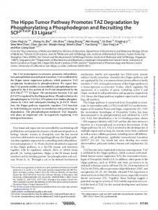
FIGURE 1. Role of Fas-dependent and Fasindependent pathways in CTL-mediated lysis of CMS4 tumor cells. A, Lytic activity by CMS4reactive CTL against CMS4 targets determined at the indicated time points. Assays were also conducted in the absence (DMSO diluent control) or the presence of CMA pretreatment of the CTL (10 M). B, Same as in A, except that the lytic assay was conducted for 18 h, in the absence or the presence not only of CMA, but also of neutralizing anti-FasL mAb (20 g/ml) or an isotype-matched control using the combinations shown in the legend box.
Statistical analysis Statistical analysis in the lung metastasis studies was determined using an unpaired, two-sided t test, with p ⬍ 0.05 considered statistically significant.
Results Role of perforin and FasL pathways in CTL-mediated cytotoxicity of CMS4 targets To better understand the potential contributions of Fas-dependent and Fas-independent pathways in tumor regression by Ag-specific CTL in vivo, we first examined the nature of the lytic mechanisms operative in vitro. To that end, CTL activity was determined in the absence and the presence of CMA, a potent inhibitor of perforinmediated lysis (20). Additionally, since optimal detection of Fasmediated lysis typically requires longer incubation periods, we examined CTL lytic activity in a longer term, 18-h assay. We reasoned that if these CTL killed CMS4 targets through Fas-dependent mechanisms, then the extent of inhibition observed with CMA under those conditions would also be diminished. In 4-h assays, we found that the perforin pathway essentially accounted for all detectable CTL activity, since CMA strongly inhibited lysis (Fig. 1A). In contrast, in 18-h assays, it appeared that CTL activity involved multiple effector mechanisms, since CMA only partially blocked lysis. Although the overall level of lysis was higher at 18 h than at 4 h at the same E:T cell ratios, it was unlikely that these enhanced lytic responses overwhelmed the inhibitory effects of CMA. For example, if the data were compared using E:T cell ratios yielding a comparable degree of lysis (e.g., 2/1 ratio at 4 h
vs 0.5/1 ratio at 18 h), then the extent of inhibition seen with CMA at 4 h was still greater than that at 18 h. To further delineate the nature of the perforin-independent pathway observed in the 18-h assay, CTL activity was examined in the absence and the presence of CMA as well as neutralizing anti-FasL mAb (Fig. 1B). Similar to that seen in Fig. 1A, pretreatment of CTL with CMA incompletely blocked lysis. The inclusion of antiFasL mAb alone had a marginal inhibitory effect noted at three of the five E:T cell ratios, suggesting that the perforin pathway under these in vitro assay conditions was still the dominant effector mechanism. However, the combination of both CMA pretreatment of CTL and anti-FasL mAb exerted maximal inhibition of lysis compared with cultures exposed to CMA with or without isotype control Ab (Fig. 1B). Thus, under conditions in which the perforin pathway was blocked, the contribution of the CTL-directed FasL pathway was more easily revealed. Taken collectively, these data demonstrated that maximal CTL-mediated lysis of CMS4 targets predominately involved both perforin- and Fas-based effector mechanisms.
Fas expression by CMS4 tumor cells The observation that lysis proceeded via both perforin- and Fasbased lytic mechanisms implied that CMS4 targets expressed a functional Fas pathway. To directly examine Fas responsiveness, we assessed cell surface Fas expression by flow cytometry and functional Fas activity under agonistic anti-Fas mAb stimulation
The Journal of Immunology
2405
FIGURE 2. Fas expression by CMS4 tumor cells. A, CMS4 cells were analyzed for cell surface Fas expression by flow cytometry in the absence or the presence of cytokine pretreatment. CMS4 cells were pretreated overnight with nothing (dotted line), IFN-␥ (solid thin line), TNF-␣ (dashed line), or both cytokines (solid thick line). Isotype-matched Ab staining for IFN-␥- plus TNF-␣-pretreated tumor cells is shown as the shaded area. B, Fas-mediated lysis of CMS4 cells, as measured by 51Cr release under the indicated cytokine pretreatment conditions, which match those in A. Untreated or cytokine-pretreated cells were incubated in the presence of anti-Fas mAb (under agonistic conditions) or an isotype-matched Ab. C, Fas expression by CMS4 cells after incubation with cytokine-containing supernatants produced by Ag-stimulated CTL. Cell-free supernatants were collected from CTL that had been cocultured with nothing (dotted line), CMS4 cells (solid thick line), or P815 cells (solid thin line). CMS4 cells pretreated with both IFN-␥ and TNF-␣ were included as a positive control (dashed line). These supernatants (25%, v/v) were then added separately to fresh cultures of CMS4 cells for a 24-h incubation. Cell surface Fas expression after the different treatments was assessed by flow cytometry. Isotype-matched Ab staining for tumor cells pretreated with CTL/CMS4-derived supernatants is shown as the shaded area. D, Fas-mediated lysis of CMS4 cells, as measured by 51Cr release under the indicated supernatant pretreatment conditions, which match those in C. Untreated or supernatant (sn)-pretreated tumor cells were incubated under anti-Fas stimulation conditions, as in B. E, Anti-IFN-␥ and anti-TNF-␣ mAb inhibited Fas up-regulation by CMS4 cells during incubation with CTL-derived cytokine-containing supernatants. CTL-derived supernatants, as in C, were preincubated for 60 min at 4oC with nothing (solid thin line), anti-IFN-␥ plus anti-TNF-␣ (each at 25 g/ml; solid thick line), or an isotype-matched control Ab (50 g/ml; dashed line, which overlaps with the solid thin line). Untreated CMS4 cells are shown by the dotted line. These supernatants were then transferred to fresh cultures of CMS4 cells for a 24-h incubation. Cell surface Fas expression was assessed by flow cytometry. Isotype-matched Ab staining for CMS4 cells exposed to both anti-IFN-␥- and TNF-␣-pretreated supernatants is shown as the shaded area, although similar results for isotype Ab staining were observed with the other treatment groups. Mean fluorescence intensity values of untreated and IFN-␥- plus TNF-␣-treated CMS4 cells were 52 (median, 16) and 138 (median, 85), respectively.
2406 conditions (Fig. 2). Interestingly, CMS4 targets expressed low levels of cell surface Fas (Fig. 2A) and failed to display sensitivity to Fas-mediated lysis (Fig. 2B). Under certain circumstances, the level of cell surface Fas on both mouse and human tumor cell lines can be enhanced by pretreatment with proinflammatory cytokines, namely IFN-␥ and/or TNF-␣ (21, 23–25). Therefore, we examined the effects of these cytokines on modulating Fas expression by CMS4 cells. Although pretreatment of CMS4 cells with IFN-␥ or TNF-␣ each independently increased cell surface Fas expression compared with untreated tumor cells, the combination of both cytokines enhanced Fas levels even more (Fig. 2A). Functionally, pretreatment of CMS4 cells with either IFN-␥ or TNF-␣ led to only a marginal increase in sensitivity to Fas-mediated death (Fig. 2B). However, the combination of IFN-␥ and TNF-␣ also sensitized CMS4 targets to Fas-mediated death more efficiently (Fig. 2B). Similar results were observed with PI staining as well as with recombinant human soluble FasL as an anti-Fas stimulus (data not shown). Thus, CMS4 tumor cells harbored a functional Fas pathway that was unmasked or enhanced in vitro under proinflammatory cytokine-inducible conditions. These results also suggested that for these CTL to kill CMS4 targets through the Fas pathway (Fig. 1B), cytokines such as IFN-␥ and TNF-␣ may be necessary to efficiently sensitize them to FasL-dependent interactions. Therefore, physiologically, such cytokines may be secreted by activated CTL following TCR engagement. Previously, we demonstrated that these CMS4-reactive CTL produced abundant amounts of both IFN-␥- and TNF-like activities in vitro in response to tumor-specific stimulation (9). Additionally, these CMS4-reactive CTL produced high amounts of GM-CSF, but no detectable levels of IL-4, IL-5, or IL-10 after tumor-specific stimulation (9). To examine whether such CTL-derived soluble factors containing those cytokines could modulate Fas expression by CMS4 cells, we conducted supernatant transfer experiments (Fig. 2, C and D). We collected supernatants from CTL/tumor cell cocultures after a 24-h coincubation, and then transferred them to fresh CMS4 targets for subsequent phenotypic and functional analyses. We found that CTL/ CMS4 coculture supernatants enhanced cell surface Fas levels compared with untreated tumor cells as well as the functional response of fresh CMS4 targets to Fas-mediated lysis (Fig. 2, C and D). In contrast, supernatants from CTL/P815 cocultures, which were employed as a target cell specificity control, failed to alter the Fas expression and function of fresh CMS4 targets compared with those in untreated CMS4 cells or CMS4 cells treated with supernatants from relevant CTL/CMS4 cocultures (Fig. 2, C and D). To determine whether the enhancement of cell surface Fas expression caused by these CTL/CMS4 coculture supernatants was due at least in part to IFN-␥ and TNF-␣, we conducted mAb blocking experiments. To that end, CTL-derived supernatants were divided into three groups and then incubated with nothing, a combination of both mAb, or an isotype-matched control Ab. These supernatants were then transferred to groups of fresh CMS4 cells, followed by incubation for 24 h (Fig. 2E). As shown previously (Fig. 2C), incubation of CMS4 cells with untreated supernatants increased the level of cell surface Fas expression compared with untreated CMS4 cells. Furthermore, incubation of CMS4 cells with supernatants pre-exposed to neutralizing anti-IFN-␥ and TNF-␣ mAb resulted in a reduced level of cell surface Fas expression compared with CMS4 cells exposed to untreated supernatants or supernatants treated with the isotype control Ab (Fig. 2E). Production of FasL-deficient CMS4-reactive CTL These data revealed that CMS4 targets expressed a Fas-responsive phenotype and that maximal tumor cell lysis by these Ag-specific
MECHANISMS OF CTL ADOPTIVE IMMUNOTHERAPY CTL involved both perforin- and Fas-dependent pathways. The approach taken to then investigate the potential role of FasL-dependent interactions in CTL-mediated regression of CMS4 cells in vivo was to produce tumor-specific CTL from FasL-deficient (gld) mice and then functionally compare them with wild-type (wt) CTL. To that end, CMS4-reactive CTL were produced from gld mice using the same anti-CTLA4 mAb-based strategy previously employed for generation of the wt-CTL line (9). Fig. 3A illustrates the lytic activity expressed by an established gld-CTL line compared with that of the wt-CTL line. In a 4-h assay, both CTL lines mediated comparable lytic activity against CMS4 targets. Ag specificity of lysis was revealed by the inability of both CTL lines to lyse P815 tumor cells, which was included as an irrelevant target. In an 18-h assay we noted that lytic activity by the wt-CTL line was somewhat better than that by the gld-CTL line (Fig. 3B), consistent with the effect seen with anti-FasL blocking of wt-CTL (Fig. 1B). Furthermore, CMA nearly completely inhibited lysis by the gld-CTL line (Fig. 3B), confirming that lysis by the gld-CTL line proceeded essentially via the perforin pathway. Generation and characterization of wt-CTL and gld-CTL clones To evaluate the role of FasL-dependent interactions in CTL-mediated tumor regression in vivo, we produced single-cell clones from both CTL lines and examined individual clones for antitumor reactivity both in vitro and in vivo. Although a number of clones were generated, we expanded three clones each from the wt-CTL and gld-CTL lines, based on efficient growth at limiting dilution. Fig. 3C illustrates the lytic activity expressed collectively by the six clones. Although there were some differences in lytic efficiency among clones of a given CTL line, the overall lytic patterns between wt and gld clones appeared to be essentially similar. We also examined IFN-␥ and TNF-␣ production by the six clones in response to tumor-specific stimulation (Fig. 4) as additional assessments of Ag recognition. Furthermore, the levels of IFN-␥ and TNF-␣ secretion were analyzed because they were found to be important cytokines that served to modulate Fas expression by CMS4 cells (Fig. 2). We found that all six clones secreted comparable amounts of IFN-␥ following interaction with CMS4 targets, whereas considerably less IFN-␥ was released in response to P815 targets (Fig. 4A). In regard to TNF-␣ levels, we found that the three wt-CTL clones produced comparable amounts as did the three gld-CTL clones (Fig. 4, B and C). However, when the two groups of CTL clones were compared with each other, the wt-CTL clones overall produced much less TNF-␣ than the three gld-CTL clones. Nonetheless, these data demonstrated that both wt-CTL and gld-CTL clones were functionally competent in their capacity to produce both IFN-␥ and TNF-␣ in response to tumor-specific interactions in vitro. Finally, we examined FasL and perforin expression by these wt and gld clones to substantiate important lytic properties or features at the clonal level (Fig. 5). As expected, the three wt-CTL clones up-regulated cell surface FasL in response to anti-TCR stimulation, whereas the three gld-CTL clones failed to express cell surface FasL even under the same potent activation conditions (Fig. 5A). Furthermore, consistent with the CMA blocking studies (Figs. 1 and 3), all six clones expressed perforin, as determined by intracellular flow cytometry (Fig. 5B). Antitumor efficacy of wt-CTL and gld-CTL clones by adoptive immunotherapy To determine the importance of the FasL pathway in CTL adoptive immunotherapy, we then compared antitumor activity mediated by both wt-CTL and gld-CTL clones in minimal and extensive disease paradigms in a manner similar to that described previously (9). We found that the adoptive transfer of wt-CTL or gld-CTL clones, when given at 3 ⫻ 106 cells/mouse, resulted in nearly
The Journal of Immunology
FIGURE 3. Comparison of wt-CTL to gld-CTL populations for tumor reactivity in vitro. A, Lytic activity expressed by wt-CTL or gld-CTL lines against CMS4 or P815 targets at multiple E:T cell ratios in a 4-h assay. B, Same as in A, except that the lytic assay was conducted for 18 h in the absence or the presence of CMA, as shown in the legend box. C, Lytic activity expressed by wt-CTL and gld-CTL clones. Three clones of each functional type were analyzed. Cytotoxicity was determined against CMS4 targets at multiple E:T cell ratios in a 4-h assay. Lytic activity against P815 targets was consistently ⬍10% at the highest E:T cell ratio for all clones.
2407
FIGURE 4. Cytokine production by wt-CTL and gld-CTL clones in response to tumor-specific stimulation. A, IFN-␥ production by the various CTL clones. All six CTL clones were harvested over Ficoll-Hypaque gradients on day 7 after Ag restimulation. Cells (1 ⫻ 106/well) were then reincubated for 24 h in the presence of CMS4 or P815 targets (1/3 E:T cell ratio) in 1 ml of a 24-well plate. After incubation, cell-free supernatants were collected and measured for IFN-␥ release by ELISA. Data were reported as nanograms per milliliter per 106 cells per 24 h. TNF-␣ production by the wt-CTL (B) and gld-CTL (C) clones is shown. Similar to A, except that TNF-␣ levels were determined by the Cytometric Bead Array assay. Data were reported as picograms per milliliter per 106 cells per 24 h and were representative of two separate experiments.
2408
FIGURE 5. Expression of FasL and perforin by the various CTL clones. A, Induction of FasL expression. All six CTL clones were harvested over Ficoll-Hypaque gradients on day 6 after Ag restimulation. Cells were then reincubated for 3 h in a 24-well plate at 5 ⫻ 105/well in the absence or the presence of immobilized anti-CD3 mAb (1 g/well). After incubation cells were recovered and analyzed for cell surface FasL expression by flow cytometry. Each histogram refers to a single CTL clone. The solid thick line shows FasL expression by anti-CD3-stimulated CTL; the dotted line shows FasL expression by unstimulated CTL; the shaded area shows antiCD3-stimulated CTL stained with isotype-matched Ab. Data shown are from one of two separate experiments. B, Intracellular perforin expression. All six CTL clones were harvested over Ficoll-Hypaque gradients on day 6 after Ag restimulation and analyzed for perforin expression by intracellular flow cytometry. Each histogram refers to a single CTL clone. The solid thick line shows perforin expression by CTL; the shaded area shows CTL stained with isotype-matched Ab. Data shown are from one of two separate experiments. The averages of the mean fluorescence intensity values of the two experiments for each clone are: WT3-1, 135; WT3-2, 119; WT3-4, 91; GB10-1, 79; GD8-1, 89; and GD9-1, 132.
complete inhibition of CMS4 tumor growth in the lungs in a minimal disease setting (Fig. 6; WT3-1 was not tested). In an extensive disease setting, the degree or pattern of antitumor activity mediated by the wt-CTL clones at 3 ⫻ 106 cells/mouse was less than that observed in a minimal disease setting (Fig. 6). Nevertheless, significant antitumor activity was still observed by these CTL in an extensive disease setting compared with the saline-treated controls
MECHANISMS OF CTL ADOPTIVE IMMUNOTHERAPY (Table I). Of further interest, however, the magnitude of antitumor activity mediated by the gld-CTL clones was less effective than that achieved by the wt-CTL clones in an extensive disease setting (Fig. 6). In fact, if the data for the individual six clones were reassigned into two functional CTL groups (i.e., wt vs gld), it was observed that both groups of CTL clones displayed significant antitumor effects relative to the saline-treated controls (Table I). Moreover, if the two groups of CTL clones were compared with each other, the differences in antitumor activity between them were also highly significant (Table I). Similar functional patterns were observed with wt vs gld-CTL lines in both minimal (data not shown) and extensive disease settings (Table I). If the CTL adoptive transfer was initiated at a lower concentration (of 1 ⫻ 106 cells/mouse), essentially similar patterns emerged in both types of disease settings (Fig. 7). In the minimal disease setting, both wt-CTL and gld-CTL clones mediated strong antitumor effects, albeit the gld-CTL clones at this lower CTL dose began to show less antitumor effectiveness. In the extensive disease setting the wt-CTL clones still maintained strong antitumor effects, while two of the three gld clones lost substantial antitumor capacity (Fig. 7). Nonetheless, if the data for the individual six clones were reassigned into two functional CTL groups as before, it was observed that only the wt-CTL group of clones displayed significant antitumor effects relative to the saline-treated controls. Finally, if the two groups of CTL clones were compared with each other, the differences in antitumor activity between them was highly significant (Table I). Again, similar functional patterns were observed with wt-CTL vs gld-CTL lines at lower CTL concentrations in an extensive disease setting (Table I). Lastly, we examined the levels of cell surface Fas expression by tumor cells in the lungs of mice after treatment with antitumor CTL (WT3-2 or GD9-1 CTL clones) or saline as a control (Fig. 8). Based on our in vitro studies using recombinant cytokines or CTL-derived supernatants, we found that both IFN-␥ and TNF-␣ were important for enhancing Fas expression by CMS4 tumor cells (Fig. 2). Therefore, to determine whether Fas expression by these tumor cells increased in response to interaction with Ag-specific CTL in vivo, we collected lungs from both CTL-treated and saline-treated mice in an extensive disease setting (Fig. 8). Because the CTL-mediated antitumor response in this type of disease setting was strong, yet therapeutically incomplete, we could analyze lungs containing ample amounts of tumor cells potentially undergoing these phenotypic changes. To distinguish the tumor from nontumor (i.e., leukocyte) populations in the lung preparations, we evaluated the frequency of Fas⫹CD45⫺ cells in the large cell-gated fraction by two-color flow cytometry. (By examination under the light microscope, the larger cell morphology of CMS4 tumor cells could be readily differentiated from that of other smaller infiltrating or resident lung cell types.) Using this approach, we found that the levels of cell surface Fas expression by these tumor cell-containing lung preparations, based on the percentages of Fas⫹CD45⫺ cells, were somewhat higher in groups of mice treated with WT3-2 CTL (33 ⫾ 7) or GD9-1 CTL (44 ⫾ 5) compared with saline (27 ⫾ 3; Fig. 8). The greater increase in the proportion of Fas⫹CD45⫺ cells in GD9-1 CTLtreated mice compared with WT3-2 CTL-treated mice was associated at least in part with the differential levels of TNF-␣ (perhaps in concert with IFN-␥) produced by these two clones (Fig. 4). Based on the mean (and median) fluorescence intensity values, the relative density of cell surface Fas molecules, however, was not any higher for CTL-treated mice compared with saline-treated mice. Interestingly, the mean (and median) fluorescence intensity values for Fas expression of the tumor cell-containing lung preparations of the three groups (Fig. 8) fell between those observed with untreated and IFN-␥/TNF-␣-treated CMS4 cells (see Fig. 2E
The Journal of Immunology
2409
FIGURE 6. Adoptive immunotherapy of wt-CTL and gld-CTL clones at a higher CTL concentration in both minimal and extensive disease settings. The wt-CTL and gld-CTL clones were tested in vivo in an adoptive transfer model (see Materials and Methods). CMS4 cells (2.5 ⫻ 105/mouse) were resuspended in HBSS and injected i.v. Three or 10 days later, 3 ⫻ 106 CTL/mouse (4 days following in vitro stimulation) were also prepared in HBSS and injected i.v. Two weeks following CTL adoptive transfer (i.e., on day 17 or 24 post-tumor transplant), lungs were removed and processed for enumeration of nodules. Mice receiving HBSS were euthanized on day 17 post-tumor transplant. Each data point represents total lung nodule counts from a single mouse. Although five mice per group were used for each treatment, for groups (in the day 10 setting) receiving GB10-1 two mice had died on day 21, for those receiving GD8-1 one mouse had died on day 22, and for those receiving GD9-1 two mice had died on day 22 post-tumor transplant, probably due to disease burden. Clone WT3-1 was not tested in the day 3 setting.
for example). Despite the fact that more sizeable differences in Fas expression were not detectable under these experimental conditions, it is important to emphasize that Fas expression was still demonstrable using these in vivo-isolated tissues, and that the antitumor response was therapeutically more effective with the wtCTL clone than with the gld-CTL clone.
Discussion In this study we examined the potential role of the FasL pathway in tumor regression by Ag-specific CD8⫹ CTL in vivo, which remains largely uncharacterized. One approach to understanding the mechanisms of antitumor activity is through adoptive transfer,
Table I. Comparison of wt-CTL to gld-CTL lines or clones for antitumor activity under extensive disease conditions p Value vsd Lines/Clones
Clones
Lines
a
Treatmenta
No. of CTL Transferredb
No. of Lung Nodulesc
None wt-CTL gld-CTL None wt-CTL gld-CTL None wt-CTL gld-CTL wt-CTL gld-CTL wt-CTL gld-CTL
Nonee 3 ⫻ 106 3 ⫻ 106 Nonef 1 ⫻ 106 1 ⫻ 106 None 3 ⫻ 106 3 ⫻ 106 1 ⫻ 106 1 ⫻ 106 3 ⫻ 105 3 ⫻ 105
127 ⫾ 10 (5) 32 ⫾ 5 (15) 82 ⫾ 9 (10) 173 ⫾ 36 (4) 28 ⫾ 5 (15) 111 ⫾ 17 (14) 182 ⫾ 21 (8) 11 ⫾ 2 (10) 45 ⫾ 7 (10) 26 ⫾ 3 (8) 108 ⫾ 12 (9) 75 ⫾ 12 (8) 114 ⫾ 17 (10)
No Rx
CTL
7.1 ⫻ 10⫺5 0.006
0.0002
0.016 0.176
0.0003 ⫺5
7.9 ⫻ 10 0.0002 4.1 ⫻ 10⫺6 0.011 0.0007 0.025
0.0002 9.5 ⫻ 10⫺5 0.09
Groups of BALB/c mice harboring 10-day established pulmonary metastases received either HBSS (none) or the indicated type of CTL. The individual clones of Figs. 6 and 7 were reassigned into two functional CTL groups as shown. b Concentration of CTL given i.v. per mouse. c Two weeks after CTL adoptive transfer (i.e., day 24 post-tumor transplant), lung tumor nodules of individual mice were quantified (from Figs. 6 and 7 for the clones). Mice receiving HBSS were euthanized on day 17 post-tumor transplant. Data are reported as the mean ⫾ SEM of the total number of mice (shown in parentheses) receiving all clones of a given CTL functional type or the indicated CTL line. d No Rx refers to the comparison of the CTL treatment group vs the no treatment group, while CTL refers to the comparison of the wt-CTL group vs the gld-CTL group. e Saline-treated control group for mice receiving 3 ⫻ 106 CTL/mouse. f Saline-treated control group for mice receiving 1 ⫻ 106 CTL/mouse.
2410
MECHANISMS OF CTL ADOPTIVE IMMUNOTHERAPY
FIGURE 7. Adoptive immunotherapy of wt-CTL and gld-CTL clones at a lower CTL concentration in minimal and extensive disease settings. A, The wt-CTL and gld-CTL clones were tested in vivo in an adoptive transfer model, as described in Fig. 6, except that 1 ⫻ 106 CTL/mouse were administered. Each data point represents total lung nodule counts from a single mouse. Five mice per group were used for treatment. For groups (in the day 10 setting) receiving HBSS or clone GB10-1, however, one data point from each group was not evaluated due to technical reasons encountered during lung retrieval and staining.
which involves the passive administration of effector cells or established T cell lines or clones from immunized hosts to naive, tumor-bearing recipients. Furthermore, through an understanding
of the nature of the antitumor immune mechanisms, adoptive transfer experiments may have implications for administration of the most appropriate functional subsets of ex vivo-expanded effector
FIGURE 8. Fas expression by CMS4 cells in vivo following CTL adoptive transfer. In a day 10 adoptive transfer setting, groups of BALB/c mice (three mice per group) received saline (HBSS; top panels), WT3-2 CTL clone (middle panels), or GD9-1 CTL clone (bottom panels; each at 3 ⫻ 106 CTL/mouse) as described in Fig. 6. Seven or 14 days post-tumor transplant, lungs were removed from groups of mice receiving HBSS or CTL, respectively. Single-cell suspensions of lung digests were then prepared as described in Materials and Methods and were cryopreserved after collection so that all groups could be analyzed at the same time. Tumor cell-containing lung preparations were then examined for Fas expression by flow cytometry. To distinguish tumor from nontumor (i.e., leukocyte) populations in these lung preparations, the proportion of Fas⫹CD45⫺ cells (solid thick line) in the large cell-gated fraction was analyzed. Staining with the isotype-matched Ab is shown as the shaded area. Each histogram represents the results from a single mouse, with the percentage of Fas⫹CD45⫺ cells shown in the upper right. The mean fluorescence intensity ⫾ SEM of the three separate mice receiving HBSS, WT3-2, or GD9-1 was 142 ⫾ 26 (median, 51 ⫾ 2), 102 ⫾ 8 (median, 55 ⫾ 4), and 89 ⫾ 4 (median, 53 ⫾ 2), respectively. In a parallel set of mice, total lung tumor nodules were counted from mice receiving HBSS, WT3-2, or GD9-1 (mean ⫾ SEM): 127 ⫾ 10 (n ⫽ 5), 42 ⫾ 5 (n ⫽ 5), and 83 ⫾ 19 (n ⫽ 3), respectively.
The Journal of Immunology cells in anticancer therapies. Indeed, in recent clinical studies of patients with advanced metastatic melanoma, the adoptive transfer of autologous cultured tumor-infiltrating lymphocytes mediated efficient tumor regression in several patients (26), suggesting that adoptive immunotherapy is still very much a viable and relevant anticancer strategy. Although CD8⫹ CTL play important roles in antitumor activity (26 –30), it remains to be fully understood whether optimal tumor regression by MHC-restricted, Ag-specific CD8⫹ CTL requires functional Fas/FasL interactions, and whether the disruption or disengagement of that interaction allows subpopulations of tumor cells to escape death. Several studies in humans (21, 31–33) support the idea that the loss of Fas expression or function by different cancer types associates with a more malignant phenotype and sustain the idea that the host Fas/FasL system may be important for the regulation of local tumor growth. Transfection of the cellularderived FLICE inhibitory protein gene, an inhibitor of Fas-mediated signaling (34), into syngeneic tumor cells enhanced the frequency and decreased the latency of s.c. tumor growth (35, 36). However, it remains unclear whether the overexpression of cellular-derived FLICE inhibitory protein naturally exists in mouse tumors and mechanistically contributes to the progression of the neoplastic process. Nonetheless, Winter et al. (13) argued that neither perforin nor the Fas pathway was required to mediate tumor regression in their model of adoptive immunotherapy. It is important to point out, however, that this observation was based on the use of a tumor cell line that failed to express functional Fas activity. Thus, the relative participation of Fas/FasL interactions in tumor immunity, whether in mouse or human systems, is intimately linked to the functional status of Fas on the neoplastic cell, which will probably vary among different neoplastic populations and perhaps at different stages of neoplastic development. To determine the role of the Fas/FasL system in this CTL adoptive immunotherapy model, we first examined Fas expression by CMS4 sarcoma cells at both cell surface (protein) and functional levels. Our findings revealed that CMS4 cells constitutively expressed low levels of Fas, but following treatment with IFN-␥ and TNF-␣, there was a marked enhancement in both cell surface Fas levels and functional Fas responsiveness (Fig. 2). Furthermore, maximal lysis of CMS4 targets by these CTL in vitro resulted from both Fas-dependent and -independent mechanisms, since the combination of anti-FasL mAb and CMA abrogated cytotoxicity (Fig. 1). It is conceivable that such cytokines are supplied in vivo either by the host as part of a pathogen (tumor)-induced inflammatory response or by the adoptively transferred CTL following Ag recognition and CTL activation, which warrants further investigation. In vitro studies demonstrated that these CTL produced these proinflammatory cytokines in response to tumor-specific recognition (Fig. 2) (9), consistent with that latter hypothesis. When we sought to extend these in vitro observations at least in terms of phenotypic alterations in Fas expression in vivo, we found that the levels of cell surface Fas by tumor cell-containing lung preparations based on the percentages of Fas⫹CD45⫺ cells were higher, although not significantly so, in groups of mice treated with CTL than in those treated with saline. These data were consistent in part with the in vitro observations (Fig. 2) and suggested that CTL may release proinflammatory cytokines during effector/target interactions in vivo, which, in turn, may boost the proportion of tumor cells expressing Fas. Based on the mean (and median) fluorescence intensity values, the relative density of cell surface Fas molecules, however, was not any higher for CTL-treated mice compared with saline-treated mice. Although the exact reasons why the levels of Fas expression did not change more appreciably after treatment with antitumor CTL remain unclear, they may reflect a number of
2411 biologic considerations inherent in the in vivo model. The ability to discern phenotypic changes in Fas expression in vivo might be influenced not only by extrinsic (i.e., adoptively transferred CTL) elements, but also by intrinsic host-dependent (i.e., other immune and nonimmune cell types) factors. For example, an important consideration of this paradigm is the putative role of proinflammatory cytokines in vivo that may be potentially contributed by the host. The process of tumor growth in vivo in the lung may initiate or provoke a localized inflammatory reaction, perhaps consisting of IFN-␥ and TNF-␣ production by a number of cell types within the host innate immune compartment. If so, this may potentially contribute to and therefore mask our ability to detect any additional enhancing effects caused by the adoptively transferred CTL. The observation that the mean (and median) fluorescence intensity values for Fas expression of the tumor cell-containing lung preparations of the saline-treated group (Fig. 8) were higher than those of untreated CMS4 cells (Fig. 2) lends support to that possibility. Furthermore, because of the dynamics of the Ag-specific CTLtumor interaction in vivo, the ability to precisely identify the time of induction of maximal Fas expression becomes more complex and thus will probably require detailed studies of the temporal relationship of both phenotypic and functional aspects of these immunologic parameters. We then investigated the potential role of FasL-dependent interactions occurring between Ag-specific CTL and CMS4 cells in vivo by comparing CMS4-reactive CTL clones that expressed or lacked functional FasL. To that end, CMS4-reactive CTL clones were produced from both wt and gld syngeneic BALB/c mice. Based on lymphocyte-mediated cytotoxicity and cytokine (IFN-␥) release assays as measurements of Ag recognition capability and effector functions, we demonstrated that wt-CTL and gld-CTL clones expressed comparable reactivities in vitro (Fig. 3). Furthermore, since effector/target titration experiments revealed similar degrees or patterns of tumor cell lysis by wt-CTL and gld-CTL at the population and clonal levels (Fig. 3, A and C), this suggested, at least in vitro, similar levels of sensitivity for Ag recognition. However, whether differences in TCR affinity/avidity exist among these clones in vivo remain unclear. The observation that both functional groups of CTL clones produced equivalent amounts of IFN-␥ (Fig. 4A), and that the gld-CTL clones secreted substantially higher amounts of TNF-␣ compared with the wt-CTL clones (Fig. 4, B and C) also suggested that any differences in antitumor efficacy were unlikely to reflect an altered capacity of the gld-CTL clones to produce those cytokines, at least as determined in vitro. Although these in vitro studies indicated that, except for the absence of cell surface FasL expression by gld-CTL, both populations of CTL clones were functionally alike, they do not preclude the possibility that other potential differences exist among these clones, which require further elucidation. The in vivo adoptive transfer studies were then conducted under different conditions to unmask or unravel the potential role of CTL-derived FasL from other dominant mechanisms, such as perforin, in the tumor rejection response. Using a constant tumor cell dose, but varying the effector cell dose range of both FasL-competent and FasL-incompetent CTL clones, we examined antitumor activity in 3-day (minimal) and 10-day (extensive) pulmonary metastases settings. These parameters served to examine antitumor effects under limiting conditions, such as when effector cell numbers were suboptimal (ⱕ1 ⫻ 106; based on earlier titration experiments) or when tumor burden was extensive (⬎100 metastases usually by day 10 at the time of adoptive transfer), which we thought would also better mirror the conditions seen in patients with advanced bulky or metastatic disease. We also reasoned that since physiologic E:T cell ratios at a tumor site in vivo are likely
2412 to be extremely low, the requirement or benefit for multiple effector mechanisms is probably the greatest under those conditions to maximize activation of cell death signals and to reduce the potential for tumor escape. Under effector cell dose-limiting conditions, Fas/FasL interactions may also compensate for, or dominate as, a relevant effector mechanism if the perforin pathway becomes insufficient because of inherent tumor cell threshold responses to the levels of secreted granule contents or fails to be fully executed because unique characteristics of the tumor microenvironment alter TCR signaling, leading to preferential activation of one mechanism (i.e., FasL-dependent lysis) over the other (37, 38). Although similarly potent antitumor effects were observed by both wt-CTL and gld-CTL in the minimal disease setting at the higher CTL concentration, we found that under conditions of extensive disease burden or lowered CTL concentration, the functional patterns began to diverge, in that the gld-CTL preparations began to mediate significantly less antitumor effects compared with the wtCTL (Figs. 6 and 7 and Table I). Interestingly, gld clone GB10-1 still retained efficient antitumor activity at the lower CTL concentration, consistent with the idea of functional heterogeneity among the different gld clones. The observations that both wt-CTL and gld-CTL clones competently mediated antitumor activity in a minimal disease setting also suggested that both groups of CTL migrated efficiently to a relevant site of tumor growth. Although the identity of the FasL-independent mechanisms of tumor regression remains to be fully defined in this model, one possibility includes perforin, based in part on our CMA blocking studies (Figs. 1 and 3) and extrapolation of work by others (6, 7, 18). In summary, using the experimental strategies described in this study, our findings support the hypothesis of a significant role for the FasL pathway in optimal tumor regression of experimental CMS4 lung metastases by adoptive transfer of Ag-specific CTL. Interestingly, the importance of this pathway was unmasked or unveiled under more physiologically limiting conditions, namely extensive disease burden. Under those conditions, it is likely that the collective contributions of multiple immune effector mechanisms are warranted to elicit maximal antitumor reactivity. These data also raise the opposing hypothesis that if tumor cell subpopulations emerge or develop resistance to Fas-mediated death, this may perhaps influence the potential for tumor escape and metastatic development.
References 1. Nagata, S., and P. Golstein. 1995. The Fas death factor. Science 267:1449. 2. Van Parijs, L., and A. K. Abbas. 1998. Homeostasis and self-tolerance in the immune system: turning lymphocytes off. Science 280:243. 3. Schulze-Osthoff, K., D. Ferrari, M. Los, S. Wesselborg, and M. E. Peter. 1998. Apoptosis signaling by death receptors. Eur J. Biochem. 254:439. 4. Krammer, P. H. 1999. CD95(APO-1/Fas)-mediated apoptosis: live and let die. Adv. Immunol. 71:163. 5. Siegel, R. M., F. K. Chan, H. J. Chun, and M. J. Lenardo. 2000. The multifaceted role of Fas signaling in immune cell homeostasis and autoimmunity. Nat. Immunol. 1:469. 6. Henkart, P. A. 1994. Lymphocyte-mediated cytotoxicity: two pathways and multiple effector molecules. Immunity 1:343. 7. Russell, J. H., and T. J. Ley. 2002. Lymphocyte-mediated cytotoxicity. Annu. Rev. Immunol. 20:323. 8. Sarin, A., E. K. Haddad, and P. A. Henkart. 1998. Caspase dependence of target cell damage induced by cytotoxic lymphocytes. J. Immunol. 161:2810. 9. Ryan, M. H., J. A. Bristol, E. McDuffie, and S. I. Abrams. 2001. Regression of extensive pulmonary metastases in mice by adoptive transfer of antigen-specific CD8⫹ CTL reactive against tumor cells expressing a naturally occurring rejection epitope. J. Immunol. 167:4286. 10. DeLeo, A. B., H. Shiku, T. Takahashi, M. John, and L. J. Old. 1977. Cell surface antigens of chemically induced sarcomas of the mouse. I. Mouse leukemia virus related antigens and alloantigens on cultured fibroblasts and sarcomas: description of a unique antigen on BALB/c Meth A sarcoma. J. Exp. Med. 146:720. 11. Walunas, T. L., D. J. Lenschow, C. Y. Bakker, P. S. Linsley, G. J. Freeman, J. M. Green, C. B. Thompson, and J. A. Bluestone. 1994. CTLA-4 can function as a negative regulator of T cell activation. Immunity 1:405.
MECHANISMS OF CTL ADOPTIVE IMMUNOTHERAPY 12. Leach, D. R., M. F. Krummel, and J. P. Allison. 1996. Enhancement of antitumor immunity by CTLA-4 blockade. Science 271:1734. 13. Winter, H., H. M. Hu, W. J. Urba, and B. A. Fox. 1999. Tumor regression after adoptive transfer of effector T cells is independent of perforin or Fas ligand (APO-1L/CD95L). J. Immunol. 163:4462. 14. Peng, L., J. C. Krauss, G. E. Plautz, S. Mukai, S. Shu, and P. A. Cohen. 2000. T cell-mediated tumor rejection displays diverse dependence upon perforin and IFN-␥ mechanisms that cannot be predicted from in vitro T cell characteristics. J. Immunol. 165:7116. 15. Helmich, B. K., and R. W. Dutton. 2001. The role of adoptively transferred CD8 T cells and host cells in the control of the growth of the EG7 thymoma: factors that determine the relative effectiveness and homing properties of TC1 and TC2 effectors. J. Immunol. 166:6500. 16. Hobeika, A. C., T. M. Clay, P. J. Mosca, H. K. Lyerly, and M. A. Morse. 2001. Quantitating therapeutically relevant T-cell responses to cancer vaccines. Crit. Rev. Immunol. 21:287. 17. Seki, N., A. D. Brooks, C. R. Carter, T. C. Back, E. M. Parsoneault, M. J. Smyth, R. H. Wiltrout, and T. J. Sayers. 2002. Tumor-specific CTL kill murine renal cancer cells using both perforin and Fas ligand-mediated lysis in vitro, but cause tumor regression in vivo in the absence of perforin. J. Immunol. 168:3484. 18. van den Broek, M. E., D. Kagi, F. Ossendorp, R. Toes, S. Vamvakas, W. K. Lutz, C. J. Melief, R. M. Zinkernagel, and H. Hengartner. 1996. Decreased tumor surveillance in perforin-deficient mice. J. Exp. Med. 184:1781. 19. Abrams, S. I., S. N. Khleif, E. S. Bergmann-Leitner, J. A. Kantor, Y. Chung, J. M. Hamilton, and J. Schlom. 1997. Generation of stable CD4⫹ and CD8⫹ T cell lines from patients immunized with ras oncogene-derived peptides reflecting codon 12 mutations. Cell. Immunol. 182:137. 20. Kataoka, T., N. Shinohara, H. Takayama, K. Takaku, S. Kondo, S. Yonehara, and K. Nagai. 1996. Concanamycin A, a powerful tool for characterization and estimation of contribution of perforin- and Fas-based lytic pathways in cell-mediated cytotoxicity. J. Immunol. 156:3678. 21. Bergmann-Leitner, E. S., and S. I. Abrams. 2000. Differential role of Fas/Fas ligand interactions in cytolysis of primary and metastatic colon carcinoma cell lines by human antigen-specific CD8⫹ CTL. J. Immunol. 164:4941. 22. Mule, J. J., J. C. Yang, R. Lafreniere, S. Shu, and S. A. Rosenberg. 1987. Identification of cellular mechanisms operational in vivo during the regression of established pulmonary metastases by the systemic administration of high-dose recombinant interleukin-2. J. Immunol. 139:285. 23. Moller, P., K. Koretz, F. Leithauser, S. Bruderlein, C. Henne, A. Quentmeier, and P. H. Krammer. 1994. Expression of APO-1 (CD95), a member of the NGF/TNF receptor superfamily, in normal and neoplastic colon epithelium. Int. J. Cancer 57:371. 24. Owen-Schaub, L. B., R. Radinsky, E. Kruzel, K. Berry, and S. Yonehara. 1994. Anti-Fas on nonhematopoietic tumors: levels of Fas/APO-1 and bcl-2 are not predictive of biological responsiveness. Cancer Res. 54:1580. 25. Lee, J. K., T. J. Sayers, A. D. Brooks, T. C. Back, H. A. Young, K. L. Komschlies, J. M. Wigginton, and R. H. Wiltrout. 2000. IFN-␥-dependent delay of in vivo tumor progression by Fas overexpression on murine renal cancer cells. J. Immunol. 164:231. 26. Dudley, M. E., J. R. Wunderlich, P. F. Robbins, J. C. Yang, P. Hwu, D. J. Schwartzentruber, S. L. Topalian, R. Sherry, N. P. Restifo, A. M. Hubicki, et al. 2002. Cancer regression and autoimmunity in patients after clonal repopulation with antitumor lymphocytes. Science 298:850. 27. Pardoll, D. M. 2000. Therapeutic vaccination for cancer. Clin. Immunol. 95:S44. 28. Romero, P., P. R. Dunbar, D. Valmori, M. Pittet, G. S. Ogg, D. Rimoldi, J. L. Chen, D. Lienard, J. C. Cerottini, and V. Cerundolo. 1998. Ex vivo staining of metastatic lymph nodes by class I major histocompatibility complex tetramers reveals high numbers of antigen-experienced tumor-specific cytolytic T lymphocytes. J. Exp. Med. 188:1641. 29. Greten, T. F., and E. M. Jaffee. 1999. Cancer vaccines. J. Clin. Oncol. 17:1047. 30. Cohen, P. A., L. Peng, J. Kjaergaard, G. E. Plautz, J. H. Finke, G. K. Koski, B. J. Czerniecki, and S. Shu. 2001. T-cell adoptive therapy of tumors: mechanisms of improved therapeutic performance. Crit. Rev. Immunol. 21:215. 31. Keane, M. M., S. A. Ettenberg, G. A. Lowrey, E. K. Russell, and S. Lipkowitz. 1996. Fas expression and function in normal and malignant breast cell lines. Cancer Res. 56:4791. 32. Krammer, P. H., P. R. Galle, P. Moller, and K. M. Debatin. 1998. CD95(APO1/Fas)-mediated apoptosis in normal and malignant liver, colon, and hematopoietic cells. Adv. Cancer Res. 75:251. 33. Owen-Schaub, L., H. Chan, J. C. Cusack, J. Roth, and L. L. Hill. 2000. Fas and Fas ligand interactions in malignant disease. Int. J. Oncol. 17:5. 34. Irmler, M., M. Thome, M. Hahne, P. Schneider, K. Hofmann, V. Steiner, J. L. Bodmer, M. Schroter, K. Burns, C. Mattmann, et al. 1997. Inhibition of death receptor signals by cellular FLIP. Nature 388:190. 35. Djerbi, M., V. Screpanti, A. I. Catrina, B. Bogen, P. Biberfeld, and A. Grandien. 1999. The inhibitor of death receptor signaling, FLICE-inhibitory protein defines a new class of tumor progression factors. J. Exp. Med. 190:1025. 36. Medema, J. P., J. de Jong, T. van Hall, C. J. Melief, and R. Offringa. 1999. Immune escape of tumors in vivo by expression of cellular FLICE-inhibitory protein. J. Exp. Med. 190:1033. 37. Kessler, B., D. Hudrisier, M. Schroeter, J. Tschopp, J. C. Cerottini, and I. F. Luescher. 1998. Peptide modification or blocking of CD8, resulting in weak TCR signaling, can activate CTL for Fas- but not perforin-dependent cytotoxicity or cytokine production. J. Immunol. 161:6939. 38. Esser, M. T., B. Krishnamurthy, and V. L. Braciale. 1996. Distinct T cell receptor signaling requirements for perforin- or FasL-mediated cytotoxicity. J. Exp. Med. 183:1697.