© 1999 Oxford University Press
Human Molecular Genetics, 1999, Vol. 8, No. 11 2063–2069
The gene encoding hydroxypyruvate reductase (GRHPR) is mutated in patients with primary hyperoxaluria type II Scott D. Cramer1,2,+, Patrick M. Ferree1, Kai Lin1, Dawn S. Milliner3 and Ross P. Holmes2 1Department
of Cancer Biology, 2Department of Urology, Wake Forest University School of Medicine, Winston-Salem, NC 27157, USA and 3Division of Nephrology, Mayo Clinic, Rochester, MN 55905, USA Received May 19, 1999; Revised and Accepted July 14, 1999
Primary hyperoxaluria type II (PH2) is a rare monogenic disorder that is characterized by a lack of the enzyme that catalyzes the reduction of hydroxypyruvate to Dglycerate, the reduction of glyoxylate to glycolate and the oxidation of D-glycerate to hydroxypyruvate. The disease is characterized by an elevated urinary excretion of oxalate and L-glycerate. The increased oxalate excretion can cause nephrolithiasis and nephrocalcinosis and can, in some cases, result in renal failure and systemic oxalate deposition. We identified a glyoxylate reductase/hydroxypyruvate reductase (GRHPR) cDNA clone from a human liver expressed sequence tag (EST) library. Nucleotide sequence analysis identified a 1198 nucleotide clone that encoded a 984 nucleotide open reading frame. The open reading frame encodes a predicted 328 amino acid protein with a mass of 35 563 Da. Transient transfection of the cDNA clone into COS cells verified that it encoded an enzyme with hydroxypyruvate reductase, glyoxylate reductase and D-glycerate dehydrogenase enzymatic activities. Database analysis of human ESTs reveals widespread tissue expression, indicating that the enzyme may have a previously unrecognized role in metabolism. The genomic structure of the human GRHPR gene was determined and contains nine exons and eight introns and spans ∼9 kb pericentromeric on chromosome 9. Four PH2 patients representing two pairs of siblings from two unrelated families were analyzed for mutations in GRHPR by single strand conformation polymorphism analysis. All four patients were homozygous for a single nucleotide deletion at codon 35 in exon 2, resulting in a premature stop codon at codon 45. The cDNA that we have identified represents the first characterization of an animal GRHPR sequence. The data we present will facilitate future genetic testing to confirm the clinical diagnosis of PH2. These data will also facilitate heterozygote testing and prenatal testing in families affected with PH2 to aid in genetic counseling. +To
DDBJ/EMBL/GenBank accession nos AF146018 and AF146689
INTRODUCTION Primary hyperoxaluria type II (PH2) is a rare monogenic disease with an autosomal recessive pattern of inheritance that appears to result from an absence of an enzyme with hydroxypyruvate reductase (HPR) and glyoxylate reductase (GR) activities (1). It was first reported as a disease distinct from primary hyperoxaluria type I (PH1) 30 years ago and is characterized by an elevated urinary excretion of oxalate and L-glycerate (1,2). The increased oxalate excretion can cause nephrolithiasis and nephrocalcinosis and can, in some cases, result in renal failure and systemic oxalate deposition. Previous reports have demonstrated HPR enzymatic activity in liver, kidney and peripheral blood (3–5), and this activity is apparently important in the conversion of serine to glucose in humans (6). The GR activity reduces glyoxylate to glycolate, and may be important in regulating the amount of glyoxylate that is converted to oxalate (1,3,7). The enzyme also has Dglycerate dehydrogenase (DGDH) activity. HPR and DGDH are reciprocal reduction/dehydrogenation reactions, with the equilibrium favoring the reduction reaction (8). It has been suggested that NADH/NAD+ ratios are important in the etiology of PH2 (3). However, biochemical evidence demonstrates that the GRHPR enzyme uses NADPH as a coenzyme (8), generating NADP+, which is required for a functional pentose phosphate pathway. The exact mechanism of oxalate overproduction due to an absence of the GRHPR enzyme in PH2 patients is not known (9). The bovine HPR protein has been partially purified from liver (10,11); however, no amino acid or nucleotide sequence information has been reported. The HPR cDNA has been cloned from eubacterial (12) and plant (13,14) species. The crystal structure has been resolved at 2.4 Å for the recombinant enzyme purified from Hyphomicrobium methylovorum (12). The H.methylovorum enzyme functions as a homodimer. Each subunit is 322 amino acids long and contains a consensus nicotinamide adenine dinucleotide binding site. The bacterial enzyme has a preference for NADH rather than NADPH which is preferred by mammalian HPR (8,11). Both plant and bacterial enzymes are homologous to phosphoglycerate dehydrogenase; however, the latter enzyme can be distinguished from HPR by an additional ∼200 C-terminal residues (S.D. Cramer, unpublished data). Predicted protein sequences with homology to the plant and bacterial HPR
whom correspondence should be addressed. Tel: +1 336 716 9330; Fax +1 336 716 0255; Email:
[email protected]
2064 Human Molecular Genetics, 1999, Vol. 8, No. 11
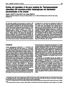
Figure 1. The predicted hGRHPR protein is homologous to HPR from other organisms. Conservation of HPR in various organisms is illustrated by the Clustal alignment (performed by the Biology Workbench: http://biology.ncsa.uiuc.edu ). Conserved residues are defined as identical or conservative residues in at least 50% of organisms (bold). Residues in upper case letters are identical to the human sequence. Dots represent gaps in the sequence. The putative co-factor binding site is boxed and shaded. The putative residues involved in substrate binding are boxed with no shading. The derived consensus sequence is represented on the bottom line and has the following code: upper case bold letters represent 100% conservation of the residue; lower case letters represent residues present in at least 50% of sequences; a dash represents a conserved site but no one residue is present in 50% of the sequences; a dot represents no conservation.
protein sequences are present in the Schizosaccharomyces pombe and Pyrococcus horikoshii genomes, although neither the genes nor the proteins have been characterized. With the addition of S.pombe (fungi) and P.horikoshii (archeae), a complete HPR coding sequence from Animalia represents the only major taxa not represented in the database. In this report we describe the identification and characterization of an expressed sequence tag (EST) from a human liver cDNA library that encodes a functional GRHPR protein. We also describe the genomic structure of the human GRHPR (hGRHPR) gene and mutations in hGRHPR in DNA from PH2 patients. RESULTS Identification and characterization of the hGRHPR cDNA Several potential hGRHPR ESTs were identified in the human EST database available at the National Center for Biotechnology Information (NCBI) web site using the Entrez
and Blast search engines. By alignment of these ESTs, an EST from a hepatic cDNA library from a normal male was selected as likely to contain the most 5′ sequence information. We obtained this clone and determined its nucleotide sequence. The entire nucleotide sequence is available from GenBank under accession no. AF146018. The size of the cDNA was determined to be 1198 nucleotides with a predicted open reading frame of 328 amino acids encoding a protein with molecular weight of 35 563 Da. The predicted hGRHPR protein sequence bears homology to known HPR protein sequences from eubacteria and plants and to the predicted HPR sequences from archaea and fungi (Fig. 1). Several absolutely conserved residues are found in the alignment, including residues thought to be involved in nicotinamide adenine dinucleotide binding (Fig. 1, shaded box) (15,16) and substrate binding (Fig. 1, boxed) (12). Using the Blast search engine at the NCBI web site, we identified hundreds of predicted proteins that aligned with high similarity to the hGRHPR protein (data not shown). Representative organisms from Archaea, Eubacteria, Fungi, Planta and
Human Molecular Genetics, 1999, Vol. 8, No. 11 2065
Table 1. PCR–SSCP primers Exon 1
2
First reaction primers (5′→3′)
Final reaction primers (5′→3′)
GGCACGAGCTGCCAGG
ACGAGCTGCCAGGTCCG
CCCAAAACTCCAAGCCTGC
CCAGCCGGCCACAAGG
GCCAGGATTCCCAGCTGG
ACAGGTGTGCGGCTCCTG
TTCCATGGGCTGACACCCG
CCACCCTCAAGTCCCCTG
3
GCCCTGAGGTGAACCCGG CCTGAATGGCCGAGGGATATG
4
5
GGCAGGCAGATCAAAGAGGG
CTCCATGGAAATGTCCCAGC
CTCCTAACCTCATGATCCGC
GGCCTCCCAAAGTGCTGG
CCGTGACCTGGAGGGTGG
CATCTTGGTCCAAGGCTGG
AGACACGTGGTGCCAGGG
CCACGCTGTGAGAGCCGG
6
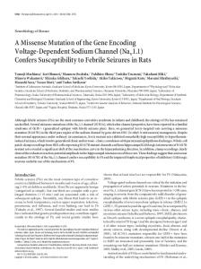
Figure 2. The hGRHPR cDNA encodes a functional protein. COS 1 cells were cotransfected with luciferase reporter vector and control expression vector (pcDNA3.1) or expression vector containing the hGRHPR EST (pHPR3.1). Enzyme activities were determined as described in Materials and Methods. HPR, hydroxypyruvate reductase activity; GR, glyoxylate reductase activity; DGDH, Dglycerate dehydrogenase activity. The bars represent the mean ± SEM (n = 3). Numbers in parentheses represent fold increase in enzyme activity relative to vector control. *Significantly different from the corresponding control vector transfected extracts, P < 0.02.
Animalia were contained in the list, suggesting a near ubiquitous expression of this enzyme. A Blast search of the human EST database using the EST that we characterized identified 114 hits, with sequences represented in libraries from many different human tissues including adrenal, heart, testis, aorta, kidney, tonsil, blood, liver, uterus, brain, lung, whole embryo, breast, ovary, parathyroid, colon, placenta, prostate, germ cell and synovial membrane. To determine whether the EST that we sequenced encoded a full-length, functional GRHPR, we expressed the cDNA in COS cells and assayed for the enzymatic activities associated with GRHPR (HPR, GR and DGDH). Figure 2 shows the results of transfection of control vector (pcDNA3.1) or hGRHPR-encoding vector (pHPR3.1) into COS cells. Each bar represents the mean ± SEM of three independent transfections for control and GRHPR-encoding constructs. All the enzymatic assays were performed on identical cell extracts. Luciferase activities were similar in all cell extracts (data not shown), indicating that transfection efficiencies were equivalent. It can be seen that the EST that we identified encodes a functional GRHPR with HPR, DGDH and GR activities. Although the relative increase in activity was roughly equivalent for each of the enzyme activities examined (∼24-fold), the absolute activity of DGDH was much lower compared with HPR and GR. Genomic structure of hGRHPR Having established that the EST that we identified encodes a fulllength hGRHPR we proceeded to determine the intron–exon structure of the hGRHPR gene. 5′ and 3′ oligonucleotide primers were designed for sequences of the hGRHPR cDNA at ∼250 bp intervals and were used to PCR amplify human genomic DNA derived from a normal laboratory volunteer. PCR products were purified and sequenced in both directions in 400–600 bp segments
GCTGTTCCGGAAATGCTGGG CAACTGGGCACAGATAGGC
7
8
9
GGCCTTCAGGAAGCATCTTGG
AGCAAGGGGCTGGTCTCC
CACCAACAACCCACGG
TCCAGATGGCAGCAGTGG
CAGCTAGAAGGAGGCAGG
TGGGCGGAGGGATCTTCG
GACAATACCAGGGCACGG
GTCCTGCAGGGCAGCAGG
CAGCTAGAAGGAGGCAGG
GCTGAAGGCTGCTGAACC
GACAATACCAGGGCACGG
GCACGGTTTGCTTCCCGG
using automated fluorescent nucleotide sequencing. Overlapping PCR products were assembled to determine the entire hGRHPR genomic structure. The gene spans ∼9 kb and contains nine exons and eight introns. All splice acceptor and donor sites conform to the consensus sequences. The complete nucleotide sequence and structural features of the gene can be obtained from GenBank (accession no. AF146689). Radiation hybrid analysis performed by the Sanger Center (http//:www.sanger.ac.uk/ ) has been used to assign a chromosomal location of a human EST that contains identical 3′ sequences to the EST that we identified. This EST maps pericentromeric on human chromosome 9 within the reference interval D9S1874–D9S273 and has been assigned the unique human sequence locator Hs.155742 (http:// inhouse.ncbi.nlm.nih.gov/cgi-bin/UniGene/clust?ORG=Hs&CID =155742 ). The human genome nomenclature committee has assigned the locus designation GRHPR to this gene (http:// www.gene.ucl.ac.uk/nomenclature/ ). Identification of mutations in HPR in PH2 patients Using the intron–exon structure of the hGRHPR gene we designed PCR primers complementary to intronic sequences flanking the intron–exon boundaries, such that the entire exon plus boundaries would be amplified (Table 1). These primers were used to develop PCR–single strand conformation polymorphism (SSCP) assays for each exon in the hGRHPR gene. PCR–SSCP was initially performed on genomic DNA from normal laboratory volunteers to determine the prevalence of normal polymorphisms. PCR–SSCP of normal DNA samples identified two putative polymorphisms (data not shown). A G-A polymorphism in exon 6 at base 579 of the cDNA lies at the third position of codon 193. Both alleles of this codon encode alanine. A second putative G-A polymorphism in exon 9 at base 963 of the cDNA lies in the
2066 Human Molecular Genetics, 1999, Vol. 8, No. 11
A
B
Figure 3. Exon 2 of hGRHPR in PH2 patients contains a single nucleotide point deletion. (A) Autoradiogram of PCR–SSCP analysis of exon 2 of hGRHPR in seven DNA samples from normal controls (Controls, lanes 1–7) and four PH2 patients (PH2 Patients, lanes 1–4). The arrow indicates a shift in band pattern in PH2 patients. (B) Nucleotide sequence analysis of exon 2 of hGRHPR in DNA from a representative normal control (Control) and a representative PH2 patient (PH2). An isolated section of the electropherogram of exon 2 sequencing in the location of the mutation is shown. Above each panel is the deduced nucleotide sequence and the codons corresponding to the hGRHPR protein. The arrow indicates the G residue in the wild-type sequence that is deleted in all four PH2 patients.
third position of codon 321. Both alleles of this codon encode proline. All exons from PH2 patients were found to have a normal PCR–SSCP pattern (data not shown), except in exon 2. Figure
3A shows the results of PCR–SSCP for exon 2. All four PH2 patients are homozygous for a divergent PCR–SSCP pattern compared with wild-type controls. DNA from each of the PH2 patients was used to PCR amplify exon 2 of HPR, and PCR
Human Molecular Genetics, 1999, Vol. 8, No. 11 2067
products were sequenced in both directions by automated fluorescent dideoxynucleotide sequencing. All four patients had identical nucleotide sequences. Figure 3B shows that all four PH2 patients are homozygous for an identical single nucleotide deletion in the first nucleotide of codon 35; this results in a frameshift beginning at codon 35. The translation of the predicted mRNA produced in these patients would result in a truncated protein of 44 amino acids. DISCUSSION In this report we describe the identification of a human liver EST with all the properties of the hGRHPR cDNA. The protein encoded by the EST that we identified catalyzed HPR, GR and DGDH enzymatic activities. Consistent with the reported coenzyme preference for NADPH versus NADP+ (8), the reduction reactions (HPR and GR) had a higher rate of catalysis than the oxidation reaction (DGDH) (Fig. 2). The predicted 328 amino acid protein has 30–37% similarity with sequences from Archeae, Eubacteria, Fungi and Plantae, with several amino acids conserved among all the sequences (Fig. 1). Aspartate residues in homologous proteins aligning with residues T183 or G184 in hHPR are characteristic of NADHbinding proteins, but absent in NADPH-binding proteins (15,16). Aside from the absence of this Asp there is no consensus that distinguishes NADH- and NADPH-binding proteins (15,16). The lack of this Asp residue in hGRHPR supports the observed preference of mammalian HPR for NADPH (8). Goldberg et al. (12) suggested that a Glu-His catalytic pair is important in the recognition of the D-isomer of glyceric acid by H.methylovorum DGDH. This pair aligns with E274 and H293 in hHPR (Fig. 1). D269 and R245 are also conserved and proposed to be important residues in the catalytic site in the recognition of D-isomers. Previously, GRHPR enzymatic activity has been recognized only in liver, kidney, fibroblasts and peripheral blood lymphocytes (3–5). A role for the enzyme in serine metabolism for gluconeogenesis and in oxalate metabolism is evident in liver and kidney, but not in leukocytes (6). A search of the human EST database suggests that the hGRHPR gene is expressed in a wide variety of human tissues. Although expression of mRNA does not necessarily imply translation into functional protein, the EST data suggest that GRHPR has functions in these previously unrecognized sites. As GRHPR utilizes NADPH and generates NADP+, its activity may possibly be linked to that of the pentose phosphate pathway. In support of the EST data, we have demonstrated hGRHPR transcripts by northern analysis and RT–PCR, and DGDH enzymatic activity using primary cultures of human prostatic epithelial cells as a model for a non-classical GRHPRexpressing tissue (R.P. Holmes and S.D. Cramer, unpublished data). Currently, it is not clear what the role of GRHPR might be in these non-classical tissues, and whether it performs essential functions. No obvious metabolic abnormalities in PH2 patients other than those associated with hepatic oxalate metabolism have been noted (2), suggesting that the expression in non-classical tissues is not an essential function or that alternative mechanisms can compensate for loss of GRHPR activity in these tissues. In addition to the fundamental basic biology questions that the expression data raise, the observation of GRHPR transcripts in many tissues may be
useful in the development of future diagnostic assays using non-invasive techniques such as buccal swabs to evaluate GRHPR enzymatic activity in patients suspected of PH2. Currently, the definitive diagnosis of PH2 versus PH1 requires liver biopsies. Analysis of GRHPR activity in buccal swabs may eliminate the necessity of liver biopsies for definitive diagnosis. The hGRHPR gene that we cloned contains nine exons spanning 9 kb pericentromeric on chromosome 9. We performed PCR–SSCP on DNA samples from seven normal individuals and identified two putative single nucleotide polymorphisms. Neither polymorphism resulted in an amino acid substitution. Given the limited number of normal individuals we have evaluated and the possibility that PCR– SSCP lacks the sensitivity to identify all alleles, there are likely to be other normal polymorphisms in the hGRHPR gene. Future analysis of PH2 patients should recognize these polymorphisms. The data that we present on the GRHPR gene in PH2 patients demonstrate that the gene that we have cloned is mutated in patients with PH2, providing strong evidence that this is the cause of the disease. The PH2 patients that we evaluated in this study represent two sets of siblings from two apparently unrelated families. There was no known consanguinity among the parents within either family, or between the two families. Both families are of Northern European descent. Surprisingly, we identified the same homozygous mutation in all four patients: a single nucleotide deletion in exon 2. Given the small number of PH2 patients that we have genotyped and the lack of definitive data on the patients’ ancestry, it is premature to make conclusions about the prevalence of this mutation in the North American population. However, we expect that other PH2 patients are likely to have additional mutations in the GRHPR gene. In conclusion, we present evidence that mutations in GRHPR are the genetic basis of PH2. The lack of this enzyme results in hyperoxaluria, which can lead to urolithiasis, nephrocalcinosis and renal failure. With the identification of a fully functional cDNA, future studies can now be directed at the development of gene therapy strategies for enzyme replacement therapy in patients with PH2. MATERIALS AND METHODS Identification of the human GRHPR EST The human GRHPR EST was identified using the Entrez search engine at the NCBI web site (http://www.ncbi.nlm.nih.gov ). The keywords ‘Glycerate Dehydrogenase’ and ‘Hydroxypyruvate Reductase’ were used to search against the non-redundant nucleotide database. Several human clones contained in the EST database were identified and analyzed further. The nucleic acid sequences downloaded from GenBank were used to assemble a contig map using the Wisconsin software package of the Genetics Computer Group (GCG). A protein translation of the contig was used to search the non-redundant protein database for homologous proteins. One EST clone was identified which appeared to have a sequence that extended furthest into the 5′ region. This EST was produced by the IMAGE consortium (17) and was derived from a human clone from a cDNA library generated from a hepatectomy specimen from a 49-year-old male. We obtained this clone from
2068 Human Molecular Genetics, 1999, Vol. 8, No. 11
the American Tissue Culture Center (ATCC, Rockville, MD) for further analysis (GenBank accession no. T72836). Characterization of the hHPR cDNA The entire nucleotide sequence of the hGRHPR cDNA EST clone described above was determined by sequencing in overlapping 400–500 bp segments in both directions using fluorescent cycle sequencing with an ABI Prism 377 DNA Sequencer at the Nucleic Acid Sequencing Facility of the Comprehensive Cancer Center of Wake Forest University (CCCWFU). Transient transfection and enzymatic activities The entire cDNA for hGRHPR contained in the EST was subcloned into the mammalian expression vector pcDNA 3.1(–) (Invitrogen, San Diego, CA) at the XbaI and KpnI restriction sites (pHPR3.1). Plasmid DNA was purified using the Wizard Plasmid kit (Promega, Madison, WI). COS cells were obtained from the Tissue Culture Core Laboratory of CCCWFU. For transfections, COS cells were routinely grown in a 37°C humidified incubator in the presence of 5% CO2. Medium was changed every 2–3 days and consisted of bicarbonate-buffered Dublecco’s modified Eagle medium supplemented with 10% fetal bovine serum. The day before transfection, the cells were trypsinized from the culture dish and plated on 60 mm culture dishes at 5 × 10⁵ cells/dish. Transfections were carried out using Lipofectamine (Gibco BRL, Grand Island, NY) according to the manufacturer’s protocol. Ten micrograms of pHPR3.1 plasmid or control vector (pcDNA 3.1) were cotransfected with 1 µg of a luciferase reporter plasmid (pGL3 control; Promega) to control for differences in transfection efficiencies between transfections. Each co-transfection was performed in triplicate. Twenty-six hours after transfection, the cells were scraped off the plates and lysed by sonication in 60 µl of cell culture lysis buffer (100 mM potassium phosphate pH 7.2, 100 mM KCl). Cell lysates were stored at –70°C in 10 µl aliquots until use. Protein concentration in extracts was determined by the BCA microassay according to the manufacturer’s protocol (Pierce, Rockford, IL). Luciferase activity was measured in 5 µl aliquots of cell lysate at 37°C using the Promega luciferase assay system in a Turner TD-20e luminometer according to the manufacturer’s protocol. D-glycerate dehydrogenase enzymatic activity was measured by the conversion of 20 mM D-glycerate to hydroxypyruvate in 20 min at 37°C in an incubation mixture containing 0.1 M Tris–HCl, 0.1 M hydrazine, 1 mM NADP+, 10 mM phenylhydrazine, pH 9. The hydroxypyruvate phenylhydrazone formed was measured in the supernatant by reverse phase HPLC as previously described (33). Hydroxypyruvate reductase and glyoxylate reductase enzymatic activities in cell extracts were determined at 22°C exactly as described previously by Van Schaftingen (8), except that 200 µM glyoxylate was used as substrate in glyoxylate reductase assays. All assays were performed in the presence of 100 µM NADPH. The rate of change in A400–A340 was monitored in a Beckman DU-640 spectrophotometer. The initial linear rate was used to determine enzymatic rate in each extract. Results of enzyme assays are expressed as V (nmol/ min/mg protein).
Determination of the hHPR genomic structure Oligonucleotide primers complementary to hHPR cDNA sequences were designed such that sets of 5′ and 3′ primers were separated from each other by ∼200 nucleotides. Primer sets were used to PCR amplify human genomic DNA isolated from peripheral blood using the QIAmp Blood kit (Qiagen, Chatsworth, CA). PCR products were separated by agarose gel electrophoresis and visualized by ethidium bromide staining. PCR products which were larger than predicted based on the location of the primers in the cDNA sequence were purified from agarose gels using the Geneclean DNA purification kit (BIO 101, Vista, CA) and subjected to automated fluorescent sequence analysis. Intron and exon boundaries were determined by sequence alignment of the deduced sequence from the PCR product with the cDNA sequence, and by the proximal two nucleotides flanking the junction (Table 1). As more sequence information became available, new oligonucleotide primers were synthesized. All PCR products were sequenced in both directions in overlapping 400–600 bp segments. The entire sequence was assembled by alignment of overlapping PCR products. PCR–SSCP Blood was collected from patients at the Mayo Clinic and shipped on ice by overnight courier to Wake Forest University School of Medicine. Total genomic DNA was isolated from samples using the QIAmp Blood kit (Qiagen). PCR–SSCP was used to identify altered exons in PH2 patients using a modification of the method of Orita et al. (18). Nested PCR was used for exons 1, 2, 4, 5, 7, 8 and 9. A single PCR reaction was used for exons 3 and 6. Oligonucleotide primer sequences are listed in Table 1. For nested reactions the first PCR reaction was performed using 25–50 ng of genomic DNA, 15 pmol of each unlabeled oligonucleotide primer, 0.65 mM dNTPs, 2 mM MgCl2, 1× Taq DNA polymerase buffer (Promega, San Diego, CA) in a final volume of 100 µl. A 2 µl aliquot of the first reaction was used for the second PCR with internally nested primers that were end-labeled with 32P using T4 polynucleotide kinase (Promega) and [γ-32P]ATP (NEN, Boston, MA) according to the manufacturer’s protocol. PCR conditions were similar to the first reaction, except that 2.25 pmol of each primer end-labeled with 32P and 4.5 pmol of each unlabelled primer were used, and the reaction volume was reduced to 20 µl. For exons 3 and 6, 25–50 ng of genomic DNA were amplified in the presence of 32P-end-labeled primers under similar conditions described for the second reaction above. Amplifications consisted of 94°C for 5 min, 80°C for 10 min [0.5 U of Taq DNA polymerase (Promega) were added at this stage], followed by 30 cycles of 94°C for 45 s, 45 s at the appropriate annealing temperature (dependent on primer pair) and 72°C for 45 s. Five microliters of each labeled PCR product was added to 35 µl of SSCP stop buffer (95% formamide, 10 mM NaOH, 0.25% bromophenol blue, 0.25% xylene cyanol). The entire mixture was heated at 95°C for 2 min and snap-cooled on ice. Two microliters of each cooled sample were fractionated on SSCP gels [0.6 × MDE (FMC Bioproducts, Rockland, ME) prepared according to the manufacturer, 5% glycerol, 0.045 M Tris–borate, 0.001 M EDTA] for 10–12 h at 15 W. Kodak XOMAT film was exposed to gels overnight at –70°C.
Human Molecular Genetics, 1999, Vol. 8, No. 11 2069
PH2 patients PH2 patients were recruited from the Mayo Clinic. Informed consent was obtained for all patients in accordance with the Institutional Review Board of the Mayo Clinic. Four patients representing two pairs of siblings from two apparently unrelated families were genotyped. These patients have been previously described elsewhere (2). All four patients had elevated urinary oxalate ranging from 0.94 to 1.85 mmol/24 h (normal is