The Impact of the Boston Ocular Surface Prosthesis on Wavefront Higher-Order Aberrations KORAY GUMUS, ANISA GIRE, AND STEPHEN C. PFLUGFELDER To evaluate the effect of the Boston Ocular Surface Prosthesis (Boston Foundation for Sight) on higher-order wavefront aberrations in eyes with keratoconus, eyes that have undergone penetrating keratoplasty, eyes that have undergone refractive surgery, and eyes with ocular surface diseases. ● DESIGN: Prospective, clinical study. ● METHODS: The study evaluated 56 eyes of 39 patients with irregular astigmatism who were treated with the Boston Ocular Surface Prosthesis when conventional treatments failed. Patients were sorted into 4 clinical groups based on the underlying cause of irregular astigmatism, including keratoconus (group 1), post–penetrating keratoplasty (group 2), post–refractive surgery (group 3), and ocular surface diseases (group 4). Another 6 eyes of 5 patients who were treated with rigid gas permeable lenses also were evaluated. Best-corrected visual acuity; topographic refractive indices, including spherical, cylindrical, spherical equivalent values; and higher-order and total wavefront aberration errors were noted at baseline and after fitting the lens. ● RESULTS: In all groups, higher-order wavefront aberration error was noted to decrease significantly in eyes wearing the Boston Ocular Surface Prosthesis (P < .001, P ! .001, P ! .002, and P ! .001, respectively). By post hoc analysis, significant differences in the level of higher-order aberrations were observed only between groups 1 and 4 (P ! .012) and groups 1 and 2 (P ! .033). In the overall group, mean correction rate of higher-order aberration error with the Boston Ocular Surface Prosthesis was 72.3%. However, in eyes with rigid gas permeable lenses, 2 eyes demonstrated increased higher-order aberration error, whereas the mean correction rate in other 4 eyes was only 42.5%. ● CONCLUSIONS: With its unique structure, the Boston Ocular Surface Prosthesis was found to be very effective in reducing higher-order wavefront aberrations in patients with irregular astigmatism resulting from a number of corneal and ocular surface conditions who had not responded satisfactorily to conventional methods of op● PURPOSE:
Accepted for publication Oct 14, 2010. From the Ocular Surface Center, Cullen Eye Institute, Department of Ophthalmology, Baylor College of Medicine, Houston, Texas (K.G., A.G., S.C.P.). Inquiries to Stephen C. Pflugfelder, Ocular Surface Center, Cullen Eye Institute, Baylor College of Medicine, 6565 Fannin, NC205, Houston, TX 77030; e-mail:
[email protected]
682
©
2011 BY
tical correction. (Am J Ophthalmol 2011;151: 682– 690. © 2011 by Elsevier Inc. All rights reserved.)
I
RREGULAR ASTIGMATISM AND HIGHER-ORDER ABERRA-
tions resulting from severe keratoconus are common causes of reduced quality of vision in most patients with that disease and in certain patients with a history of complicated refractive surgery, penetrating keratoplasty (PK), or ocular surface disease. Visual rehabilitation of patients with irregular astigmatism is challenging because conventional methods of optical correction often do not provide satisfactory quality of vision. Based on the severity of refractive errors and underlying causes, spectacles and soft and rigid gas permeable contact lenses may provide visual rehabilitation in some of the cases.1–3 Recently, some invasive procedures such as intracorneal ring segment implantation4 and corneal collagen cross-linking5 alone or in combination6 have been reported to improve vision in these challenging cases. However, these therapies are not effective in all cases, and their effects on reducing higher-order aberrations remains to be determined. The Boston Ocular Surface Prosthesis (Boston Foundation for Sight, Needham, Massachusetts, USA) is a specially designed gas-permeable scleral lens that was approved by the Food and Drug Administration in 1994. Each device is manufactured using computer-aided design and a computerized lathe to obtain strict tolerances and a precise fit.7 It has been used to improve visual function in corneal diseases with high irregular astigmatism, such as severe keratoconus that cannot be corrected adequately with spectacles or other types of contact lenses.7 The aim of this prospective clinical study was to evaluate the impact of the Boston Ocular Surface Prosthesis on refractive errors and higher-order wavefront aberrations in patients with irregular astigmatism resulting from a variety of conditions that include keratoconus, PK, refractive surgery, and ocular surface diseases.
METHODS The study included 56 eyes of 39 patients (17 female and 22 male) who were referred for treatment of reduced visual function resulting from irregular astigmatism and who did not have satisfactory im-
● STUDY POPULATION:
ELSEVIER INC. ALL
RIGHTS RESERVED.
0002-9394/$36.00 doi:10.1016/j.ajo.2010.10.027
TABLE. Change of Study Parameters in Patients before and after Fitting the Boston Ocular Surface Prosthesis Group
Parameter
Baseline
With the BOSP
Change
P Value
Group 1 (n ! 18)
BCVA ZS error ZC error SE HO error Total error BCVA ZS error ZC error SE HO error Total error BCVA ZS error ZC error SE HO error Total error BCVA ZS error ZC error SE HO error Total error
0.59 " 0.46 %20.2 " 12.2 7.94 " 6.15 %16.2 " 10.8 1.89 " 0.83 7.97 " 3.59 0.76 " 0.29 %12.6 " 6.10 7.87 " 3.09 %8.71 " 5.40 1.06 " 0.59 5.21 " 1.80 0.48 " 0.40 %5.65 " 6.22 3.40 " 1.81 %3.95 " 5.45 1.32 " 0.71 3.47 " 1.98 0.52 " 0.51 %1.87 " 4.36 3.08 " 2.73 %0.33 " 3.76 0.98 " 0.84 2.98 " 2.18
0.09 " 0.10 %0.25 " 0.55 0.97 " 0.88 0.24 " 0.64 0.38 " 0.29 0.98 " 0.64 0.14 " 0.17 %0.47 " 0.70 0.70 " 0.19 %0.12 " 0.66 0.25 " 0.16 0.65 " 0.23 0.04 " 0.08 %0.50 " 0.72 0.45 " 0.40 %0.27 " 0.73 0.33 " 0.24 0.68 " 0.33 0.14 " 0.16 %0.63 " 1.03 0.58 " 0.40 %0.34 " 0.99 0.22 " 0.14 0.64 " 0.39
# 4 lines (1 to 10) %19.9 " 12.2 6.97 " 6.26 %16.5 " 11.1 77.1% (32.0 to 96.2) 86.8% (75.0 to 95.3) # 5 lines (0 to 8) %12.2 " 5.74 7.17 " 3.08 %8.59 " 5.08 72.7% (41.9 to 91.5) 86.8% (78.2 to 93.3) # 4 lines (0 to 10) %5.15 " 6.55 2.95 " 1.77 %3.67 " 5.86 69.2% (16.3 to 93.5) 74.9% (45.2 to 92.2) # 3 lines (0 to 7) %1.24 " 4.20 2.50 " 2.57 0.01 " 3.64 70.1% (42.9 to 89.8) 69.2% (35.1 to 96.8)
$.001 $.001 $.001 $.001 $.001 $.001 .001 $.001 $.001 $.001 .001 $.001 .006 .035 .001 .079 .002 .002 .001 .229 .001 .987 .001 $.001
Group 2 (n ! 10)
Group 3 (n ! 10)
Group 4 (n ! 18)
BCVA ! best-corrected visual acuity; BOSP ! Boston Ocular Surface Prosthesis; HO ! higher-order; SE ! standard error; ZC error ! cylindrical error; ZS error ! spherical error. Group 1 ! keratoconus; group 2 ! post penetrating keratoplasty; group 3 ! post refractive procedure; group 4 ! ocular surface disease. BCVA was converted to logarithm of the minimal angle of resolution values. P values were obtained using the paired samples t test analysis.
provement in vision from conventional treatments that included spectacles and soft or rigid gas permeable (RGP) corneal contact lenses. Eyes were stratified into 4 groups based on the cause of irregular astigmatism. The groups included 18 eyes with keratoconus (group 1), 10 eyes with a history of PK (group 2), 10 eyes with a history of refractive corneal surgery (3 radial keratotomy, 2 radial keratotomy and laser in situ keratomileusis (LASIK), 4 corneal rings, 1 LASIK; group 3), and 18 eyes with a variety of cornea and ocular surface diseases, such as Sjögren syndrome, neurotrophic corneal scarring, postherpetic corneal scarring, and dysfunctional tear syndrome (group 4). For further analysis, 6 eyes of 5 patients wearing RGP corneal contact lenses also were evaluated for study parameters. RGP corneal lenses included spherical, keratoconus, and postgraft designs. Fitting of the Boston Ocular Surface Prosthesis and training on its use was performed by the same person (A.G.). During the consultation, a trial lens was inserted with and without front surface eccentricity. If eccentricity was determined to improve vision, then varying amounts of eccentricity were attempted (0.3, 0.6, and 0.8) until there was no further improvement in measured visual VOL. 151, NO. 4
EFFECTIVE CORRECTION
OF
acuity or the patient did not report an appreciable difference in quality of vision. Of the 56 Boston Ocular Surface Prostheses fit, eccentricity was added to 16 devices. ● VISUAL ACUITY: Best-corrected visual acuity (BCVA) was measured under standardized room and chart illumination with the best manifest spectacle refraction or previously worn contact lens at baseline and with the Boston Ocular Surface Prostheses using the Marco Epic 5100 (Marco, Jacksonville, Florida, USA). BCVA values were converted to logarithm of the minimal angle of resolution values for statistical analysis. ● HIGHER-ORDER ABERRATIONS: Higher-order wavefront aberrations were measured with the NIDEK OPDScan II (Optical Path Difference Scanning System ARK1000, software version 2.10c; Nidek Co, Ltd., Jacksonville, Florida, USA) before and while wearing the Boston Ocular Surface Prosthesis after fitting. All measurements were performed by the same examiner (A.G.) with the optical measurement zone set at 4 mm and the radial order set at 4. Three images were recorded for each eye across a natural (undilated) pupil. In addition to spherical and
HO ABERRATIONS
WITH THE
BOSP
683
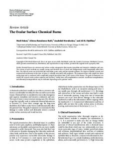
FIGURE 1. Box plot representing the distribution of higher-order (HO) aberration types after wearing the Boston Ocular Surface Prosthesis in 4 clinical groups, including the keratoconus (group 1), post–penetrating keratoplasty (group 2), post–refractive surgery (group 3), and ocular surface disease (group 4). It includes the most extreme values (*), outliers (X), maximum and minimum values, and the median value of the data set. The horizontal line on the graph is the cutoff point (0.5 "m) for clinically significant HO aberration. Post hoc Tukey P values between the groups were: coma (logarithmic), not significant; trefoil, 0.041 (between groups 1 and 4); tetrafoil (logarithmic), not significant; spherical, not significant; HO astigmatism, 0.050 (between group 1 and 3). RMS ! root mean square.
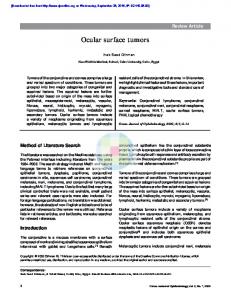
FIGURE 2. Box plot representing the distribution of higher-order (HO) aberration types after wearing the Boston Ocular Surface Prosthesis in 4 clinical groups, including keratoconus (group 1), post–penetrating keratoplasty (group 2), post–refractive surgery (group 3), and ocular surface disease (group 4). It includes the most extreme values (*), outliers (X), maximum and minimum values, and the median value of the data set. The middle line on the graph is the cutoff point (0.5 "m) for clinically significant HO aberration. Post hoc Tukey P values between the groups were: coma (logarithmic), 0.023 (between groups 1 and 4); trefoil, not significant; tetrafoil (logarithmic), not significant; spherical, not significant; HO astigmatism, not significant. RMS ! root mean square.
684
AMERICAN JOURNAL
OF
OPHTHALMOLOGY
APRIL
2011
FIGURE 3. Box plot graph presenting the change in higher-order (HO) root mean square (RMS) error values at baseline and while wearing the Boston Ocular Surface Prosthesis (BOSP) in all groups. It includes the most extreme values (*), outliers (X), maximum and minimum values, and the median value of the data set. P < .001, P < .001, P < .002, and P < .001 for each of the 4 groups, respectively.
cylindrical refractive errors, both total and higher-order aberration error values were recorded in each measurement. The OPD-Scan optical path scanning system generates a wavefront higher-order aberration map that displays specific higher-order aberration components, extracted from the total wavefront map. Moreover, this map illustrates the location and degree of higher-order aberrations in the eye. ● STATISTICAL ANALYSIS: SPSS software version 15.0 for Windows evaluation version (LEAD Technologies, Inc, Chicago, Illinois, USA) was used for statistical analysis. Differences between the groups in terms of age and sex distribution were analyzed using a 1-way analysis of variance and chi-square tests, respectively. Pairedsamples t tests were performed to evaluate the differences in study parameters before and after wearing the Boston Ocular Surface Prosthesis. A repeated-measures of analysis of variance was carried out to compare the difference in study parameters at baseline and while wearing the Boston Ocular Surface Prosthesis between the groups. A P value ! .05 was considered to be statistically significant.
VOL. 151, NO. 4
EFFECTIVE CORRECTION
OF
RESULTS IN THE OVERALL PATIENT GROUP, THE MEAN AGE OF 39
patients was 46.9 years, ranging from 23 to 75 years. There was no statistically significant difference between the 4 groups in terms of age and sex distribution (P & .05). When the patients were analyzed as a whole, statistically significant differences in all study parameters, including logarithmic BCVA, total and higher-order wavefront errors, spherical errors, cylindrical refractive errors, and spherical equivalent were noted before and after wearing the Boston Ocular Surface Prosthesis. Results of study parameters in 4 groups at baseline and while wearing the Boston Ocular Surface Prosthesis are provided in Table 1. BCVA increased significantly in all groups with the Boston Ocular Surface Prostheses ($ 0.001, 0.001, 0.006, 0.001, respectively). The mean improvement in BCVA was at least 3 lines. There was a statistically significant reduction in spherical error in eyes wearing the Boston Ocular Surface Prosthesis in all groups ($ 0.001, $ 0.001, 0.035, respectively) except for group 4 (P ! .229). A significant reduction in cylindrical errors was observed in all groups (all P ! .001). Only groups 1
HO ABERRATIONS
WITH THE
BOSP
685
FIGURE 4. Box plot graph presenting the change of total spherical aberration root mean square (RMS) error at baseline and while wearing the Boston Ocular Surface Prosthesis (BOSP) in 2 groups with and without eccentricity. It includes the most extreme values (*), outliers (X), maximum and minimum values, and the median value of the data set. P ! .05 between these 2 groups.
and 2 had a significant change (P $ .001) in spherical equivalent. The graphs showing which types of higher-order aberrations were predominant at baseline and while wearing the Boston Ocular Surface Prosthesis in both groups are provided in Figures 1 and 2. Mean values of trefoil and coma were more than 0.5 'm in the keratoconus group (P & .05), and trefoil was above this threshold in the post-PK and post–refractive surgery groups. At baseline, the higher-order aberrations accounted for 32.4% of the total aberration in the entire group. In the 4 different disease groups, these values were 25.1%, 20.7%, 44.1%, and 39.6%, respectively. The change in higher-order error values is provided in Figure 3. The ratio of higher-order error reduction that could be achieved by the Boston Ocular Surface Prosthesis wearing was the highest in keratoconus eyes, as much as 77.1%. Details of both higher-order and total aberration error reduction are provided in Table 1. In a separate analysis, we evaluated whether there was any difference in the magnitude change in wavefront aberrations with the Boston Ocular Surface Prosthesis among the 4 different disease groups. Based on the post hoc analysis, there were statistically significant differences in terms of higher-order error only between groups 1 and 4 (P ! .012) and between groups 1 and 2 (P ! .033). For total error, the difference reached statistical significance between group 1 and groups 2, 3, and 4 (group 1 vs group 686
AMERICAN JOURNAL
2, P ! .041; group 1 vs group 3, P ! .001; and group 1 vs group 4, P $ .001). Another analysis was performed to determine if there were differences between eyes fit with lenses with eccentricity compared with those without. No statistically significant difference was found in BCVA (P ! .745), total (P ! .176), or higher-order aberration error values (P ! .993) between these 2 groups. However, a significantly higher reduction in total spherical aberration was observed in the group with eccentricity (correction, 0.32 " 0.36) compared with those without eccentricity (correction, 0.12 " 0.33; P ! .05; Figure 4). For comparative purposes, higher-order and total aberration errors were measured in eyes wearing RGP corneal contact lenses. In 6 eyes wearing RGP corneal contact lenses, 2 eyes revealed increased higher-order aberrations, whereas the mean correction rate in the other 4 eyes was found to be 42.5% (range, 33.3% to 48.2%). However, the 6 eyes wearing RGP corneal contact lenses revealed a 73.9% reduction (range, 47.7% to 92.6%) in total aberration.
DISCUSSION THIS STUDY EVALUATED THE IMPACT OF THE BOSTON
Ocular Surface Prosthesis on visual acuity, spherical and cylindrical refractive errors, and total and higher-order OF
OPHTHALMOLOGY
APRIL
2011
aberrations in challenging patients with regular and irregular astigmatism resulting from corneal or ocular surface diseases or after corneal surgery that were refractory to conventional means of optical correction. In all these groups, the Boston Ocular Surface Prosthesis was found to be effective in providing satisfactory visual and refractive outcomes, including significantly improved BCVA and decreased total and higher-order wavefront aberration errors. The integrity and smoothness of the ocular surface are important factors for providing high-quality visual acuity.8,9 Some disorders such as keratoconus,10 severe ocular surface disease,11 and some undesirable postoperative outcomes after penetrating keratoplasty12 or corneal refractive surgery13,14 may result in corneal surface irregularity, which in turn results in increased higher-order wavefront aberrations. Keratoconic eyes were found to have higher total higher-order aberrations in both photopic and scotopic conditions.10 Previous aberrometry studies have reported that coma is the dominant higher-order aberration in keratoconic eyes resulting from topographic abnormalities of the anterior corneal surface.10,15 Additionally, increases in trefoil, tetrafoil, and secondary astigmatism were observed in keratoconic eyes. Consistent with the previously reported studies, we observed trefoil and coma as the most elevated higher-order aberrations in keratoconic eyes, with nonsignificantly higher mean values of trefoil than coma. The finding of higher levels of coma in the previous studies perhaps can be attributed to different methodologies (dynamic skiascopy vs Hartmann-Shack), pupil size, patient age, and location of the cones. Most patients who have undergone PK report extreme visual distortion.12 These patients, typically have excessive amounts of against-the-rule and asymmetric astigmatism. Moreover, this asymmetry generally results in large amounts of coma higher-order aberration.16 In another recent study, the authors concluded that post-PK eyes had approximately 5.5 times more higher-order aberrations, such as spherical aberration and coma, than normal eyes.15 However, they found that the dominant higher-order aberration in eyes after PK was trefoil, probably the result of suture tension.15 Similarly, trefoil was the most common higher-order aberration in post-PK eyes in the current study. Although most people are satisfied with the improvement of visual acuity after refractive surgery and they do not require spectacles or contact lens for their daily activities, some may experience bothersome visual symptoms such as decreased night vision, glare, halos, or monocular diplopia. Others may have visual problems resulting from dry eye or ectasia after LASIK. These symptoms were found to be associated with increased, measured higher-order wavefront aberrations resulting from corneal surface irregularities,14,17,18 postsurgical wound healing,19 and decentered laser treatments.20 It has been documented that refractive surgeries may cause inVOL. 151, NO. 4
EFFECTIVE CORRECTION
OF
creased spherical-like aberrations that correlate with the amount of refractive correction.21 In our study, although the spherical aberration was higher than measured in the other clinical groups, trefoil was found to be the predominant higher-order aberration in the eyes that had undergone refractive surgery. Ocular surface disorders accompanied by changes in tear volume and tear fluid dynamics can induce higher-order aberrations even if the corneal shape is completely normal.11,22,23 For instance, Lin and associates concluded that disruption of the tear film increased anterior corneal higher-order aberrations in normal eyes and more rapidly in dry eyes.22 In the current study, ocular surface disease group was found to have increased third-order higher-order aberrations such as coma and trefoil. In the above-mentioned cornea and ocular surface conditions, treatments aimed at minimizing or eliminating both high irregular astigmatism and increased higher-order wavefront aberrations is still a frustrating task for clinicians. Conventional treatment strategies, such as spectacles and soft contact lenses, may improve vision in such individuals; however, they become less tolerated and satisfactory as the disease progresses. In severe progressive keratoconus and post-LASIK ecstatic corneas, although new invasive treatment methods including collagen crosslinking, intracorneal ring implantations, or both have shown promise,5,6 noninvasive treatment strategies should be considered initially. Small-diameter RGP corneal contact lenses, largediameter inverse geometry RGPs, corneoscleral lenses, scleral lenses, or hybrid lenses with spherical or toric optics are highly indicated in patients with high irregular astigmatism and higher-order aberrations.16,24,25 These contact lenses with the underlying tear film can optically neutralize the anterior cornea, which is responsible most of the higher-order aberrations, and therefore can counteract the optically degrading corneal surface irregularity.26 However, these lenses are not always effective in eyes with higher degrees of regular and irregular astigmatism. Moreover, not all patients can tolerate the discomfort associated with rigid cornealbearing contact lenses. RGP corneal contact lens can be considered in many patients with moderate and severe keratoconus and irregular astigmatism resulting from other conditions. However, although anterior corneal aberrations can be almost extinguished by RGP corneal contact lenses in keratoconic eyes, wavefront higher-order aberrations derived from the posterior surface of the cornea may be affected or may be unmasked.27,28 This is supported by our finding of increased higher-order aberrations in 2 keratoconic eyes wearing RGP corneal contact lenses in our study. Patients who have improved vision but cannot tolerate the discomfort of an RGP corneal contact lens may benefit from hybrid lenses, such as Synergeyes (SynergEyes, Carls-
HO ABERRATIONS
WITH THE
BOSP
687
bad, California, USA).16,29 When visual performance is taken into account, the Boston Ocular Surface Prosthesis also is an excellent option, because they do not touch the cornea and they have excellent centration and no blinkassociated lens movement. Because of the less-than-perfect correction of higherorder aberrations with soft and rigid contact lenses in some cases, the concept of correcting higher-order aberrations with custom-made soft and rigid contact lenses has begun to gain interest. For instance, Tan and colleagues evaluated the efficacy of custom-made RGP contact lenses to treat increased wavefront aberrations after LASIK correction of myopia, and they concluded that although total higher-order aberrations were significantly reduced with lens wear and at least a 70% reduction in third- and fourth-order aberration was observed, only a 33% reduction in fifth-order aberrations could be achieved.25 In another series of anterior surface higher-order aberration– correcting hydrogel lenses used in keratoconic eyes, the authors concluded that these lenses could reduce higherorder aberration error by an average factor of 3, and yielded an improvement in BCVA of approximately 2 lines compared with standard spherocylindrical hydrogel lenses.30 In another study that compared the vision of 3 keratoconic eyes wearing wavefront-customized hydrogel lenses with those with their habitual RGP corneal contact lenses, custom wavefront-guided soft contact lenses were demonstrated to provide favorable results.31 In a more recent study by Katsoulos and associates, visual performance of custom hydrogel contact lenses for keratoconus incorporating correction for vertical coma aberration was investigated.32 The authors concluded that these lenses could improve both monocular and binocular visual performance in keratoconic eyes. They also found that total higherorder aberration root mean square values were reduced from 0.86 " 0.15 to 0.57 " 0.17 and vertical coma aberration was reduced from %0.64 " 0.21 to %0.29 " 0.23 for a 4-mm pupil.32 In the current study, we evaluated patients who decided to wear the Boston Ocular Surface Prosthesis for visual correction because they had not responded satisfactorily to other treatments. Previously, the safety and efficacy of the Boston Ocular Surface Prosthesis has been reported in different patient groups including keratoconus, StevensJohnson syndrome, severe dry eye, chronic ocular graftversus-host disease, and eyes that have undergone PK and LASIK.7,33–37 In the literature, there are limited data about the impact of the Boston Ocular Surface Prosthesis on higher-order aberrations in challenging cases with severe keratoconus or ocular surface disease, in eyes that have undergone PK, or in eyes with corneal irregularity occurring after refractive surgery. When the data were evaluated either in the entire group or in the 4 disease groups, a statistically significant
688
AMERICAN JOURNAL
decrease in the wavefront error was noted in eyes wearing the Boston Ocular Surface Prostheses. Correction rates of higher-order aberration errors with the Boston Ocular Surface Prosthesis were 77.1%, 72.7%, 69.2%, and 70.1% in the 4 different clinical groups, respectively. Moreover, this rate was noted to be as high as 96.2% in some cases. In contrast, 2 eyes wearing conventional RGP lenses showed increased higher-order aberration errors compared with baseline, whereas the mean correction in the other 4 RGP-wearing eyes was only 42.5%. Potential reasons for this imperfect correction with RGP lenses perhaps can be attributed to lens rotation, decentration with blinking, and less than uniform postlens tear film. Regarding BCVA, the Boston Ocular Surface Prosthesis revealed statistically significant improvement in acuity of at least 3 lines in all patient groups. Magnitude of improvement in BCVA was similar to that reported by Stason and associates.7 They concluded that the Boston Ocular Surface Prosthesis significantly improved both visual acuity and visual function in patients with corneal ectasia, irregular astigmatism, and ocular surface disease for whom conventional therapies had failed.7 Aspheric rigid lenses are used to align better the base curve-to-cornea fitting relationship, to decrease spherical aberration, and to correct for residual astigmatism. The degree of asphericity often is characterized in terms of an eccentricity or “e” value. The higher the eccentricity, the more quickly the lens flattens from the center to the periphery.38 A spherical lens has an “e” value of 0. The Boston Ocular Surface Prosthesis is available with eccentricity values of 0.3, 0.6, and 0.8. Adding eccentricity was attempted during fitting to improve visual acuity and to decrease spherical aberrations. In the current study, it was documented that eyes with the Boston Ocular Surface Prosthesis containing eccentricity had higher magnitudes of total spherical aberration correction (86%) compared with those without eccentricity (66%). However, no statistically significant difference in BCVA or total and higher-order error values were noted between eyes fit with the Boston Ocular Surface Prosthesis containing eccentricity and those without. This may be attributed to the lower numbers of eyes fit with eccentricity. In another study by Gemoules and Morris, the correction of total spherical aberration using semiscleral RGP lenses was found to be 82% in highly symptomatic patients who had undergone refractive surgery.39 The Boston Ocular Surface Prosthesis should be considered as an effective tool for minimizing irregular astigmatism and higher-order aberrations and for significantly improving BCVA in eyes with corneal surface irregularity. The relative merits of the Boston Ocular Surface Prosthesis compared with other lens designs for treatment of these conditions remains to be determined by randomized, controlled clinical trials.
OF
OPHTHALMOLOGY
APRIL
2011
PUBLICATION OF THIS ARTICLE WAS SUPPORTED BY GRANT EY11915 (S.C.P.) FROM THE NATIONAL INSTITUTES OF HEALTH, Bethesda, Maryland; an unrestricted grant from Research to Prevent Blindness, Inc, New York, New York; the Oshman Foundation, Houston, Texas; the William Stamps Farish Fund, Houston, Texas; Hamill Foundation, Houston, Texas; and the Scientific and Technological Research Council of Turkey, Ankara, Turkey (KG). Dr Pflugfelder is a consultant for Alcon, Allergan, and Glaxo SmithKline. He serves on the speakers bureaus of Allergan and Alcon and receives research grants from Allergan, Inspire, and Glaxo SmithKine. The other authors have no financial interests to disclose. Involved in Design and conduct of study (S.C.P., K.G.); Collection of data (S.C.P., K.G., A.G.); Management (K.G., S.C.P.), analysis (K.G., S.C.P.), and interpretation (K.G., S.C.P., A.G.) of the data; and Preparation, review, and approval of the manuscript (K.G., S.C.P.). This study was approved by the Baylor College of Medicine, Institutional Review Board. The research protocol adhered to the tenets of the Declaration of Helsinki for clinical research. Written informed consent was obtained from all the participants after explanation of the purpose and possible consequences of the study. The authors have had full access to all the data in the study and take responsibility for the integrity of the data and the accuracy of the data analysis.
REFERENCES 1. Villa-Collar C, Gonzalez-Meijome JM, Gutierrez-Ortega R. Objective evaluation of the visual benefit in contact lens fitting after complicated LASIK. J Refractive Surg 2009; 25(7):591–598. 2. Jacobs DS. Update on scleral lenses. Curr Opin Ophthalmol 2008;19(4):298 –301. 3. Erdurmus M, Yildiz EH, Abdalla YF, Hammersmith KM, Rapuano CJ, Cohen EJ. Contact lens related quality of life in patients with keratoconus. Eye Contact Lens 2009;35(3):123– 127. 4. Pinero DP, Alio JL, Uceda-Montanes A, El Kady B, Pascual I. Intracorneal ring segment implantation in corneas with post-laser in situ keratomileusis keratectasia. Ophthalmology 2009;116(9):1665–1674. 5. Vinciguerra P, Albe E, Trazza S, Seiler T, Epstein D. Intraoperative and postoperative effects of corneal collagen cross-linking on progressive keratoconus. Arch Ophthalmol 2009;127(10):1258 –1265. 6. Kamburoglu G, Ertan A. Intacs implantation with sequential collagen cross-linking treatment in postoperative LASIK ectasia. J Refract Surg 2008;24(7):726 –729. 7. Stason WB, Razavi M, Jacobs DS, et al. Clinical benefits of the Boston Ocular Surface Prosthesis. Am J Ophthalmol 2010;149(1):54 – 61. 8. Kaido M, Dogru M, Ishida R, Tsubota K. Concept of functional visual acuity and its applications. Cornea 2007; 26(9 Suppl 1):29 –35. 9. Montes-Mico R. Role of tear film in the optical quality of the human eye. J Cataract Refractive Surg 2007;33(9):1631–1635. 10. Maeda N, Fujikado T, Kuroda T, et al. Wavefront aberrations measured with Hartmann-Shack sensor in patients with keratoconus. Ophthalmology 2002;109(11):1996 –2003. 11. Goto E, Ishida R, Kaido M, et al. Optical aberrations and visual disturbances associated with dry eye. Ocul Surf 2006; 4(4):207–213. 12. Karabatsas CH, Cook SD, Sparrow JM. Proposed classification for topographic patterns seen after penetrating keratoplasty. Br J Ophthalmol 1999;83(4):403– 409. 13. Rabinowitz YS. Ectasia after laser in situ keratomileusis. Curr Opin Ophthalmol 2006;17(5):421– 426. 14. Jabbur NS, Sakatani K, O’Brien TP. Survey of complications and recommendations for management in dissatisfied patients seeking a consultation after refractive surgery. J Cataract Refract Surg 2004;30(9):1867–1874. 15. Pantanelli S, MacRae S, Jeong TM, Yoon G. Characterizing the wave aberration in eyes with keratoconus or penetrating
VOL. 151, NO. 4
EFFECTIVE CORRECTION
OF
16.
17.
18.
19.
20.
21.
22.
23.
24.
25.
26.
27.
28.
keratoplasty using a high-dynamic range wavefront sensor. Ophthalmology 2007;114(11):2013–2021. Costas K, Nick V, Lefteris K, Theodore M. Fitting the post-keratoplasty cornea with hydrogel lenses. Cont Lens Anterior Eye 2009;32(1):22–26. Thompson KP, Staver PR, Garcia JR, Burns SA, Webb RH, Stulting RD. Using InterWave aberrometry to measure and improve the quality of vision in LASIK surgery. Ophthalmology 2004;111(7):1368 –1379. Chalita MR, Chavala S, Xu M, et al. Wavefront analysis in post-LASIK eyes and its correlation with visual symptoms, refraction, and topography. Ophthalmology 2004;111(3): 447– 453. Melki SA, Azar DT. LASIK complications: etiology, management, and prevention. Surv Ophthalmol 2001;46(2):95– 116. Yamane N, Miyata K, Samejima T, et al. Ocular higher-order aberrations and contrast sensitivity after conventional laser in situ keratomileusis. Invest Ophthalmol Vis Sci 2004; 45(11):3986 –3990. Oshika T, Miyata K, Tokunaga T, et al. Higher order wavefront aberrations of cornea and magnitude of refractive correction in laser in situ keratomileusis. Ophthalmology 2002;109(6):1154 –1158. Lin YY, Carrel H, Wang IJ, Lin PJ, Hu FR. Effect of tear film break-up on higher order aberrations of the anterior cornea in normal, dry, and post-LASIK eyes. J Refract Surg 2005; 21(5):525–529. Koh S, Maeda N, Kuroda T, et al. Effect of tear film break-up on higher-order aberrations measured with wavefront sensor. Am J Ophthalmol 2002;134(1):115–117. Lim L, Siow KL, Sakamoto R, Chong JS, Tan DT. Reverse geometry contact lens wear after photorefractive keratectomy, radial keratotomy, or penetrating keratoplasty. Cornea 2000;19(3):320 –324. Tan G, Chen X, Xie RZ, et al. Reverse geometry rigid gas permeable contact lens wear reduces high-order aberrations and the associated symptoms in post-LASIK patients. Curr Eye Res 2010;35(1):9 –16. Dorronsoro C, Barbero S, Llorente L, Marcos S. On-eye measurement of optical performance of rigid gas permeable contact lenses based on ocular and corneal aberrometry. Optom Vis Sci 2003;80(2):115–125. Marsack JD, Parker KE, Pesudovs K, Donnelly W, Applegate RA. Uncorrected wavefront error and visual performance during RGP wear in keratoconus. Optom Vis Sci 2007; 84(6):463– 470. Negishi K, Kumanomido T, Utsumi Y, Tsubota K. Effect of higher-order aberrations on visual function in keratoconic
HO ABERRATIONS
WITH THE
BOSP
689
29. 30.
31. 32.
33. 34.
35. Rosenthal P, Cotter JM, Baum J. Treatment of persistent corneal epithelial defect with extended wear of a fluidventilated gas-permeable scleral contact lens. Am J Ophthalmol 2000;130(1):33– 41. 36. Rosenthal P, Croteau A. Fluid-ventilated, gas-permeable scleral contact lens is an effective option for managing severe ocular surface disease and many corneal disorders that would otherwise require penetrating keratoplasty. Eye Contact Lens 2005;31(3):130 –134. 37. Takahide K, Parker PM, Wu M, et al. Use of fluid-ventilated, gas-permeable scleral lens for management of severe keratoconjunctivitis sicca secondary to chronic graft-versus-host disease. Biol Blood Marrow Transplant 2007;13(9):1016 – 1021. 38. Gatinel D, Haouat M, Hoang-Xuan T. A review of mathematical descriptors of corneal asphericity. J Fr Ophtalmol 2002;25(1):81–90. 39. Gemoules G, Morris KM. Rigid gas-permeable contact lenses and severe higher-order aberrations in postsurgical corneas. Eye Contact Lens 2007;33(6 Pt 1):304 –307.
eyes with a rigid gas permeable contact lens. Am J Ophthalmol 2007;144(6):924 –929. Abdalla YF, Elsahn AF, Hammersmith KM, Cohen EJ. SynergEyes lenses for keratoconus. Cornea 2010;29(1):5– 8. Sabesan R, Jeong TM, Carvalho L, Cox IG, Williams DR, Yoon G. Vision improvement by correcting higher order aberrations with customized soft contact lenses in keratoconic eyes. Opt Lett 2007;32(8):1000 –1002. Marsack JD, Parker KE, Applegate RA. Performance of wavefront-guided soft lenses in three keratoconus subjects. Optom Vis Sci 2008;85(12):1172–1178. Katsoulos C, Karageorgiadis L, Vasileiou N, Mousafeiropoulos T, Asimellis G. Customized hydrogel contact lenses for keratoconus incorporating correction for vertical coma aberration. Ophthalmic Physiol Opt 2009;29(3):321–329. Jacobs DS, Rosenthal P. Boston scleral lens prosthetic device for treatment of severe dry eye in chronic graft-versus-host disease. Cornea 2007;26(10):1195–1199. Romero-Rangel T, Stavrou P, Cotter J, et al. Gas-permeable scleral contact lens therapy in ocular surface disease. Am J Ophthalmol 2000;130(1):25–32.
690
AMERICAN JOURNAL
OF
OPHTHALMOLOGY
APRIL
2011
Biosketch Koray Gumus, MD, FEBO, received a medical degree with high honors from Hacettepe University Medical Faculty in Ankara, Turkey, and completed his residency there in Ophthalmology. He joined the faculty of the Department of Ophthalmology at Erciyes University Medical Faculty, Kayseri, Turkey. He is board certified by the European Board of Ophthalmology, Turkish Board of Ophthalmology and International Council of Ophthalmology. He currently works as a postdoc fellow in Ocular Surface Center at Baylor College of Medicine, Houston, Texas. His main research interests include corneal and ocular surface diseases, contact lens, cataract and refractive surgery.
VOL. 151, NO. 4
EFFECTIVE CORRECTION
OF
HO ABERRATIONS
WITH THE
BOSP
690.e1
Biosketch Stephen C. Pflugfelder, MD, joined the faculty of the Cullen Eye Institute of Baylor College of Medicine as a Professor and Director of the Ocular Surface Center in July 2000. He has published over 200 peer-reviewed articles and over 45 book chapters and monographs, primarily in the field of cornea diseases and surgery. Dr Pflugfelder’s research interests include the role of inflammation in dry eye and corneal bioengineering.
690.e2
AMERICAN JOURNAL
OF
OPHTHALMOLOGY
APRIL
2011