Science of the Total Environment 541 (2016) 977–985
Contents lists available at ScienceDirect
Science of the Total Environment journal homepage: www.elsevier.com/locate/scitotenv
The impacts of pharmaceutical drugs under ocean acidification: New data on single and combined long-term effects of carbamazepine on Scrobicularia plana Rosa Freitas a,⁎, Ângela Almeida a, Vânia Calisto b, Cátia Velez a, Anthony Moreira a, Rudolf J. Schneider c, Valdemar I. Esteves b, Frederick J. Wrona d, Etelvina Figueira a, Amadeu M.V. M. Soares a a
Department of Biology & CESAM, University of Aveiro, Aveiro, Portugal Department of Chemistry & CESAM, University of Aveiro, Aveiro, Portugal c BAM Federal Institute for Materials Research and Testing, Richard-Willstaetter Str. 11, Berlin, Germany d Department of Geography, University of Victoria, National Water Research Institute, STN CSC, Victoria, BC, Canada b
H I G H L I G H T S
G R A P H I C A L
A B S T R A C T
• Clams developed mechanisms to prevent oxidative damages when under pH 7.1 • Higher mortality was observed when clams were exposed to CBZ or CBZ + pH7.1. • The toxicity of CBZ increased under seawater acidification conditions (CBZ + pH 7.1) • Oxidative stress was enhanced when clams were exposed to CBZ + pH 7.1
a r t i c l e
i n f o
Article history: Received 21 July 2015 Received in revised form 26 September 2015 Accepted 26 September 2015 Available online xxxx Editor: D. Barcelo Keywords: Ocean acidification Pharmaceuticals Biomarkers Oxidative stress Clams Long-term exposures
a b s t r a c t Ocean acidification and increasing discharges of pharmaceutical contaminants into aquatic systems are among key and/or emerging drivers of environmental change affecting marine ecosystems. A growing body of evidence demonstrates that ocean acidification can have direct and indirect impacts on marine organisms although combined effects with other stressors, namely with pharmaceuticals, have received very little attention to date. The present study aimed to evaluate the impacts of the pharmaceutical drug Carbamazepine and pH 7.1, acting alone and in combination, on the clam Scrobicularia plana. For this, a long-term exposure (28 days) was conducted and a set of oxidative stress markers was investigated. The results obtained showed that S. plana was able to develop mechanisms to prevent oxidative damage when under low pH for a long period, presenting higher survival when exposed to this stressor compared to CBZ or the combination of CBZ with pH 7.1. Furthermore, the toxicity of CBZ on S. plana was synergistically increased under ocean acidification conditions (CBZ + pH 7.1): specimens survival was reduced and oxidative stress was enhanced when compared to single exposures. These findings add to the growing body of evidence that ocean acidification will act to increase the toxicity of CBZ to marine organisms, which has clear implications for coastal benthic ecosystems suffering chronic pollution from pharmaceutical drugs. © 2015 Elsevier B.V. All rights reserved.
⁎ Corresponding author at: Departamento de Biologia, Universidade de Aveiro, Campus Universitário de Santiago, 3810-193 Aveiro, Portugal. E-mail address:
[email protected] (R. Freitas).
http://dx.doi.org/10.1016/j.scitotenv.2015.09.138 0048-9697/© 2015 Elsevier B.V. All rights reserved.
978
R. Freitas et al. / Science of the Total Environment 541 (2016) 977–985
1. Introduction Carbon dioxide (CO2) emitted to the atmosphere by human activities (namely fossil fuel combustion and deforestation) is absorbed by the oceans, causing their acidification (Caldeira and Wickett, 2003; Feely et al., 2009; Orr et al., 2005). The potential impacts of lowering seawater pH has been assessed from the analysis of carefully-controlled laboratory experiments and field studies, where the performance of different species under present and predicted pH conditions have been determined (for review see Nikinmaa, 2013). In fact, among other findings, studies have shown that exposure to acidified seawater caused reductions in growth of the mussel Mytilus galloprovincialis, associated with a reduction in metabolic rate (Michaelidis et al., 2005); decreased survival, development, and growth in hard clams (Mercenaria mercenaria), bay scallops (Argopecten irradians), and eastern oysters (Crassostrea virginica) (Talmage and Gobler, 2010); immune response damage in Chamelea gallina and M. galloprovincialis (Matozzo et al., 2013). Large variation in bivalve sensitivity between species and even between populations of the same species has also been reported (Range et al., 2013). However, Hendriks et al. (2015) recently showed that in highly variable pH environments calcifying organisms have developed the capacity to alter the pH of their calcifying environment, specifically within critical tissues where calcification occurs, achieving homeostasis. In addition to ocean acidification, the increased consumption of pharmaceuticals has also led to great environmental concern. In fact, due to their presence in the aquatic systems and their potential to impact wildlife, pharmaceutical drugs have become an important environmental issue (Fent et al., 2006). To date, most attention has been focused on identification, fate and distribution of active pharmaceutical compounds in the aquatic environment, which are commonly found at very low concentrations, ranging from ng/L to g/L (Fent et al., 2006). Among the most common pharmaceuticals in the environment is carbamazepine (CBZ), an antiepileptic drug (Bahlmann et al., 2012, 2009; Calisto et al., 2011a; Metcalfe et al., 2003). Carbamazepine presents low elimination efficiency in wastewater treatment plants (WWTPs) (below 10%) (Zhang et al., 2008), low resistance to biodegradation (Martín-Diaz et al., 2009), poor attachment onto sludge therefore remaining in the aqueous phase (Zhang et al., 2008; Ternes et al., 2004). For these reasons CBZ frequently occurs in water bodies, namely in WWTP influents and effluents, surface waters, groundwaters and even in treated drinking water, with concentrations ranging from 0.03 to 6.3 μg/L (Ternes, 1998; Sacher et al., 2001; Ferrari et al., 2003; Metcalfe et al., 2003; Bahlmann et al., 2009, 2012; Calisto et al., 2011a). Also, due to CBZ characteristics, several authors have been studying the effects of this drug in aquatic organisms, revealing its capacity to induce oxidative metabolism in trout (Oncorhynchus mykiss) (Gagné et al., 2006); decrease hemocyte lysosome membrane stability in mussel M. galloprovincialis (Martín-Diaz et al., 2009); destabilize lysosomal membrane hemocytes in Ruditapes philippinarum (AguirreMartínez et al., 2013); and cause biochemical alterations in clams Ruditapes decussatus and R. philippinarum (Almeida et al., 2014). To assess the impacts caused in aquatic organisms by natural and anthropogenic environmental changes biomarkers can provide an indication of the sub-lethal impacts of stressors, as well as the underlying biochemical mechanisms, and an “early warning” of possible population impacts. Several stressors induce the production of Reactive Oxygen Species (ROS), namely the superoxide radical, hydrogen peroxide (H2O2) and the hydroxyl radical, all of which mainly affect lipids, proteins, carbohydrates, and nucleic acids. Antioxidant enzymes (e.g. superoxide dismutase, SOD and catalase, CAT) act to prevent oxidative stress by scavenging ROS. Superoxide radicals that are generated are converted to H2O2 by the action of SOD, and the accumulation of H2O2 is prevented in the cell by CAT. When the antioxidant enzymes fail or are insufficient, an increase of ROS production may originate the oxidative degradation of lipids, known as lipid peroxidation (LPO). Although
several studies showed that organic and inorganic contaminants (including metals, organic matter, PAHs, PCBs) are often involved in oxidative stress (among others, Carregosa et al., 2014a, 2014b; Figueira et al., 2012; Freitas et al., 2012; Geret and Bebianno, 2004), few studies used oxidative stress related markers to evaluate the effects of pH decrease and CBZ exposure in marine species (Matozzo et al., 2012; Almeida et al., 2014, respectively). Currently, most studies concerning the impacts on marine organisms caused by ocean acidification and especially pharmaceutical drugs are based on short-term acute toxicity tests (Crane et al., 2006; Halling-Sørensen et al., 1998), which can provide valuable information. However, recent studies suggest that long-term exposures may provide a deeper insight into toxicity, indicating that there is a need for this type of toxicity tests to, more accurately, predict ecological consequences (Crane et al., 2006; Fent et al., 2006; Santos et al., 2010). Furthermore, most studies concerning ocean acidification or pharmaceutical drugs are conducted with single stressors (e.g. Almeida et al., 2014; Aguirre-Martínez et al., 2013; Antunes et al., 2013; Gagné et al., 2006; Gonzalez-Rey and Bebianno, 2012; Martín-Diaz et al., 2009; Murray et al., 2013; Range et al., 2012) and very few adopted the combination of stressors (Campbell et al., 2014; Gobler et al., 2014; Ivanina et al., 2014; Matoo et al., 2013; Matozzo et al., 2013), especially the exposure to pharmaceuticals under ocean acidification conditions. Thus, the aim of the present work was to evaluate the impacts of pH decrease and CBZ, acting alone and in combination, on the marine clam Scrobicularia plana (da Costa, 1778), after a under long-term exposure. For this, the physiological and biochemical alterations induced in clams were assessed. 2. Methodology 2.1. Field sampling and experiment set up Scrobicularia plana was collected in the Ria de Aveiro lagoon (Portugal), in September 2014 to avoid the clams reproductive period. S. plana is an infaunal bivalve commonly inhabiting the intertidal soft bottoms of Northeast Atlantic estuaries, from the Norwegian Sea to the Mediterranean and south to Senegal (Tebble, 1966). Organisms were transported to the laboratory and placed in containers filled with artificial seawater, and acclimated for eight days. During this period temperature was 19 ± 1.0 °C, salinity was maintained at 22 ± 1 g/L, pH ranged between 7.70–7.75, with a photoperiod of 12 h light:12 h dark. Organisms were fed with Algamac Protein Plus (150,000 cells/L/animal) every 2–3 days. After acclimatization, organisms were placed in different glass 20 L aquaria, each one corresponding to a different condition: A — Control (pH 7.8, without carbamazepine), B — Carbamazepine exposure (3.00 μg/L, pH 7.8), C — low pH (pH 7.1, without carbamazepine), D — combining stressors (pH 7.1 and carbamazepine 3.00 μg/L). For each condition 15 individuals were used (5 per container, 3 containers per condition). Only clams with similar size (length 3.7–3.9 cm, width 2.8–3.0 cm) were used for the experiments. Aquaria were filled with sediment (6 L) and artificial seawater (salinity 28 ± 1 g/L) (1:3 v/v). Salinity was adjusted by the addition of artificial sea salt (Tropic Marin® SEA SALT from Tropic Marine Center) to deionized water. The CBZ concentration tested was chosen as representative of those detected in different aquatic systems (Ternes, 1998; Sacher et al., 2001; Ferrari et al., 2003; Metcalfe et al., 2003; Bahlmann et al., 2009, 2012; Calisto et al., 2011a) and because previous studies identified clams response to CBZ at this concentration (Almeida et al., 2014). According to Calisto et al. (2011b), CBZ was detected in the Ria de Aveiro surface water as well as in wastewaters with concentrations ranging between 0.1 and 0.7 μg/L. The tested pH reflected the values found in the Ria de Aveiro lagoon (between 8.0 and 7.6) and the worst predicted scenario of climate change for 2100 (ΔpH = − 0.4). During experiments each aquarium was recirculated and filtered with individual power filters, organisms
R. Freitas et al. / Science of the Total Environment 541 (2016) 977–985
were fed every other day with Algamac protein plus (150,000 cells/L/ animal) and pH continuously monitored and controlled by a pHstat system (Aquamedic AT Controler). The pH was manipulated by introducing CO2 gas bubbles into individual acidified tanks, to achieve testing pH conditions. Also, prior to the experiment initiation, each tank was progressively acidified 0.2 pH units per day, until testing pH value was reached. During the acclimatization (8 days) and exposure (28 days) periods, water in aquaria was completely renewed weekly and CBZ concentration was restored after water renewal. After the exposure period organisms were frozen (−80 °C) for biochemical analysis and CBZ quantification. Salinity, pH and temperature were monitored on a daily basis using specific probes (Hanna Instruments) for each parameter. Along the exposure, water samples were collected from each tank at the beginning and before each water renewal operation. These samples were used to quantify total alkalinity by potentiometric titration, following Gran (1952). Total alkalinity (TA), pH, temperature and salinity determined values were used to determine CO2 partial pressure (pCO2), bicarbonate 2− (HCO− 3 ) and carbonate (CO3 ) ion concentrations, and the saturation states of calcite (ΩCal) and aragonite (ΩAg) for each tank, using CO2SYS software (Robbins et al., 2010) and Mehrbach et al. (1973) refitted by Dickson and Millero (1987) K1 and K2 carbonate dissociation constants, and KSO4 from Dickson (1990). Physico-chemical water parameters and the associated variation for each condition are presented in Table 1. 2.2. Laboratory analysis 2.2.1. Carbamazepine quantification 2.2.1.1. Quality control. To assess the possible losses of CBZ during exposure (namely adsorption of CBZ onto the sediment, photodegradation or adsorption onto aquaria), this drug was quantified in water samples (without clams) with and without sediment (0.30 μg/L of CBZ), collected at the beginning of the experiment and before each water renewal operation. 2.2.1.2. Sample preparation. Carbamazepine was also quantified in clams (S. plana) after a 28 day exposure period to different conditions (CBZ and CBZ + pH). For this quantification, a supernatant of each individual was obtained by extraction of pulverized clam whole soft tissue (0.5 g fresh weight) with deionized water (1:2, w/v). Supernatants were analyzed as they were, with no other treatment (Almeida et al., 2014). During the exposure assay, the concentration of CBZ in water of exposure aquaria was also determined in order to assess the variation of CBZ along the exposure duration. 2.2.1.3. Immunoassay procedure and calibration curve. Carbamazepine was quantified through the application of a direct competitive ELISA (Enzyme-Linked Immunosorbent Assay), following the procedure developed by Bahlmann et al. (2009) and optimized by Calisto et al. (2011b) for water samples collected in the environment. The application of this assay to directly quantify CBZ in clam whole soft tissue is described in Almeida et al. (2014). 2.2.2. Elements quantification Total concentrations of sodium (Na) and potassium (K) were measured in clam soft tissue after 28 days exposure, following the procedure described in Carregosa et al. (2014a). 2.2.3. Physiological and biochemical parameters After exposure (28 days), specimens from each condition were pulverized after being frozen with liquid nitrogen. For each biochemical analysis 0.5 g of soft tissue was used. Extraction was performed with a
979
Table 1 Carbonate system physicochemical parameters for each testing tank. Mean values of measured pH and determined total alkalinity (At), in water samples collected from each tank at the beginning and before each water renewal operation (temperature 19 ± 1 °C and salinity 28 ± 1 g/L). Partial CO2 pressure (pCO2), bicarbonate (HCO− 3 ) and carbonate ion concentrations (CO2− 3 ), and saturation states of calcite (ΩCal) and aragonite (ΩAra), calculated with CO2SYS software (Robbins et al., 2010). Mean ± std. pH at
At at (μmol kg−1)
ƿCO2 at (μatm)
HCO− 3 at (μmol kg-1)
CO32− at (μmol kg−1)
ΩCal at
ΩAra at
Control CBZ pH 7.1 pH 7.1 + CBZ Control CBZ pH 7.1 pH 7.1 + CBZ Control CBZ pH 7.1 pH 7.1 + CBZ Control CBZ pH 7.1 pH 7.1 + CBZ Control CBZ pH 7.1 pH 7.1 + CBZ Control CBZ pH 7.1 pH 7.1 + CBZ Control CBZ pH 7.1 pH 7.1 + CBZ
7.82 ± 0.04 7.82 ± 0.04 7.03 ± 0.06 7.05 ± 0.04 1787 ± 182 1786 ± 124 2220 ± 258 1940 ± 159 650 ± 143 582 ± 114 5252 ± 63 4391 ± 458 1647 ± 186 1480 ± 128 2191 ± 252 1913 ± 283 68.9 ± 3.0 62.0 ± 5.2 14.7 ± 2.7 13.6 ± 3.0 1.78 ± 0.05 1.61 ± 0.11 0.38 ± 0.08 0.36 ± 0.07 1.11 ± 0.04 1.00 ± 0.09 0.24 ± 0.05 0.22 ± 0.05
specific buffer for each physiological and biochemical parameter (Carregosa et al., 2014b). 2.2.3.1. Energy-related parameters. Total protein (PROT) content was determined according to the Biuret spectrophotometric method (Robinson and Hogden, 1940), using bovine serum albumin (BSA) as standard (0–40 mg/mL). The absorbance was read at 540 nm. Results were expressed in mg per g of fresh weight (FW). Following the procedure described by Yoshikawa (1959), glycogen (GLY) was quantified by the phenol–sulfuric acid method. Absorbance was measured at 492 nm. Results were expressed in mg per g of FW. The electron transport activity (ET) was measured based on King and Packard (1975) and modifications performed by De Coen and Janssen (1997). The supernatants were extracted in homogenizing buffer (0.1 M Tris–HCl pH 8.5, 15% (w/v) PVP, 153 mM magnesium sulfate (MgSO4) and 0.2% (v/v) Triton X-100. 35.7 μL of supernatant was incubated on a microplate with 107 μL of buffered substrate solution (0.13 M Tris–HCl, 0.3% (v/v) Triton X-100, pH 8.5), 35.7 μL of NAD(P)H (1.7 mM NADH and 250 μM NADPH). The reaction was started by adding 71.4 μL of 8 mM p-IodoNitroTetrazolium. The absorbance was read at 490 nm during 10 min in 25 s intervals. The amount of formazan formed was calculated using Ɛ = 15,900 M−1cm−1 and results expressed in nmol/min/g FW. 2.2.3.2. Indicators of cellular damage. Lipid peroxidation (LPO) was measured by the quantification of malondialdehyde (MDA), a by-product of lipid peroxidation, according to the method described by Ohkawa et al. (1979) and the modifications referred to by Carregosa et al. (2014b). Absorbance was read at 535 nm (ε = 156 mM− 1 cm− 1). Lipid peroxidation levels were expressed in nmol of MDA formed per g of FW. Reduced (GSH) and oxidized (GSSG) glutathione contents were determined according to Rahman et al. (2006). Absorbance of GSH and GSSG
980
R. Freitas et al. / Science of the Total Environment 541 (2016) 977–985
was read at 412 nm. GSH and GSSG were expressed in μmol per g of FW. The GSH/GSSG ratio was determined. 2.2.3.3. Antioxidant and biotransformation enzymes. Catalase (CAT) activity was quantified according to Johansson and Borg (1988). The standard curve was determined using formaldehyde standards (0– 150 μM). Samples were incubated for 20 min in a shaker, at room temperature. Formaldehyde formation with purpald was measured at 540 nm. The results were expressed in U of g FW. One unit of enzyme (U) is defined as the amount of enzyme that caused the formation of 1.0 nmol formaldehyde, per min, under the assay conditions. Superoxide dismutase (SOD) activity was determined based on the method of Beauchamp and Fridovich (1971). The standard curve was performed with SOD standards (0.25–60 U/mL). SOD activity was measured spectrophotometrically at 560 nm, at room temperature and expressed in U per g of FW. One unit of enzyme (U) corresponds to a reduction of 50% of nitroblue tetrazolium (NBT). The activity of glutathione S-transferase enzymes (GSTs) was determined following an adaptation of the method described by Habig et al. (1974). These enzymes catalyze the conjugation of the substrate 1chloro-2,4-dinitrobenzene (CDNB) with glutathione, forming a thioether. The absorbance was determined at 340 nm, at room temperature, in intervals of 10 s during 5 min. For the enzyme activity quantification the time interval (5 min) was selected, during which the activity was linear. The activity of GSTs was determined using an extinction coefficient of 9.6 mM−1 cm−1 for CDNB. Results were expressed in U per g of FW. One unit of enzyme (U) corresponds to the amount of enzyme that caused the formation of 1 mmol of dinitrophenyl thioether per min under the assay conditions.
Table 2 Carbamazepine (CBZ) concentration determined by ELISA, in water samples (μg/L) from quality control (QC), in water (μg/L) from aquaria where the clams were exposed, and in clams (μg/kg). Samples were obtained after 0 h and before each water renewal (presented as mean values for weekly water changes, during 28 days). For each condition, significant differences (p ≤ 0.05) between exposure periods are presented with letter (a).
Quality control Water Clams
Matrix
0h
Weekly water changes
Water Water + sediment CBZ CBZ + pH CBZ CBZ + pH
2.9 (±0.5)a 3.2 (±0.1)a 3.4 (±0.2)a 3.3 (±0.3)a – –
2.8 (±0.4)a 2.5 (±0.4)a 3.4 (±0.5)a 3.0 (±0.6)a 2.1 (±0.3) 2.0 (±0.4)
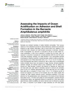
The bioconcentration factor (BCF) determined for each exposure condition after 28 days was 0.7 ± 0.1 for both CBZ and CBZ + pH 7.1, revealing no significant differences between conditions. 3.2. Na and K quantification After a 28 day exposure period, clams exposed to pH 7.1 presented significantly lower Na concentration, when compared to the control condition (Fig. 1A). However, under CBZ and CBZ + pH 7.1 clams presented significantly higher Na concentration than in control (Fig. 1A). A similar pattern was obtained for the concentration of K in clams, although no significant differences were observed between CBZ + pH 7.1 and control (Fig. 1B). 3.3. Physiological and biochemical responses
2.3. Data analysis In order to evaluate CBZ accumulation in clams, the bioconcentration factor (BCF) was calculated in clams exposed to CBZ and CBZ + pH 7.1, dividing the concentration of CBZ present in clam tissue by the spiked CBZ concentration (Gobas and Morrison, 2000). The physiological and biochemical descriptors were submitted to hypothesis testing using permutational multivariate analysis of variance, employing the PERMANOVA+ add-on in PRIMER v6 (Anderson et al., 2008). For each descriptor, significant differences (p ≤ 0.05) among exposure conditions were identified with letters. A matrix gathering the physiological and biochemical descriptors per condition was used to calculate the Euclidean distance similarity matrix. This matrix was simplified through the calculation of the distance among centroids based on the condition (CTL, CBZ, pH 7.1, CBZ + pH 7.1 for 28 days of exposure), which was then submitted to ordination analysis performed by Principal Coordinates (PCO). Pearson correlation vectors of physiological and biochemical descriptors (correlation N0.75) were provided as supplementary variables being superimposed on the PCO graph. 3. Results 3.1. Carbamazepine quantification The CBZ concentration measured in quality control and water samples from the assay are presented in Table 2. Quantifications were done at the beginning of the experiment (0 h) and before each water renewal operation. Regarding the quality control the results showed that no CBZ loss occurred along the exposure both for water and water + sediment samples (Table 2). Similar findings were obtained for water samples from the exposed aquaria, with no losses of CBZ concentration along the exposure (Table 2). The results for CBZ quantification in organism tissue (Table 2) revealed that clams exposed for 28 days did not present significant differences between conditions (CBZ and CBZ + pH 7.1).
3.3.1. Mortality When exposed to CBZ 33% of clams did not survive after a 28 day assay, 11% of them recorded after 96 h of exposure. When organisms were exposed to low pH, 22% did not survive, although in this case the mortality was recorded up to 96 h. Under CBZ + pH 7.1 33% of mortality was observed after 28 days, with no mortality before 96 h of exposure. In the control no mortality was observed during the entire experiment (28 days). 3.3.2. Glycogen, protein content and electron transport activity No significant differences were found in GLY content between control, CBZ and pH 7.1 conditions after 28 days of exposure (cf. Fig. 2A). Clams exposed to CBZ + pH 7.1 significantly decreased the GLY content compared to the remaining conditions. The PROT levels showed that after 28 days of exposure organisms significantly increased the PROT content in all conditions (CBZ, pH 7.1 and CBZ + pH 7.1) when compared to control (Fig. 2B), with no significant differences among CBZ, pH 7.1 and CBZ + pH 7.1 conditions. The ET activity (Fig. 2C) revealed a decrease in all conditions compared to control, with no significant differences among CBZ, pH 7.1 and CBZ + pH 7.1 conditions, although a 30% decrease in ET activity was observed between CBZ and CBZ + pH 7.1. 3.3.3. Indicators of cellular damage The results obtained showed that clams presented lower LPO levels in control than when exposed to pH 7.1, CBZ or the combination of both stressors (Fig. 3A), with significant differences between control and the remaining conditions. After a 28 day exposure period, significantly higher LPO levels were found when organisms were exposed to the combination of CBZ and low pH (cf. Fig. 3A). Although the GSH/GSSG ratio was lower when clams were exposed to different stressful conditions compared to control, no significant differences were found among the tested conditions after a 28 day exposure (Fig. 3B).
R. Freitas et al. / Science of the Total Environment 541 (2016) 977–985
981
Fig. 1. Concentration of (A) Na; and (B) K (mg/g) in Scrobicularia plana. Significant differences (p ≤ 0.05) among conditions are presented with letters (a–c).
3.3.4. Antioxidant enzymes Regarding SOD activity, although a slight increase was noticed in clams exposed to CBZ and CBZ + pH 7.1, clams presented no significant differences among tested conditions (Fig. 4A). CAT activity revealed a significant decrease between control and the remaining conditions (Fig. 4B) with no significant differences among clams exposed to CBZ, pH 7.1 and CBZ + pH 7.1. 3.3.5. Biotransformation enzymes GSTs activity was significantly higher in all exposure conditions compared to control (Fig. 4C), but no significant differences were found among clams exposed to CBZ, pH 7.1 and CBZ + pH 7.1. 3.3.6. Overall analysis The results from PCO analysis (Fig. 5), based on the biochemical parameters, revealed that the first principal component (PCO1), which accounted for 52.9% of total variability, showed a clear distinction between the control (at the negative side) and the exposure conditions CBZ, pH 7.1 and CBZ + pH 7.1 (at the positive side) (Fig. 5). The PCO2 explained 24% of total variance among conditions, with CBZ at the positive side and pH 7.1 at the negative side. The biochemical descriptors (PROT, GLY, LPO, CAT, SOD, GSTs and GSH/GSSG ratio) superimposed on the PCO showed that: organisms under control conditions were characterized by higher ET, CAT and GSH/GSSG levels; organisms exposed to CBZ and pH 7.1 were correlated with higher and lower Na and K levels respectively; organisms exposed to the combination of both stressors were mainly characterized by higher PROT content, and higher LPO and GSTs levels. 4. Discussion Organisms in marine systems are exposed to a combination of multiple stressors creating a range of associated environmental and
Fig. 2. Energy related parameters: (A) GLY, Glycogen content (mg/g fresh weight, FW); (B) PROT, total protein (mg/g fresh weight, FW); and (C) ET, electron transport activity (nmol/min/g FW) mean values (±standard deviation) in Scrobicularia plana. Significant differences (p ≤ 0.05) among conditions are presented with letters (a–b).
ecotoxicological risks. Examples of stressors include alterations in the range and variability of physical and chemical conditions related to alterations in pH and the magnitude and duration of exposure to chemical pollutants arising from human activities (e.g., near-shore municipal/industrial point-source discharges) (Doney et al., 2012; Fabbri, 2015). However, significant scientific uncertainties remain in understanding and ultimately predicting the effects arising from exposure to multiple stressors at relevant levels of biological and ecological organization. In fact, although there is a growing body of evidence that climate change will have broad negative impacts on the behavior and fate of environmental contaminants (Götzea et al., 2014; Schiedek et al., 2007; MacDonald et al., 2005; Wrona et al., 2006), the interactive effects on marine organisms are poorly understood. Therefore, the primary objective of this study was to assess and quantify the biochemical responses of the clam S. plana when exposed to environmentally relevant CBZ concentrations and pH values projected to occur in the near future, alone and in combination. The species S. plana has been used to assess the effects of contaminants in benthic organisms (among others, Solé et al., 2009; Boldina-Cosqueric et al., 2010; Ahmad et al., 2011; Silva et al., 2012; Tankoua et al., 2012) but little is known regarding its tolerance to seawater acidification and pharmaceutical drugs. Most of the
982
R. Freitas et al. / Science of the Total Environment 541 (2016) 977–985
Fig. 5. Centroid ordination diagram (PCO). Pearson correlation vectors, representing biochemical data, are superimposed as supplementary variables(r N 0.75).
Fig. 3. Indicators of celular damage: (A) LPO, lipid peroxidation (nmol/g fresh weight, FW); and (B) GSH/GSSG ratio, mean values (± standard deviation) in Scrobicularia plana. Significant differences (p ≤ 0.05) among conditions are presented with letters (a–c).
Fig. 4. Antioxidant and biotransformation enzymes: (A) SOD, superoxide dismutase (U/g fresh weight, FW); (B) CAT, catalase (U/g fresh weight, FW); and (C) GSTs, glutathione S-transferase (U/g fresh weight, FW) in Scrobicularia plana. Significant differences (p ≤ 0.05) among conditions are presented with letters (a–b).
literature on climate change effects, namely ocean acidification, or pharmaceutical impacts is based on acute exposures that may not be sufficient to allow acclimatization to a new environment or may not induce alterations. Consequently, acute exposures may give a misleading picture of the real impacts of climate change and drugs in the environment. Thus, in the present study, the impacts of pH decrease and CBZ was assessed submitting S. plana to a long-term exposure. The results obtained revealed that after 28 days of exposure clams seem to have acquired ability to adapt to pH 7.1 since mortality was recorded only up to 96 h of exposure. Bamber (1990) reported that the critical acid tolerances of marine bivalves, after 30 days exposure, could range from pH 6.6 in Mytilus edulis to pH 6.0 in the oyster Crassostrea gigas, which are values lower than the pH tested in the present study. As shown by Stumpp et al. (2012) and Thomsen et al. (2013) the impact of elevated pCO2 in an organism will reflect the magnitude and duration of exposure and the physiological capacity of that organism to regulate tissue pCO2. In fact, whilst exposure to elevated pCO2 has been reported to cause negative impacts in key marine invertebrates (Pörtner, 2008) others have shown that some species can thrive in the field in unfavorable pH environments (Thomsen et al., 2013). Species that might be resilient to elevated pCO2 have been predicted to be metabolically active to compensate acid–base disturbances (Fabry et al., 2008; Widdicombe and Spicer, 2008). These species have the capacity to compensate the increase of H+ concentration, by excretion of acid equivalents (e.g. H+) or by increasing the intracellular buffering capacity via the import of buffers (e.g. HCO3−) to compensate for the intracellular acidosis. Acid–base equivalent transport is facilitated through primary active transporters (e.g. V-type H+-ATPase) or ion transporters such as Na+/H+ exchangers (NHEs) and Na+-dependent HCO3− transporters of the SLC4 transporter family (Boron, 2004; Romero et al., 2004). The activity of these transporters lowers H+ concentration with the concomitant increase of Na+. After 28 days of exposure clams exposed to pH 7.1 decreased the Na+ concentration, indicating that organisms may achieve the acid–base stability which is in accordance with mortality data (22% of mortality up to 96 h of exposure and 0% of mortality afterwards). Maintenance of intracellular pH is essential for countless cellular functions and regulations (Putnam and Roos, 1997) and, in general, intracellular pH is regulated at 0.5–0.8 pH units below extracellular pH (Pörtner et al., 2004). Decreases in intracellular pH appear to be almost fully compensated for after 24– 48 h exposure in a wide range of marine invertebrates (Lindinger et al., 1984). Recent studies also confirmed that calcifying organisms can biologically control the environment of carbonate deposition controlling the pH in extracellular fluid, or by controlling deposition in a regulated, intracellular environment (Hendriks et al., 2015). Exposure to CBZ (essentially at pH 7.8) led to the increase in Na+ concentration, indicating that this drug may affect the cellular acid– base homeostasis. In fact, acidosis was reported as a side-effect of CBZ
R. Freitas et al. / Science of the Total Environment 541 (2016) 977–985
overdose (Hughes et al., 1985). Higher mortality obtained when clams were exposed to CBZ and especially to CBZ + pH 7.1 compared with pH 7.1 supports the indication that these clams are capable of adapting to low pH. Furthermore, since under CBZ + pH 7.1 mortality was only recorded after 96 h of exposure, our work may suggest that under the most stressful condition (CBZ + pH 7.1, where clams showed higher LPO levels) clams may have the ability to develop response mechanisms at the beginning of the exposure which are not sufficient to protect clams from the impacts after a longer exposure period. Recently Almeida et al. (2014) showed that, although CBZ may induce physiological and biochemical alterations in organisms exposed to environmentally relevant concentrations, no mortality was induced in R. philippinarum and R. decussatus after a 96 h exposure period. Thus, our findings may also indicate that longer exposures to CBZ may cause stronger impacts and this might cause higher mortality rates. Nevertheless, no significant mortality was recorded in the freshwater clam Corbicula fluminea exposed to CBZ (50 μg/L), during 21 days (Aguirre-Martínez et al., 2015). The present study showed that under CBZ + pH 7.1 the glycogen content was significantly lower when compared to the remaining conditions, but clams exposed to CBZ and low pH tended to maintain this energy reserve when compared to control conditions, indicating that when under these conditions clams have the ability to preserve their energy reserves as a defense mechanism. Previous works revealed that bivalves exposed to stressful conditions during short periods tend to close their valves, reducing oxygen consumption and, consequently, reducing energy expense. Range et al. (2013) demonstrated that seawater acidification had a negative effect on the feeding and digestive behavior of R. decussatus decreasing clearance rate and ingestion rate in clams exposed to low pH (7.4) during 7 days. Studying the effects of an acute exposure of CBZ on R. philippinarum and R. decussatus, Almeida et al. (2014) also demonstrated that the glycogen content, in both species, increased when compared to control conditions after a 96 h exposure assay, which could be associated to clams valve closure and, consequently, clearance rate decrease. Nevertheless, our findings may indicate that clams under the most stressful condition (CBZ + pH 7.1), for a long period (28 days), were not able to keep this defense mechanism leading to the use of their energy reserves. Regarding protein content, our findings showed that values were significantly higher at all exposure conditions compared to control. It is known that if compensation of acid–base imbalance is not achieved, some species may depress their metabolism (Pörtner et al., 1998; Guppy and Withers, 1999). In fact, metabolic suppression is considered a strategy for acid–base homeostasis which is typically achieved by shutting down expensive processes, mainly protein synthesis (Pörtner et al., 2005). However, results obtained in the present work may indicate that the suppression of proteins was not a mechanism used by S. plana to achieve acid–base homeostasis since the protein content significantly increased in all exposure conditions compared to control. The increase in protein content in clams under stressful conditions may be related to the higher rate of enzyme synthesis (namely antioxidant and biotransformation enzymes) used to prevent oxidative damage. In fact, our study revealed that the GSTs activity increased at all exposure conditions compared to the control which may result from higher protein production. Furthermore, although our results showed that CAT and SOD activities did not increase in clams exposed to CBZ, pH 7.1 and CBZ + pH 7.1 this does not mean that the amount of these enzymes was not enhanced. Increased protein content was also found in R. decussatus after a 96 h exposure to CBZ (Almeida et al., 2014). The exposure to contaminants can increase energy demands, with the concomitant increase in electron transport activity in mitochondria, for the maintenance of homeostasis, survival and reproduction (Gagné et al., 2006). However, in our study, after a long exposure period the ET decreased in all conditions compared to the control which can indicate that clams presented a lower metabolic rate as a defense mechanism. This may explain why clams exposed to stressful conditions did not strongly reduce their energy reserves, i.e. glycogen.
983
In accordance with ET, glycogen and protein content results, our study further revealed that clams showed higher LPO when under pH 7.1, CBZ and especially when exposed to CBZ + pH 7.1 compared to control. In previous studies lipid peroxidation has been used as a biomarker for increased oxidative stress due to CBZ and low pH. An induction of LPO levels was found in rainbow trout (O. mykiss) hepatocytes (Gagné et al., 2006), in all tissues of the crab Carcinus maenas (Aguirre-Martínez et al., 2013), and in Corbicula fluminea (Chen et al., 2014) exposed to CBZ. Concerning the effects of elevated CO2 on the lipid peroxidation our study revealed that although presenting higher levels than in control, clams showed lower LPO at pH 7.1 than under CBZ and CBZ + pH 7.1. These results corroborate the idea that after longer exposure periods clams may have the capacity to adapt to low pH conditions. Similar findings were obtained by Matoo et al. (2013) that showed that when exposed to hypercapnia conditions (i.e., elevated CO2) the MDA in the clam M. mercenaria significantly increased when compared to control conditions, despite the fact that MDA levels decreased after 8 and 15 weeks of hypercapnia exposure compared to 2 weeks of experiment. Our findings further demonstrated that, when exposed to different stressors, S. plana maintained or significantly decreased the activity of antioxidant enzymes SOD and CAT, respectively. These results may explain the higher LPO levels obtained, when S. plana was exposed to CBZ, low pH and especially when under the combination of both stressors compared to control. Previous studies also demonstrated that antioxidant enzymes were not activated or even decreased their activity after exposure to CBZ, as was the case of the O. mykiss trout (Li et al., 2009, 2010). These authors showed significantly lower CAT activity compared to control after a 21 day exposure period and with a prolonged exposure (42 days), SOD activity was seriously inhibited. Almeida et al. (2014) also found that, in R. decussatus, CAT activity decreased at 3.0 μg/L of CBZ relative to control. Studies with other pharmaceutical drugs revealed similar results, including the decrease of SOD activity in R. philippinarum exposed to the non-steroidal anti-inflammatory drug ibuprofen (Milan et al., 2013); and an inhibition tendency in CAT activity over time in the mussel M. galloprovincialis exposed to the same drug (Gonzalez-Rey and Bebianno, 2012). Concerning impacts of ocean acidification, previous studies conducted by Matoo et al. (2013) showed that prolonged exposure (15 weeks) to elevated CO2 levels led to a significant decrease of total antioxidant capacity in M. mercenaria tissues. Regarding GSTs, the results obtained are in accordance with other studies, where an increased activity of these enzymes was demonstrated in R. philippinarum exposed to paracetamol for 96 h (Antunes et al., 2013) and in R. decussatus exposed to CBZ during the same period (Almeida et al., 2014). In conclusion, the present findings indicate that the interactions between climate-related abiotic change and pharmaceuticals may alter the organisms sensitivity and/or may enhance the toxicity of the pharmaceutical drug CBZ. The present study further demonstrated that long-term exposures may allow bivalves to develop mechanisms to tolerate low pH conditions which may not be sufficient to prevent oxidative damage in organisms exposed to CBZ and especially CBZ under seawater acidification conditions.
Acknowledgments This work was supported by European Funds through COMPETE and by National Funds through the Portuguese Science Foundation (FCT) within project PEst-C/MAR/LA0017/2013. Rosa Freitas and Vânia Calisto benefited from post-doc grants (SFRH/BPD/92258/2013 and SFRH/BPD/ 78645/2011, respectively) funded by the Portuguese Fundação para a Ciência e Tecnologia (FCT). Cátia Velez and Anthony Moreira benefited from PhD grants (SFRH/BD/86356/2012 and SFRH/BD/93107/2013, respectively) funded by the FCT.
984
R. Freitas et al. / Science of the Total Environment 541 (2016) 977–985
References Aguirre-Martínez, G.V., Del Valls, T.A., Martín-Díaz, M.L., 2013. Early responses measured in the brachyuran crab Carcinus maenas exposed to carbamazepine and novobiocin: application of a 2-tier approach. Ecotoxicol. Environ. Saf. 97, 47–58. Aguirre-Martínez, G.V., Del Valls, T.A., Martin-Diaz, M.L., 2015. Yes, caffeine, ibuprofen, carbamazepine, novobiocin and tamoxifen have an effect on Corbicula fluminea (Müller, 1774). Ecotoxicol. Environ. Saf. 120, 142–154. Ahmad, I., Mohmood, I., Mieiro, C.L., Coelho, J.P., Pacheco, M., Santos, M.A., Duarte, A.C., Pereira, E., 2011. Lipid peroxidation vs. antioxidant modulation in the bivalve Scrobicularia plana in response to environmental mercury–organ specificities and age effect. Aquat. Toxicol. 103, 150–158. Almeida, A., Calisto, V., Esteves, V., Soares, A.M.V.M., Figueira, E., Freitas, R., 2014. Presence of carbamazepine in coastal systems: effects on bivalves. Aquat. Toxicol. 156, 74–87. Anderson, M.J., Gorley, R.N., Clarke, K.R., 2008. PERMANOVA+ for PRIMER: Guide to Software and Statistical Methods. Primer-e, Plymouth, UK, pp. 1–214. Antunes, S.C., Freitas, R., Figueira, E., Gonçalves, F., Nunes, B., 2013. Biochemical effects of acetaminophen in aquatic species: edible clams Venerupis decussata and Venerupis philippinarum. Environ. Sci. Pollut. Res. 20 (9), 6658–6666. Bahlmann, A., Carvalho, J.J., Weller, M.G., Panne, U., Schneider, R.J., 2012. Immunoassays as high-throughput tools: monitoring spatial and temporal variations of carbamazepine, caffeine and cetirizine in surface and wastewaters. Chemosphere 89 (11), 1278–1286. Bahlmann, A., Weller, M.G., Panne, U., Schneider, R.J., 2009. Monitoring carbamazepine in surface and wastewaters by an immunoassay based on a monoclonal antibody. Anal. Bioanal. Chem. 395 (6), 1809–1820. Bamber, R.N., 1990. The effects of acidic sea water on three species of lamellibranch mollusks. J. Exp. Mar. Biol. Ecol. 143, 181–191. Beauchamp, C., Fridovich, I., 1971. Superoxide dismutase: improved assays and an assay applicable to acrylamide gels. Anal. Biochem. 44, 276–287. Boldina-Cosqueric, I., Amiard, J.C., Amiard-Triquet, C., Dedourge-Geffard, O., Métais, I., Mouneyrac, C., Moutel, B., Berthet, B., 2010. Biochemical, physiological and behavioural markers in the endobenthic bivalve Scrobicularia plana as tools for the assessment of estuarine sediment quality. Ecotoxicol. Environ. Saf. 73, 1733–1741. Boron, W.F., 2004. Regulation of intracellular pH. Adv. Physiol. Educ. 28 (1–4), 160–179. Caldeira, K., Wickett, M.E., 2003. Anthropogenic carbon and ocean pH. Nature 425, 365-365. Calisto, V., Domingues, M.R.M., Esteves, V.I., 2011a. Photodegradation of psychiatric pharmaceuticals in aquatic environments — kinetics and photodegradation products. Water Res. 45 (18), 6097–6106. Calisto, V., Domingues, M.R.M., Erny, G.L., Esteves, V.I., 2011b. Direct photodegradation of carbamazepine followed by micellar electrokinetic chromatography and mass spectrometry. Water Res. 45, 1095–1104. Campbell, A.L., Mangan, S., Ellis, R.P., Lewis, C., 2014. Ocean acidification increases copper toxicity to the early life history stages of the polychaete Arenicola marina in artificial seawater. Environ. Sci. Technol. 48 (16), 9745–9753. Carregosa, V., Figueira, E., Gil, A.M., Pereira, S., Pinto, J., Soares, A.M.V.M., Freitas, R., 2014a. Tolerance of Venerupis philippinarum to salinity: osmotic and metabolic aspects. Comp. Biochem. Physiol. A 171, 36–43. Carregosa, V., Velez, C., Soares, A.M.V.M., Figueira, E., Freitas, R., 2014b. Physiological and biochemical responses of three veneridae clams exposed to salinity changes. Comp. Biochem. Physiol. B 177-178, 1–9. Chen, H., Zha, J., Liang, X., Li, J., Wang, Z., 2014. Effects of the human antiepileptic drug carbamazepine on the behavior, biomarkers, and heat shock proteins in the Asian clam Corbicula fluminea. Aquat. Toxicol. 155, 1–8. Crane, M., Watts, C., Boucard, T., 2006. Chronic aquatic environmental risks from exposure to human pharmaceuticals. Sci. Total Environ. 367, 23–41. De Coen, W.M., Janssen, C.R., 1997. The use of biomarkers in Daphnia magna toxicity testing. IV. Cellular energy allocation: a new methodology to assess the energy budget of toxicant-stressed Daphnia populations. J. Aquat. Ecosyst. Stress. Recover. 6, 43–55. Dickson, A.G., 1990. Standard potential of the reaction AgCls + 1/2 H2 = Ags + HClAq and the standard acidity constant of the ion HSO4 − in synthetic sea-water from 273.15-K to 318.15-K. J. Chem. Thermodyn. 22, 113–127. Dickson, A.G., Millero, F.J., 1987. A comparison of the equilibrium-constants for the dissociation of carbonic-acid in seawater media. Deep-Sea Res. 34, 1733–1743. Doney, S.C., Ruckelshaus, M., Duffy, J.E., Barry, J.P., Chan, F., English, C.A., Galindo, H.M., Grebmeier, J.M., Hollowed, A.B., Knowlton, N., Polovina, J., Rabalais, N.N., Sydeman, W.J., Talley, L.D., 2012. Climate change impacts on marine ecosystems. Ann. Rev. Mar. Sci. 4, 11–37. Fabbri, E., 2015. Pharmaceuticals in the environment: expected and unexpected effects on aquatic fauna. Ann. N. Y. Acad. Sci. 1340, 20–28. Fabry, V.J., Seibel, B.A., Feely, R.A., Orr, J.C., 2008. Impacts of ocean acidification on marine fauna and ecosystem processes. ICES J. Mar. Sci. 65, 414–432. Feely, R.A., Doney, S.C., Cooley, S.R., 2009. Ocean acidification: present conditions and future changes in a high-CO2 world. Oceanography 22, 36–47. Fent, K., Weston, A.A., Caminada, D., 2006. Ecotoxicology of human pharmaceuticals. Aquat. Toxicol. 76 (2), 122–159. Ferrari, B., Paxéus, N., Giudice, R.L., Pollio, A., Garric, J., 2003. Ecotoxicological impact of pharmaceuticals found in treated wastewaters: study of carbamazepine, clofibric acid, and diclofenac. Ecotoxicol. Environ. Saf. 55, 359–370. Figueira, E., Cardoso, P., Freitas, R., 2012. Ruditapes decussatus and Ruditapes philippinarum exposed to cadmium: toxicological effects and bioaccumulation patterns. Comp. Biochem.Physiol. C Toxicol. Pharmacol. 156 (2), 80–86. Freitas, R., Costa, E., Velez, C., Santos, J., Lima, A., Oliveira, C., Rodrigues, A.M., Quintino, V., Figueira, E., 2012. Looking for suitable biomarkers in benthic macroinvertebrates inhabiting coastal areas with low metal contamination. Ecotoxicol. Environ. Saf. 75, 109–118.
Gagné, F., Blaise, C., Andre, C., 2006. Occurrence of pharmaceutical products in a municipal effluent and toxicity to rainbow trout (Oncorhynchus mykiss) hepatocytes. Ecotoxicol. Environ. Saf. 64, 329–336. Geret, F., Bebianno, M.J., 2004. Does zinc produce reactive oxygen species in Ruditapes decussatus? Ecotoxicol. Environ. Saf. 57 (3), 399–409. Gobas, F.A.P.C., Morrison, H.A., 2000. Bioconcentration and biomagnification in the aquatic environment. In: Boethling, R.S., Mackay, D. (Eds.), Handbook of Property Estimation Methods for Chemicals. Lewis Publishers, Boca Raton, pp. 189–231. Gobler, C.J., DePasquale, E.L., Griffith, A.W., Baumann, H., 2014. Hypoxia and acidification have additive and synergistic negative effects on the growth, survival, and metamorphosis of early life stage bivalves. PLoS One 9 (1), 83648. Gonzalez-Rey, M., Bebianno, M.J., 2012. Does non-steroidal anti-inflammatory (NSAID) ibuprofen induce antioxidant stress and endocrine disruption in mussel Mytilus galloprovincialis? Environ. Toxicol. Pharmacol. 33 (2), 361–371. Götzea, S., Matoo, O.B., Beniash, E., Saborowski, R., Sokolova, I.M., 2014. Interactive effects of CO2 and trace metals on the proteasome activity and cellular stress response of marine bivalves Crassostrea virginica and Mercenaria mercenaria. Aquat. Toxicol. 2014 (149), 65–82. Gran, G., 1952. Determination of the equivalence point in potentiometric titrations, part II. Analyst 77, 661–671. Guppy, M., Withers, P., 1999. Metabolic depression in animals: physiological perspectives and biochemical generalizations. Biol. Rev. 74, 1–40. Habig, W.H., Pabst, M.J., Jakoby, W.B., 1974. Glutathione S-transferases. The first enzymatic step in mercapturic acid formation. J. Biol. Chem 249, 7130–7139. Halling-Sørensen, B., Nielsen, S.N., Lanzky, P.F., Ingerslev, F., Holten Lützhøft, H.C., Jørgensen, S.E., 1998. Occurrence, fate and effects of pharmaceutical substances in the environment — a review. Chemosphere 36 (2), 357–393. Hendriks, I.E., Duarte, C.M., Olsen, Y.S., Steckbauer, A., Ramajo, L., Moore, T.S., Trotter, J.A., McCulloch, M., 2015. Biological mechanisms supporting adaptation to ocean acidification in coastal ecosystems. Estuar. Coast. Shelf Sci. 152 (5), 1–8. Hughes, H.W., Mamlok, R.J., Dodge, W.F., Snodgrass, W.R., 1985. Metabolic acidosis, hyperglycemia, and ketonuria in carbamazepine overdose. Pediatr. Res. 19, 142A-142A. Ivanina, A.V., Hawkins, C., Sokolova, I.M., 2014. Immunomodulation by the interactive effects of cadmium and hypercapnia in marine bivalves Crassostrea virginica and Mercenaria mercenaria. Fish Shellfish Immunol. 37, 299–312. Johansson, L.H., Borg, L.A., 1988. A spectrophotometric method for determination of catalase activity in small tissue samples. Anal. Biochem. 174, 331–336. King, F.D., Packard, T.T., 1975. Respiration and the activity of the repiratory electron transport system in marine zooplankton. Limnol. Oceanogr. 20, 849–854. Li, Z.H., Velisek, J., Zlabek, V., Grabic, R., Machova, J., Kolarova, J., Randak, T., 2010. Hepatic antioxidant status and hematological parameters in rainbow trout, Oncorhynchus mykiss, after chronic exposure to carbamazepine. Chem. Biol. Interact. 183, 98–104. Li, Z.H., Zlabek, V., Velisek, J., Grabic, R., Machova, J., Randak, T., 2009. Responses of antioxidant status and Na+/K +-ATPase activity in gill of rainbow trout, Oncorhynchus mykiss, chronically treated with carbamazepine. Chemosphere 77, 1476–1481. Lindinger, M.I., Lauren, D.J., McDonald, D.G., 1984. Acid–base balance in the sea mussel, Mytilus edulis. III. Effects of environmental hypercapnia on intra- and extra- cellular acid–base balance. Mar. Biol. Lett. 5, 371–381. Macdonald, R.W., Harner, T., Fyfe, J., 2005. Recent climate change in the Arctic and its impacts on contaminant pathways and interpretation of temporal trends data. Sci. Total Environ. 342 (1–3), 5–86. Martín-Diaz, L., Franzellitti, S., Buratti, S., Valbonesi, P., Capuzzo, A., 2009. Fabbri, E, Effects of environmental concentrations of the antiepilectic drug carbamazepine on biomarkers and cAMP-mediated cell signaling in the mussel Mytilus galloprovincialis. Aquat. Toxicol. 94 (3), 177–185. Matoo, O.B., Ivanina, A.V., Ullstad, C., Beniash, E., Sokolova, I.M., 2013. Interactive effects of elevated temperature and CO2 levels on metabolism and oxidative stress in two common marine bivalves (Crassostrea virginica and Mercenaria mercenaria). Comp. Biochem. Physiol. A 164, 545–553. Matozzo, V., Chinellato, A., Munari, M., Bressan, M., Marin, M.G., 2013. Can the combination of decreased pH and increased temperature values induce oxidative stress in the clam Chamelea gallina and the mussel Mytilus galloprovincialis? Mar. Pollut. Bull. 72, 34–40. Matozzo, V., Chinellato, A., Munari, M., Finos, L., Bressan, M., Marin, M.G., 2012. First evidence of immunomodulation in bivalves under seawater acidification and increased temperature. PLoS ONE 7 (3), 33820. Mehrbach, C., Culberso, C.H., Hawley, J.E., Pytkowic, R.M., 1973. Measurement of apparent dissociation-constants of carbonic-acid in seawater at atmospheric-pressure. Limnol. Oceanogr. 18, 897–907. Metcalfe, C.D., Koenig, B.G., Bennie, D.T., Servos, M., Ternes, T.A., Hirsch, R., 2003. Occurrence of neutral and acidic drugs in the effluents of Canadian sewage treatment plants. Environ. Toxicol. Chem. 22 (12), 2872–2880. Michaelidis, B., Ouzounis, C., Paleras, A., Pörtner, H.O., 2005. Effects of long-term moderate hypercapnia on acid–base balance and growth rate in marine mussels Mytilus galloprovincialis. Mar. Ecol. Prog. Ser. 293, 109–118. Milan, M., Pauletto, M., Patarnello, T., Bargelloni, L., Marin, M.G., Matozzo, V., 2013. Gene transcription and biomarker responses in the clam Ruditapes philippinarum after exposure to ibuprofen. Aquat. Toxicol. 126, 17–29. Murray, F., Widdicombe, S., McNeill, C.L., Solan, M., 2013. Consequences of a simulated rapid ocean acidification event for benthic ecosystem processes and functions. Mar. Pollut. Bull. 73, 435–442. Nikinmaa, M., 2013. Climate change and ocean acidification—interactions with aquatic toxicology. Aquat. Toxicol. 126, 365–372. Ohkawa, H., Ohishi, N., Yagi, K., 1979. Assay for lipid peroxides in animal tissues by thiobarbituric acid reaction. Anal. Biochem. 95, 351–358.
R. Freitas et al. / Science of the Total Environment 541 (2016) 977–985 Orr, J.C., Fabry, V.J., Aumont, O., Bopp, L., Doney, S.C., Feely, R.A., Gnanadesikan, A., Gruber, N., Ishida, A., Joos, F., Key, R.M., Lindsay, K., Maier-Reimer, E., Matear, R., Monfray, P., Mouchet, A., Najjar, R.G., Plattner, G.-K., Rodgers, K.B., Sabine, C.L., Sarmiento, J.L., Schlitzer, R., Slater, R.D., Totterdell, I.J., Weirig, M.-F., Yamanaka, Y., Yool, A., 2005. Anthropogenic ocean acidification over the twenty-first century and its impact on calcifying organisms. Nature 437, 681–686. Pörtner, H.O., 2008. Ecosystem effects of ocean acidification in times of ocean warming: a physiologist's view. Mar. Ecol. Prog. Ser. 373, 203–217. Pörtner, H.O., Langenbuch, M., Michaelidis, B., 2005. Synergistic effects of temperature extremes, hypoxia and increases in CO2 on marine animals: from earth history to global change. J. Geophys. Res. 110, C09-S09. Pörtner, H.O., Langenbuch, M., Reipschläger, A., 2004. Biological impact of elevated ocean CO2 concentrations: lessons from animal physiology and earth history? J. Oceanogr. 60, 705–718. Pörtner, H.O., Reipschläger, A., Heisler, N., 1998. Metabolism and acid–base regulation in Sipunculus nudus as a function of ambient carbon dioxide. J. Exp Biol. 201, 43–55. Putnam, R.W., Roos, A., 1997. Intracellular pH regulation. In: Hoffman, J.F., Jamieson, J.D. (Eds.), Handbook of Physiology, Cell Physiology. Oxford University Press, New York, pp. 389–440. Rahman, I., Kode, A., Biswas, S.K., 2006. Assay for quantitative determination of glutathione and glutathione disulfide levels using enzymatic recycling method. Nat. Protoc. 1, 3159–3165. Range, P., Chícharo, M.A., Ben-Hamadou, R., Piló, D., Fernandez-Reiriz, M.J., Labarta, U., Marin, M.G., M.G., Bressan, M., Matozzo, V., Chinellato, A., Munari, M., El Menif, N.T., Dellali, M., Chícharo, L., 2013. Impacts of CO2-induced seawater acidification on coastal Mediterranean bivalves and interactions with other climatic stressors. Reg. Environ. Chang. 14 (1), 19–30. Range, P., Piló, D., Ben-Hamadou, R., Chícharo, M.A., Matias, D., D., Joaquim, S., Oliveira, A.P., Chícharo, L., 2012. Seawater acidification by CO2 in a coastal lagoon environment: effects on life history traits of juvenile mussels Mytilus galloprovincialis. J. Exp. Mar. Biol. Ecol. 424–425, 89–98. Robbins, L.L., Hansen, M.E., Kleypas, J.A., Meylan, S.C., 2010. CO2calc — a user-friendly seawater carbon calculator for Windows, Max OS X, and iOS (iPhone). U.S. Geol. Surv. Open File Rep. 1280 (17 pp.). Robinson, H.W., Hogden, C.G., 1940. The biuret reaction in the determination of serum proteins. J. Biol. Chem 135, 707–725. Romero, M.F., Fulton, C.M., Boron, W.F., 2004. The SLC4 family of HCO3 − transporters. Pflugers Arch. 447 (5), 495–509. Sacher, F., Lange, F.T., Brauch, H., Blankenhorn, I., 2001. Pharmaceuticals in groundwaters: analytical methods and results of a monitoring program in Baden-Württemberg, Germany. J. Chromatogr. A 938, 199–210.
985
Santos, L.H.M.L.M., Araújoa, A.N., Fachini, A., Pena, A., Delerue-Matos, C., Montenegroa, M.C.B.S.M., 2010. Ecotoxicological aspects related to the presence of pharmaceuticals in the aquatic environment. J. Hazard. Mater. 175, 45–95. Schiedek, D., Sundelin, B., Readman, J.W., Macdonald, R.W., 2007. Interactions between climate change and contaminants. Mar. Pollut. Bull. 54, 1845–1856. Silva, C., Mattioli, M., Fabbri, E., Yáñez, E., DelValls, T.A., Martín-Díaz, M.L., 2012. Benthic community structure and biomarker responses of the clam Scrobicularia plana in a shallow tidal creek affected by fish farm effluents (Rio San Pedro, SW Spain). Environ. Int. 47, 86–98. Solé, M., Kopecka-Pilarczyk, J., Blasco, J., 2009. Pollution biomarkers in two estuarine invertebrates, Nereis diversicolor and Scrobicularia plana, from a Mars ecosystem in SW Spain. Environ. Int. 35, 523–531. Stumpp, M., Hu, M.Y.-A., Melzner, F., Gutowska, M., Dorey, N., Himmerkus, N., Holtmann, W.C., Dupont, S.T., Thorndyke, M.C., Bleich, M., 2012. Acidified seawater impacts sea urchin larvae pH regulatory systems relevant for calcification. Proc. Natl. Acad. Sci. 109 (44), 18192–18197. Talmage, S.C., Gobler, C.J., 2010. Effects of past, present, and future ocean carbon dioxide concentrations on the growth and survival of larval shellfish. Proc. Natl. Acad. Sci. 107 (14), 17246–17251. Tankoua, O.F., Buffet, P.E., Amiard, J.C., Amiard-Triquet, C., Méléder, V., Gillet, P., Mouneyrac, C., Berthet, B., 2012. Intersite variations of a battery of biomarkers at different levels of biological organisation in the estuarine endobenthic worm Nereis diversicolor (Polychaeta, Nereididae). Aquat. Toxicol. 114–115, 96–103. Tebble, N., 1966. British Bivalve Seashells: A Handbook for Identification. H.M.S.O, Edinburgh. Ternes, T.A., 1998. Occurrence of drugs in German sewage treatment plants and rivers. Water Res. 32, 3245–3260. Ternes, T.A., Herrmann, N., Bonerz, M., Knacker, T., Siegrist, H., Joss, A., 2004. A rapid method to measure the solid–water distribution coefficient for pharmaceuticals and musk fragrances in sewage sludge. Water Res. 38 (19), 4075–4084. Thomsen, J., Casties, I., Pansch, C., Körtzinger, A., Melzner, F., 2013. Food availability outweighs ocean acidification effects in juvenile Mytilus edulis: laboratory and field experiments. Glob. Chang. Biol. 19, 1017–1027. Widdicombe, S., Spicer, J.I., 2008. Predicting the impact of ocean acidification on benthic biodiversity: what can animal physiology tell us? J. Exp. Mar. Biol. Ecol. 366, 187–197. Wrona, F.J., Prowse, T.D., Reist, J.D., Hobbie, J., Lévesque, L., Macdonald, R., Vincent, W.F., 2006. Effects of ultraviolet radiation and contaminant-related stressors on Arctic freshwater ecosystems. Ambio 35 (7), 388–401. Yoshikawa, H., 1959. Glycogen. Rinsho Ikagaku Kyodoisho, Tokyo, pp. 150–152. Zhang, Y., Geissen, S., Gal, C., 2008. Carbamazepine and diclofenac: removal in wastewater treatment plants and occurrence in water bodies. Chemosphere 73 (8), 1151–1161.