0021-972X/05/$15.00/0 Printed in U.S.A.
The Journal of Clinical Endocrinology & Metabolism 90(3):1655–1661 Copyright © 2005 by The Endocrine Society doi: 10.1210/jc.2004-0785
The in Vitro Effects of Triiodothyronine on Epidermal Growth Factor-Induced Trophoblast Function K. J. Barber, J. A. Franklyn, C. J. McCabe, F. L. Khanim, J. N. Bulmer, G. S. J. Whitley, and M. D. Kilby Divisions of Reproductive and Child Health (K.J.B., M.D.K.) and Medical Sciences (C.J.M., F.L.K., J.A.F.), The Medical School, University of Birmingham, Edgbaston, Birmingham B15 2TT, United Kingdom; Department of Biochemistry and Immunology (G.S.J.W.), St. George’s Hospital Medical School, London SW17 0RE, United Kingdom; and School of Clinical and Laboratory Science (J.N.B.), University of Newcastle, Newcastle upon Tyne NE1 4LP, United Kingdom The development of the human placenta involves a complex process of tightly regulated proliferation and invasion by extravillous trophoblast into the uterine decidua. Inadequate placentation is a feature of intrauterine growth restriction and other gestational pathology. There is some evidence that T3 plays a role in the regulation of these processes and that T3 may act synergistically with epidermal growth factor (EGF). The aim of this study was to define the expression of thyroid hormone receptors in extravillous trophoblast, elucidate the effects of T3 on both proliferation and differentiation of human trophoblast cells of varying origins, and define the potential interaction between EGF and T3 on these processes. Using immunohistochemistry, specific thyroid hormone receptor isoforms were localized in extravillous trophoblast in
E
ARLY HUMAN PLACENTATION involves proliferation of cytotrophoblast cells from the tips of anchoring chorionic villi to form cytotrophoblast columns from which extravillous trophoblast cells invade the decidualized endometrium and myometrium (1, 2). The uterine spiral arteries are transformed by endovascular trophoblast that migrates in a retrograde direction, the end result being relatively large, dilated, low-capacitance vessels with loss of elastin and medial smooth muscle and the deposition of fibrinoid (3, 4). The mechanisms leading to the switching of extravillous trophoblast from a proliferative phenotype in cytotrophoblast columns to an invasive phenotype within uterine tissues and spiral arteries must be tightly regulated but are largely unknown. Inadequate trophoblast invasion of spiral arteries results in malplacentation and abnormalities in the uteroplacental circulation, which predispose toward preeclampsia (5), intrauterine growth restriction (6), preterm labor (7), and preterm rupture of membranes (8). Invasion by human trophoblast is controlled both temporally and spatially, but the factors responsible are largely unknown. Various studies have suggested that cytokines and growth factors produced First Published Online December 14, 2004 Abbreviations: EGF, Epidermal growth factor; EVT, extravillous trophoblast; FCS, fetal calf serum; MTT, methyltetrazoleum assay; TBS, Tris-buffered saline; TR, thyroid hormone receptor. JCEM is published monthly by The Endocrine Society (http://www. endo-society.org), the foremost professional society serving the endocrine community.
first- and second-trimester placental bed biopsies, indicating potential sensitivity to T3. In studies of human trophoblastderived cell lines and primary cultures of cytotrophoblast cells in vitro, T3 and EGF exerted an antiproliferative effect on an extravillous-like cell line (SGHPL-4) but stimulated proliferation in JEG-3 choriocarcinoma cells. EGF enhanced survival of nonproliferative term primary cytotrophoblast cells and significantly enhanced invasion of fibrin gels by SGHPL-4 cells, an effect attenuated by T3. Both T3 and EGF also significantly enhanced SGHPL-4 motility. These results suggest that EGF and T3 may act synergistically to regulate both proliferation and differentiated function of human trophoblast. (J Clin Endocrinol Metab 90: 1655–1661, 2005)
by autocrine and paracrine mechanisms play an important regulatory role (9). Epidermal growth factor (EGF) stimulates the production of human chorionic gonadotropin and human placental lactogen by human trophoblast (10) and choriocarcinoma cells (11). It is a potent growth factor that is known to be a mitogen in numerous cell types. In isolated human cytotrophoblasts in vitro, EGF also induces pseudosyncytialization of isolated cells in both first- and third-trimester human cytotrophoblast (12). In addition, in murine pregnancy, EGF directly stimulates trophoblast outgrowth in vitro (13). Compared with many growth factors, EGF produces marked morphological changes and has also been demonstrated to increase the invasiveness of cultured cytotrophoblast cells derived from first-trimester placenta (14). EGF receptors, which are transmembrane glycoproteins, have been demonstrated on isolated placental syncytiotrophoblast membranes using both radiolabeled EGF and immunohistochemistry (15, 16). Additional immunohistochemical studies localized EGF receptors to villous syncytiotrophoblast, extravillous trophoblast within maternal uterine tissues, and the chorion laeve (17). In addition, JEG-3 choriocarcinoma cells and normal placental trophoblasts immunostain for EGF receptors (18). Immunohistochemical studies in human primary trophoblast localized EGF and receptors to villous cytotrophoblast at 4 –5 wk gestation, whereas at 6 –12 wk, both were found in villous syncytiotrophoblast. This suggests that EGF has an autocrine role in the first trimester. In contrast, in second- and third-trimester placenta, EGF was detected in cytotrophoblast,
1655
1656
J Clin Endocrinol Metab, March 2005, 90(3):1655–1661
whereas EGF receptors were found predominantly in syncytiotrophoblast, implying a paracrine role (19). Early evidence indicated that placental tissue and decidua have a high nuclear binding capacity for radiolabeled T3 (20), consistent with the placenta being a thyroid hormoneresponsive tissue. Both syncytiotrophoblast and cytotrophoblast in first- and third-trimester human placentae have been reported to express specific thyroid hormone receptor (TR) isoforms, demonstrated using RT-PCR and immunohistochemistry identifying TR␣ and - mRNA transcripts and proteins (21). Maruo et al. (22) described stimulatory effects of maternal T3 and T4 on trophoblast endocrine functions and demonstrated that T3 enhances cytotrophoblast production of EGF, especially in the first trimester of pregnancy. In addition, recent data indicate that T3 down-regulates Fas and fas-ligand expression and caspase-3 activity in human extravillous trophoblast (23). The findings described above have led to the hypothesis that T3 may work in synergy with EGF to regulate trophoblast growth and function (24). The aims of the present study were to investigate the expression of specific TR isoforms in extravillous trophoblast in human placental bed biopsies and elucidate the effects on trophoblast of T3 alone and in combination with EGF. We investigated villous trophoblast cell proliferation/survival using primary cytotrophoblast cultures. Complementary studies were performed using the immortalized JEG 3 choriocarcinoma cell line; SGHPL-4 cells (an immortalized extravillous trophoblast-like cell line) were also used to investigate effects on not only proliferation but also invasion and motility of extravillous lineage cells. Materials and Methods Placental bed biopsies Transcervical placental bed biopsies were obtained, using biopsy forceps, under ultrasound guidance after surgical termination of pregnancy in the first and second trimester (7–18 wk) (n ⫽ 16) at the Royal Victoria Infirmary, Newcastle upon Tyne, UK (25, 26). A preceding ultrasound confirmed gestation, fetal viability, and placental site. Informed consent was obtained, and the study had ethical approval from the Joint Ethics Committee of Newcastle upon Tyne Health Authority and the University of Newcastle (provided by Professor S. C. Robson). All samples were fixed in 10% neutral buffered formalin for 24 h and embedded in paraffin wax using standard techniques. Each specimen was stained with hematoxylin and eosin for histological assessment. The presence of extravillous trophoblast and spiral arteries was assessed by immunostaining for cytokeratin (trophoblast) and desmin (smooth muscle). Only biopsies that included at least one decidual or myometrial spiral artery were examined.
TR␣1, TR␣2, and TR1 localization in placental bed biopsies determined by immunohistochemistry Placental bed biopsies were sectioned at 3 m and immunostained using an avidin-biotin peroxidase technique (Vectastain Elite, Vector Laboratories, Peterborough, UK) as described previously (27). Briefly, after dewaxing in xylene and rehydration, the sections were heated in 0.1 mol/liter sodium citrate buffer (pH 6.0) in a pressure cooker for 60 sec for antigen retrieval. The slides were then incubated with 1.6% hydrogen peroxide in methanol to block endogenous peroxidase activity. After washing in 0.1 m Tris/0.05 m saline (pH 7.6) (TBS), the slides were overlain with the supplied blocking serum diluted in TBS for 10 min. The sections were then incubated for 45 min at room temperature with specific rabbit polyclonal antibodies against human TR␣1, TR␣2, and TR1 (Cambridge Biosciences Ltd., Cambridge, UK) diluted 1:500, 1:200, and 1:200 in TBS, respectively, as described previously (28). For
Barber et al. • The in Vitro Effects of Thyroid Hormone on Trophoblast
negative controls the primary antibody was replaced by nonimmune serum. After three 5-min washes in TBS, the sections were then incubated with biotinylated antirabbit secondary antibody for 30 min, followed by three additional TBS washes by the avidin-biotin-peroxidase complex, prepared as per the kit instructions. The reaction was developed by incubation in 3,3⬘-diaminobenzidine (Sigma Chemical Co., Poole, UK) for 5–10 min. The reaction was monitored by microscopy and was stopped by washing in excess tap water. Sections were lightly counterstained with Mayer’s hematoxylin, dehydrated, cleared, and mounted in synthetic resin. Placental sections were assessed and five randomly selected high-powered fields examined. Immunostaining was subjectively graded as 0 for no staining (as assessed with preincubation with immunizing peptide) to 2⫹⫹ for intense staining (28).
SGHPL-4 cell culture SGHPL-4 cells had been derived from primary extravillous trophoblasts transfected with the early region of Simian virus 40 (29). This is a nonimmortal line, characterized by an extravillous-like phenotype, expressing cytokeratin-7 and HLA-G. SGHPL-4 cells were cultured in Ham’s F10 (Sigma) containing 2 mm l-glutamine, penicillin (100 U/ml), streptomycin (0.1 mg/ml), and 0.5% fetal calf serum (FCS) as in previous studies (30).
JEG 3 cell culture JEG 3 choriocarcinoma cells were cultured in DMEM (Life Technologies, Inc., Paisley, UK) containing 10% charcoal-stripped (and thus depleted of steroid hormones) FCS (First Link UK Ltd., Midlands, UK) and 1% 0.6 g benzylpenicillin, 1 g streptomycin sulfate, and 2.92 g l-glutamine in 100 ml ultrafiltered water).
Primary cytotrophoblast isolation and culture Cytotrophoblast cells were isolated from term placentae obtained at elective cesarean section by a method based on that of Kliman et al. (31) and previously published by our group (32). Placental villous material was dissected into small pieces and digested three times with trypsinDNase at 37 C in Hank’s balanced salt solution containing 0.25% trypsin, 0.2 mg/ml deoxyribonuclease I type IV, 25 mm HEPES (pH 7.4) in a shaking water bath. The resulting cell pellets were fractionated by centrifugation on a discontinuous 10 –70% Percoll gradient. Cytotrophoblasts were isolated from the middle layers of the gradient (35–55% Percoll). Culture medium consisted of DMEM with 25 mm HEPES; 44% F12-Ham’s 1% 0.6 g benzylpenicillin, 1 g streptomycin sulfate, and 2.92 g l-glutamine in 100 ml ultrafiltered water; 0.1% gentamicin sulfate; and 10% charcoal-stripped FCS (Life Technologies). Trophoblast cells were simultaneously cultured on sterile cover slips and fixed for immunocytochemical staining. Cytokeratin-7, a cytoskeletal element, is the only keratin filament type that is expressed in all trophoblast populations but that is not expressed in placental mesenchymal cells (33, 34). Placental mesenchymal cells express Vimentin, also a cytoskeletal filament that is not expressed by trophoblast cells (35). Dual immunocytochemistry was performed using cytokeratin-7 and vimentin (DakoCytomation, Ely, UK) immunostaining of trophoblast isolations to ascertain their purity as previously described (32). Trophoblast cultures were routinely more than 95% pure cytotrophoblast cells with less than 5% vimentin-positive cells.
Measurement of cell survival/proliferation All cell culture was carried out in 96-well plates. Cells of each type were seeded at a previously optimized density in serum-depleted medium and allowed to adhere overnight. EGF and/or T3 was added to each well 16 h later. Control wells were treated with medium plus vehicle alone. Cell number was determined after 24, 48, and 72 h using the methyltetrazoleum assay (MTT), a quantitative colorimetric assay for mammalian cell survival and proliferation (36). One hundred microliters 3-(4,5-dimethyldiazol-2-yl)-2,5 diphenyl tetrazolium bromide (MT) (Sigma-Aldrich) (5 mg/ml) in PBS was added to each well after culture medium had been carefully aspirated with a 20-ml syringe and needle (to minimize loss of formazan crystals). Cells were incubated for 4 h at 37 C. Lysis was achieved by the addition of 100 l dimethylsulf-
Barber et al. • The in Vitro Effects of Thyroid Hormone on Trophoblast
J Clin Endocrinol Metab, March 2005, 90(3):1655–1661
1657
oxide (Sigma) to each well. The plates were then read on a multiwell scanning spectrophotometer. Initial experiments examined MTT incorporation into SGHPL-4 and JEG 3 cells, as well as primary cytotrophoblasts, at 24, 48, and 72 h after plating. In addition, dose-response experiments with EGF and T3 alone were conducted in advance of the combined experiments. Each condition was replicated eight times within an experiment, and each experiment was performed in triplicate.
Invasion of fibrin gels by SGHPL-4 cells In keeping with their extravillous trophoblast phenotype, when adherent to gelatin-coated microcarrier beads, SGHPL-4 cells produce finger-like invasive processes when embedded in fibrin gel. The bead can be used as a reference point from which the distance of invasion by SGHPL-4 cells can be measured. Trophoblast invasion was thus determined using a modification of the method described by Cartwright et al. (37). Sterile gelatin-coated microcarrier beads (Sigma) were prepared at 10 mg/ml in PBS. SGHPL-4 cells were suspended at a density of 5 ⫻ 105 cells/ml in 5 ml of Ham’s F10 containing 10% FCS and 200 l of the microcarrier bead suspension (1500 microcarrier beads) and incubated at 37 C for 30 min. Fibrin gels were prepared by dissolving fibrinogen (bovine fibrinogen ⬎ 95% of protein clottable, Sigma) at 2.5 mg/ml in PBS. Trasylol (aprotinin, Bayer, Inc., Chicago, IL) was added at 200 U/ml. Cell-coated microcarrier beads and thrombin (0.625 U/ml) were added to 35-mm plates and incubated at 37 C for 15 min, allowing the gel to set. The gel was overlaid with 1 ml of the culture medium with 0.5% (vol/vol) FCS for 1 h to allow the gel to equilibrate. This medium was then replaced with 1 ml fresh culture medium for 24 h and aspirated. The stimuli (EGF 10 –50 ng/ml and/or T3 1–10 nm) were then added in medium with 0.5% (vol/vol) FCS. Control plates were treated with medium plus vehicle alone. After 72 h incubation, digital images of 20 microcarrier beads per plate were captured using a 1 ⫻ 50 microscope (Olympus, Tokyo, Japan). The length and number of invasive processes were analyzed using Image Pro-Plus software (Media Cybernetics, Silver Spring, MD). Each condition was replicated three times within an experiment, and the experiment was performed three times.
SGHPL-4 trophoblast cell motility Motility assays were carried out using previously described methods (37). SGHPL-4 cells (5–7 ⫻ 103 cells/well of 6-well plates) were allowed to adhere for 24 h in Ham’s F10 containing 10% (vol/vol) FCS. Medium was replaced with serum-depleted medium (0.5%) FCS for 24 h. Cells were stimulated with EGF (10 and 50 ng/ml) and/or T3 (1 and 10 nm) in medium with 0.5% (vol/vol) FCS. Control wells were treated with medium plus vehicle alone. Images of the cells were captured every 15 min over a 6-h period using a motorized stage and charge-coupled device camera connected to an Olympus IX70 microscope at 37 C in an atmosphere of 5% CO2 in air as previously described (37, 38). At least 20 cells in a field of view were chosen at random, and the distances moved over the 6-h period were analyzed using Image Pro-Plus software (Media Cybernetics). Each condition was replicated three times per experiment, and the experiments were performed in triplicate.
Statistical analysis Statistical significance was assessed by a Kruskal-Wallis one-way ANOVA on ranks followed by post hoc pairwise multiple comparison procedures (Dunn’s method). Two treatments were considered to be statistically different when P ⬍ 0.05.
Results TR␣1, TR␣2, and TR1 immunostaining within the placental bed
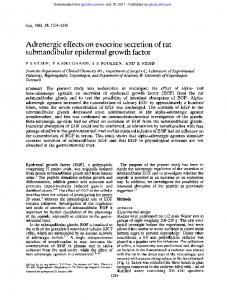
The presence of TR␣1, TR␣2, and TR1 in endovascular and interstitial extravillous trophoblast was evidenced by immunohistochemical staining in placental bed biopsies from 7 to 18 wk gestation. There was also immunostaining seen within decidua and myometrium. A representative il-
FIG. 1. Immunohistochemical staining of spiral arteries of the placental bed with TR␣1 antibodies in a representative sample harvested in the first trimester. SA, Spiral artery; L, lymphocytes; D, maternal decidua. Magnification, ⫻200.
lustration of TR␣1 immunostaining in a first-trimester placental bed biopsy is shown in Fig. 1. Table 1 summarizes the cellular localization of specific TR isoforms in placental bed samples in which decidual and/or myometrial spiral arteries were present. There were no qualitative differences in immunolocalization of TRs evident between first- and early second-trimester placental bed biopsies. All TRs showed more pronounced nuclear than cytoplasmic staining in extravillous trophoblast cells. Proliferation of different trophoblast cell preparations
Preliminary experiments demonstrated ongoing proliferation of SGHPL-4 and JEG 3 cells at 24, 48, and 72 h after plating cells (Fig. 2). JEG 3 cells proliferated in medium containing 10% stripped FCS (Fig. 2) but not in medium with 0.5% FCS (data not shown); these cells were therefore cultured in 10% stripped FCS for subsequent experiments. In contrast to SGHPL-4 and JEG 3 cells, an absence of proliferation, as determined in the MTT assay, was evident for primary cytotrophoblasts (Fig. 2). In subsequent experiments, the effects of treatment with T3 and/or EGF were evaluated at 72 h. SGHPL-4 cell proliferation and effects of T3 and EGF
In this extravillous cell line, a significant antiproliferative effect was noted when SGHPL-4 cells were treated with either EGF or T3 (Fig. 3). The largest antiproliferative effect was observed at lower concentrations of both EGF (1 and 10 TABLE 1. Relative expression and localization of TR isoforms in first and early second trimester placental bed biopsies determined by immunocytochemistry Placental bed cell population
TR␣1
TR␣2
TR1
Interstitial trophoblast Endovascular trophoblast Decidual stromal cell Decidual leukocytes Endometrial epithelium Endothelium Myometrium
⫹⫹/⫹⫹⫹ ⫹⫹ ⫹ ⫹⫹⫹ ⫹⫹ ⫹/⫺ ⫹ weak
⫹⫹⫹ ⫹⫹⫹ ⫹ ⫹⫹ ⫹⫹ ⫹⫹ ⫹⫹
⫹⫹⫹ ⫹⫹ ⫹⫹ ⫹⫹⫹ ⫹⫹⫹ ⫹⫹⫹ ⫹⫹
⫹, Weak staining; ⫹⫹, intermediate staining; ⫹⫹⫹, intense staining; ⫺, no staining.
1658
J Clin Endocrinol Metab, March 2005, 90(3):1655–1661
Barber et al. • The in Vitro Effects of Thyroid Hormone on Trophoblast
FIG. 3. Proliferation of SGHPL-4 cells at 72 h after treatment with EGF and/or T3 as determined using an MTT assay. *, P ⬍ 0.05, **, P ⬍ 0.001, compared with control.
Term cytotrophoblast cells in primary culture and effects of T3 and EGF
Term cytotrophoblast cells are not proliferative in vitro but do differentiate to form a pseudosyncytium (29). They were easily cultured in both stripped serum medium (10% FCS vol/vol) and reduced serum medium (0.5% FCS). The results from four primary placental cultures were combined and demonstrated that treatment with EGF significantly enhanced cytotrophoblast incorporation of MTT at all concentrations. Treatment with T3, however, had no effect on MTT incorporation. Coincubation of EGF 10 ng/ml and T3 10 nm showed no effect on EGF enhancement of MTT incorporation (Fig. 5). Because term cytotrophoblasts do not proliferate in vitro, EGF stimulation of MTT incorporation is likely to reflect enhanced cytotrophoblast survival; an effect of cytotrophoblast differentiation into a psuedosyncytium on this measure cannot be excluded. Invasion of SGHPL-4 cells into fibrin gel
EGF significantly enhanced both the length and number of invasive processes produced by SGHPL-4 cells (Fig. 6). T3 alone had no significant effect on invasion but significantly attenuated the number of invasive processes from each mi-
FIG. 2. A, MTT incorporation at 24, 48, and 72 h after plating in SGHPL-4 cells maintained in 0.5% FCS. B, MTT incorporation into JEG 3 cells maintained in 10% stripped FCS. C, MTT incorporation into primary cytotrophoblasts cultured in 0.5% stripped FCS.
ng/ml) and T3 (1 and 10 nm). The concentrations of EGF and T3 used for coincubation experiments were chosen to be 10 ng/ml and 10 nm, respectively. Simultaneous incubation with both treatments did not give rise to an additive effect. JEG 3 cell proliferation and effects of T3 and EGF
Both EGF and T3 exerted a significant proproliferative effect on JEG 3 cells at higher concentrations. There was no additive effect of T3 and EGF treatments (Fig. 4).
FIG. 4. Proliferation of JEG 3 cells at 72 h maintained in 10% stripped FCS after treatment with EGF and/or T3 as determined using an MTT assay. *, P ⬍ 0.05, compared with control.
Barber et al. • The in Vitro Effects of Thyroid Hormone on Trophoblast
FIG. 5. MTT incorporation into term cytotrophoblasts cultured in 0.5% FCS and treated with EGF and/or T3. *, P ⬍ 0.05; **, P ⬍ 0.001, compared with control.
crocarrier bead produced after stimulation by EGF at 10 and 50 ng/ml. The length of these EGF-induced invasive processes was also significantly attenuated by coincubation with T3 but only at a concentration of 1 nm T3. Motility of SGHPL-4 cells in vitro
It was apparent that EGF significantly enhanced SGHPL-4 motility (Fig. 7). T3 also enhanced cell motility significantly but to a lesser degree. In coincubation experiments, EGF 50
FIG. 6. A, Length of SGHPL-4 cell invasive processes 24 h after treatment with EGF and/or T3. *, P ⬍ 0.05, compared with control. B, Number of SGHPL-4 cell invasive processes per microcarrier bead 24 h after treatment with EGF and/or T3. *, P ⬍ 0.05, compared with control.
J Clin Endocrinol Metab, March 2005, 90(3):1655–1661
1659
FIG. 7. SGHPL-4 motility after treatment with EGF and/or T3. *, P ⬍ 0.05; **, P ⬍ 0.001, compared with control.
mg/ml, combined with T3 at 1 and 10 nm concentrations, significantly enhanced EGF-stimulated cell migration. Discussion
In the present study, we demonstrate that TR␣1, TR␣2, and TR1 isoforms can be localized immunohistochemically in both interstitial trophoblast and extravillous trophoblast (EVT). EVT, which is crucial during the invasive component of placentation, thus has the cellular apparatus to be thyroid hormone responsive (39), a finding relevant to the report that untreated hypothyroidism is associated with a 2-fold increase in spontaneous abortion and stillbirth rates. Furthermore, in a study of 16 pregnancies in untreated hypothyroid women, considerably higher rates of placental complications (preeclampsia, placental abruption, and postpartum hemorrhage) were observed (40). We have previously shown using immunohistochemistry that TRs are present within the nuclei of villous cyto- and syncytiotrophoblast within the villous placenta itself (21). Thyroid hormone deiodinases (41), which inactivate T3 and therefore regulate its effect within the tissues, have also been localized within villous syncytiotrophoblast cells (42). In the present study, we have shown that different trophoblast subtypes (apart from term primary cytotrophoblast cultures) all exhibit thyroid hormone responsiveness. The EVTderived cell line SGHPL-4 becomes less proliferative when treated with T3, contrasting with the choriocarcinoma line JEG 3 that shows increased proliferation. In addition, we have shown that T3 has a modulating effect on EVT migration and invasion stimulated by EGF. This role in the differentiated function of trophoblast supports earlier work showing that T3 causes enhanced human chorionic gonadotropin/ human placental lactogen production by trophoblast (22, 43, 44), which may suggest a role for T3 in differentiated function in the nonproliferative EVT and villous syncytiotrophoblast, as opposed to proliferation in villous cytotrophoblast. Our data are consistent with the view that EGF significantly enhances primary cytotrophoblast survival, a finding consistent with earlier work (45, 46). EGF is known to reduce apoptosis of trophoblast, and our finding that it prolongs term cytotrophoblast survival in vitro is in accord with this. In addition, recent data indicate that T3 may abrogate apoptosis in human EVT in vitro (23). In other cell types of ectodermal origin, EGF is recognized for its profound effects
1660
J Clin Endocrinol Metab, March 2005, 90(3):1655–1661
on differentiation and mitogenesis. A recent study of firsttrimester explant cultures of chorionic villi, using heparinbinding EGF, noted enhanced EVT differentiation and invasion (47). We have demonstrated striking changes produced in the phenotype of EVT-like SGHPL-4 cells when treated with EGF, these cells having been previously demonstrated to be a suitable model for EVT function in vitro (48). They exhibit increased motility and invasive characteristics when subjected to stimulation with EGF, which concurs with earlier mouse embryo observations (13). Interestingly cotreatment with T3 attenuates the changes in differentiated function in response to EGF but does not influence EGF-induced changes in proliferation. In summary, the present study indicates that EGF exerts an effect on cytotrophoblast proliferation/survival, motility, and invasion, investigated in a variety of human trophoblast cell models, and that local T3 action modifies the influence of this growth factor on trophoblast function. These findings in turn have implications for human placental development and for the pathogenesis of malplacentation syndromes such as preeclampsia and intrauterine growth restriction. Acknowledgments Geetha Balarajah provided technical assistance. Received April 28, 2004. Accepted December 7, 2004. Address all correspondence and requests for reprints to: Professor Mark Kilby, Department of Maternal and Fetal Medicine, Division of Reproductive and Child Health, 3rd Floor, Birmingham Women’s Hospital, Edgbaston, Birmingham B15 2TG, United Kingdom. E-mail:
[email protected]. This work was supported by The University of Birmingham Medical School Scientific Projects Committee and the Mason Trust. K.J.B. was funded by an Entry-Level Fellowship from the Wellcome Trust.
References 1. Craven CM, Zhao L, Ward K 2000 Lateral placental growth occurs by trophoblast cell invasion of decidual veins. Placenta 21:160 –169 2. Nanaev AK, Kosanke G, Reister F, Kemp B, Frank HG, Kaufmann P 2000 Pregnancy-induced de-differentiation of media smooth muscle cells in uteroplacental arteries of the guinea pig is reversible after delivery. Placenta 21: 306 –312 3. Benirschke P, Kaufmann P 1995 Pathology of the human placenta. 3rd ed. New York: Springer-Verlag New York Inc. 4. Pijnenborg R, Bland JM, Robertson WB, Brosens I 1983 Uteroplacental arterial changes related to interstitial trophoblast migration in early human pregnancy. Placenta 4:397– 413 5. Meekins JW, Pijnenborg R, Hanssens M, McFadyen IR, van Asshe A 1994 A study of placental bed spiral arteries and trophoblast invasion in normal and severe pre-eclamptic pregnancies. Br J Obstet Gynaecol 101:669 – 674 6. Sheppard BL, Bonnar J 1999 Uteroplacental hemostasis in intrauterine fetal growth retardation. Semin Thromb Hemost 25:443– 446 7. Kim YM, Bujold E, Chaiworapongsa T, Gomez R, Yoon BH, Thaler HT, Rotmensch S, Romero R 2003 Failure of physiologic transformation of the spiral arteries in patients with preterm labor and intact membranes. Am J Obstet Gynecol 189:1063–1069 8. Kim YM, Chaiworapongsa T, Gomez R, Bujold E, Yoon BH, Rotmensch S, Thaler HT, Romero R 2002 Failure of physiologic transformation of the spiral arteries in the placental bed in preterm premature rupture of membranes. Am J Obstet Gynecol 187:1137–1142 9. Bischof P, Meisser A, Campana A 2000 Paracrine and autocrine regulators of trophoblast invasion—a review. Placenta 21(Suppl A):S55–S60 10. Maruo T, Matsuo H, Oishi T, Hayashi M, Nishino R, Mochizuki M 1987 Induction of differentiated trophoblast function by epidermal growth factor: relation of immunohistochemically detected cellular epidermal growth factor receptor levels. J Clin Endocrinol Metab 64:744 –750 11. Huot RI, Foidart JM, Nardone RM, Stromberg K 1981 Differential modulation of human chorionic gonadotropin secretion by epidermal growth factor in normal and malignant placental cultures. J Clin Endocrinol Metab 53:1059 – 1063
Barber et al. • The in Vitro Effects of Thyroid Hormone on Trophoblast
12. Morrish DW, Bhardwaj D, Dabbagh LK, Marusyk H, Siy O 1987 Epidermal growth factor induces differentiation and secretion of human chorionic gonadotropin and placental lactogen in normal human placenta. J Clin Endocrinol Metab 65:1282–1290 13. Machida T, Taga M, Minaguchi H 1995 Effects of epidermal growth factor and transforming growth factor ␣ on the mouse trophoblast outgrowth in vitro. Eur J Endocrinol 133:741–746 14. Bass KE, Morrish D, Roth I, Bhardwaj D, Taylor R, Zhou Y, Fisher SJ 1994 Human cytotrophoblast invasion is up-regulated by epidermal growth factor: evidence that paracrine factors modify this process. Dev Biol 164:550 –561 15. Maruo T, Mochizuki M 1987 Immunohistochemical localization of epidermal growth factor receptor and myc oncogene product in human placenta: implication for trophoblast proliferation and differentiation. Am J Obstet Gynecol 156:721–727 16. Richards RC, Beardmore JM, Brown PJ, Molloy CM, Johnson PM 1983 Epidermal growth factor receptors on isolated human placental syncytiotrophoblast plasma membrane. Placenta 4:133–138 17. Bulmer JN, Thrower S, Wells M 1989 Expression of epidermal growth factor receptor and transferrin receptor by human trophoblast populations. Am J Reprod Immunol 21:87–93 18. Cao H, Lei ZM, Bian L, Rao CV 1995 Functional nuclear epidermal growth factor receptors in human choriocarcinoma JEG-3 cells and normal human placenta. Endocrinology 136:3163–3172 19. Ladines-Llave CA, Maruo T, Manalo AS, Mochizuki M 1991 Cytologic localization of epidermal growth factor and its receptor in developing human placenta varies over the course of pregnancy. Am J Obstet Gynecol 165:1377– 1382 20. Banovac K, Ryan EA, O’Sullivan MJ 1986 Triiodothyronine (T3) nuclear binding sites in human placenta and decidua. Placenta 7:543–549 21. Kilby MD, Verhaeg J, Gittoes N, Somerset DA, Clark PM, Franklyn JA 1998 Circulating thyroid hormone concentrations and placental thyroid hormone receptor expression in normal human pregnancy and pregnancy complicated by intrauterine growth restriction (IUGR). J Clin Endocrinol Metab 83:2964 – 2971 22. Maruo T, Matsuo H, Mochizuki M 1991 Thyroid hormone as a biological amplifier of differentiated trophoblast function in early pregnancy. Acta Endocrinol (Copenh) 125:58 – 66 23. Laoag-Fernandez JB, Matsuo H, Murakoshi H, Hamada AL, Tsang BK, Maruo T 2004 3,5,3⬘-Triiodothyronine down-regulates Fas and Fas ligand expression and suppresses caspase-3 and poly (adenosine 5⬘-diphosphate-ribose) polymerase cleavage and apoptosis in early placental extravillous trophoblasts in vitro. J Clin Endocrinol Metab 89:4069 – 4077 24. Matsuo H, Maruo T, Murata K, Mochizuki M 1993 Human early placental trophoblasts produce an epidermal growth factor-like substance in synergy with thyroid hormone. Acta Endocrinol (Copenh) 128:225–229 25. Lyall F, Bulmer JN, Kelly H, Duffie E, Robson SC 1999 Human trophoblast invasion and spiral artery transformation: the role of nitric oxide. Am J Pathol 154:1105–1114 26. Robson SC, Simpson H, Ball E, Lyall F, Bulmer JN 2002 Punch biopsy of the human placental bed. Am J Obstet Gynecol 187:1349 –1355 27. Simpson H, Robson SC, Bulmer JN, Barber A, Lyall F 2002 Transforming growth factor  expression in human placenta and placental bed during early pregnancy. Placenta 23:44 –58 28. Gittoes NJ, McCabe CJ, Verhaeg J, Sheppard MC, Franklyn JA 1997 Thyroid hormone and estrogen receptor expression in normal pituitary and nonfunctioning tumors of the anterior pituitary. J Clin Endocrinol Metab 82:1960 –1967 29. Choy MY, St Whitley G, Manyonda IT 2000 Efficient, rapid and reliable establishment of human trophoblast cell lines using poly-l-ornithine. Early Pregnancy 4:124 –143 30. Cartwright JE, Tse WK, Whitley GS 2002 Hepatocyte growth factor induced human trophoblast motility involves phosphatidylinositol-3-kinase, mitogenactivated protein kinase, and inducible nitric oxide synthase. Exp Cell Res 279:219 –226 31. Kliman HJ, Nestler JE, Sermasi E, Sanger JM, Strauss III JF 1986 Purification, characterization, and in vitro differentiation of cytotrophoblasts from human term placentae. Endocrinology 118:1567–1582 32. Driver PM, Kilby MD, Bujalska I, Walker EA, Hewison M, Stewart PM 2001 Expression of 11 -hydroxysteroid dehydrogenase isozymes and corticosteroid hormone receptors in primary cultures of human trophoblast and placental bed biopsies. Mol Hum Reprod 7:357–363 33. Blaschitz A, Weiss U, Dohr G, Desoye G 2000 Antibody reaction patterns in first trimester placenta: implications for trophoblast isolation and purity screening. Placenta 21:733–741 34. Haigh T, Chen C, Jones CJ, Aplin JD 1999 Studies of mesenchymal cells from 1st trimester human placenta: expression of cytokeratin outside the trophoblast lineage. Placenta 20:615– 625 35. Vettenranta K, von Koskull H, Heikinheimo M, Raivio KO 1986 Cytoskeletal markers and specific protein production in cells cultured from human first and third trimester placentae. In Vitro Cell Dev Biol 22:100 –106 36. Mosmann T 1983 Rapid colorimetric assay for cellular growth and survival: application to proliferation and cytotoxicity assays. J Immunol Methods 65: 55– 63
Barber et al. • The in Vitro Effects of Thyroid Hormone on Trophoblast
37. Cartwright JE, Holden DP, Whitley GS 1999 Hepatocyte growth factor regulates human trophoblast motility and invasion: a role for nitric oxide. Br J Pharmacol 128:181–189 38. Tse WK, Whitley GS, Cartwright JE 2002 Transforming growth factor-1 regulates hepatocyte growth factor-induced trophoblast motility and invasion. Placenta 23:699 –705 39. Matsuo H, Maruo T, Hayashi M, Mochizuki M 1991 [Modification of endocrine function of trophoblasts by thyroid hormone]. Nippon Sanka Fujinka Gakkai Zasshi 43:1533–1538 40. Davis LE, Leveno KJ, Cunningham FG 1988 Hypothyroidism complicating pregnancy. Obstet Gynecol 72:108 –112 41. Bates JM, St. Germain DL, Galton VA 1999 Expression profiles of the three iodothyronine deiodinases, D1, D2, and D3, in the developing rat. Endocrinology 140:844 – 851 42. Chan S, Kachilele S, Hobbs E, Bulmer JN, Boelaert K, McCabe CJ, Driver PM, Bradwell AR, Kester M, Visser TJ, Franklyn JA, Kilby MD 2003 Placental iodothyronine deiodinase expression in normal and growth-restricted human pregnancies. J Clin Endocrinol Metab 88:4488 – 4495 43. Adachi T, Maeda M, Nomoto T, Tsukahara F, Sakamoto S, Takeda Y 1986
J Clin Endocrinol Metab, March 2005, 90(3):1655–1661
44.
45.
46.
47.
48.
1661
In vitro study on thyroid hormone metabolism of human placenta in early pregnancy. Asia Oceania J Obstet Gynaecol 12:385–393 Matsuo H, Maruo T, Murata K, Mochizuki M 1993 Human early placental trophoblasts produce an epidermal growth factor-like substance in synergy with thyroid hormone. Acta Endocrinol (Copenh) 128:225–229 Levy R, Smith SD, Chandler K, Sadovsky Y, Nelson DM 2000 Apoptosis in human cultured trophoblasts is enhanced by hypoxia and diminished by epidermal growth factor. Am J Physiol Cell Physiol 278:C982–C988 Garcia-Lloret MI, Yui J, Winkler-Lowen B, Guilbert LJ 1996 Epidermal growth factor inhibits cytokine-induced apoptosis of primary human trophoblasts. J Cell Physiol 167:324 –332 Leach RE, Kilburn B, Wang J, Liu Z, Romero R, Armant DR 2004 Heparinbinding EGF-like growth factor regulates human extravillous cytotrophoblast development during conversion to the invasive phenotype. Dev Biol 266:223– 237 Cartwright JE, Kenny LC, Dash PR, Crocker IP, Aplin JD, Baker PN, Whitley GS 2002 Trophoblast invasion of spiral arteries: a novel in vitro model. Placenta 23:232–235
JCEM is published monthly by The Endocrine Society (http://www.endo-society.org), the foremost professional society serving the endocrine community.