John B. C. FINDLAY and Darryl J. C. PAPPIN. Department of Biochemistry, University of ...... Breton, 1979; Cooper & Hogan, 1976). In the absence or reduced ...
Biochem. J. (1986) 238, 625-642 (Printed in Great Britain)
625
REVIEW ARTICLE The opsin family of proteins John B. C. FINDLAY and Darryl J. C. PAPPIN Department of Biochemistry, University of Leeds, Leeds LS2 9JT, U.K.
Introduction The purpose of this review is to compare the structural and functional properties of a group of proteins that share the feature of being able to respond to light energy by way of a bound retinoid chromophore. It may be useful, by way of introduction, to place these systems into a wider biological context. The carotenoids and their bisected relatives, the retinoids, comprise one of several important groups of substances which share an important metabolic ancestor, isopentenyl pyrophosphate. The prenyl moiety of this compound also finds its way into an incredible variety of natural substances ranging from many of the plant-based oils, spices, toxins, rubber and hormones to animal products such as pheromones and steroid hormones. They include the polyprenyl alcohols so important to all living organisms as carriers in the process of glycosylation. The carotenoid/retinoid group is usually identified with the utilization of light in some way, principally because the former are often responsible for the bright colouration exhibited by plants and animals while the latter play an important role in vision, and both are involved in capture of light energy. In most cases, the biological roles of the carotenoids and retinoids are mediated through proteins to which they bind with varying affinities. Their resonating conjugated double bond structure readily allows the absorption of energy in the 300-400 nm region and part of their biological usefulness lies in the ease with which absorbance maxima can be shifted by association with protein. Two instructive examples of this can be seen in a-crustacyanin and in mammalian vision. In the first case, the blueish pigment of lobster carapace arises from the association of the carotenoid astaxanthin with the protein ac-crustacyanin, which is accompanied by a shift in the absorbance maximum from 470 nm to 632 nm (Zagalski, 1983). The reverse process, i.e. dissociation of chromophore and protein, can be dramatically appreciated in conversion of lobsters, on cooking, to their more widely recognized red colouration. In general, the utilization of carotenoids for pigmentation purposes is very common, particularly amongst marine invertebrates (Britton et al., 1982). The retinoid, 1 1-cis-retinal, or its dehydro derivative, is the primary chromophore in probably all forms of vision and it is widely accepted that at least part of the mechanism by which colour is perceived lies in a family of proteins, the rhodopsins, whose differing structures confer different absorbance maxima on the retinal-protein complexes. The ability of these compounds to absorb light energy has also been efficiently commandeered for the purposes
of biological generation of chemical energy. Carotenoids found in large amounts in the photosynthetic membranes of photosynthetic bacteria, algae and plants. Their role is still obscure for, although they combine with proteins in the photosynthetic apparatus with greater or lesser avidity and can undergo shifts in their absorbance maxima, it seems that they are involved less with the core photochemical processes and more with the harvesting of lightand with photoprotection (for review see SiefermannHarms, 1985). The characterization of these binding proteins (Cogdell, 1985; Brunisholz et al., 1984) is, however, far short of that for bacteriorhodopsin (bR), the retinal-protein complex in Halobacterium halobium responsible for the absorption of incident light and, in part, for its conversion into chemical energy. The specific chromophore we will be concerned with here, retinal, arises by complex metabolic conversions starting, in mammals, by the generation of two molecules of retinol (vitamin A) from carotene. It is important to appreciate that vitamin A and its derivatives also play vital roles in important processes other than vision, e.g. gene regulation, differentiation and reproduction. For all these purposes, there exist enzymes and binding proteins which ensure effective utilization of this hydrophobic effector. Some of their structures are now known in considerable detail and, even though they are not involved in light-capture, they may nevertheless provide important insights for our understanding of the opsins (Newcomer et al., 1984; Sawyer et al., 1985). It will be even more intriguing to see whether the disparate systems which exhibit spectral shifts and are involved in light capture exhibit similarities in their chromophore-binding sites. are
Retinoid proteins of Halobacteria The discovery of bR as the principle light-utilizing pigment of Halobacteria when growing under conditions of high salt and high light intensity (Oesterhelt & Stoeckenius, 1971, 1973) has provoked enormous international interest, both in the protein itselfand in this class of organism. Careful work with mutants has now revealed that there are at least a further two retinoidbinding pigments [termed halorhodopsin (hR) and slow or sensory rhodopsin (sR)] in the bacterial membrane. Each possesses distinctive photochemical and functional properties but they all appear to employ the same isomers of retinal linked to the amino group of a lysine side chain via a Schiff's base which, from resonance Raman spectroscopy, is unusual in being protonated (Aton et al., 1977; Terner et al., 1979). As well as shifting the absorbance maximum of the chromophore on association with protein, all the light-sensitive systems we are mainly concerned with in
Abbreviations used: bR, bacteriorhodopsin; hR, halorhodopsin, sR, sensory rhodopsin; vR, visual rhodopsin.
Vol. 238
626
J. B. C. Findlay and D. J. C. Pappin
(a)
rebi
alba
(b)
aItais refina
(4
C=NH
,-C
H
5ps
retnal
1 3-cis retinal
NHC H
*trans retia
1 3-is
retnW
H,CNH
1 3-cs retnal 1 3-cs retWa
(c)
(13
(d)
*CNI11-cs retnal
H
\V7R498 3hv Opsin >5 m] (Meta RX465)
MetaR 1 380
(hypsoR430) e.100ps Batho R548 all trans retinal {I40 ns
LWTiR497
200 rnsZZZMeta R I478
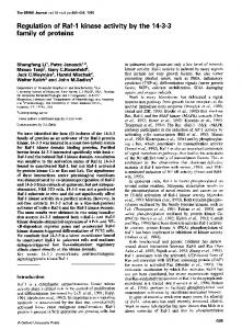
Fig. 1. The photocycles (a) Bacteriorhodopsin [adapted from Stoeckenius et al. (1979) and Stoeckenius & Bogomolni (1982)]. Intermediate designations are shown in brackets; J,K' and M' have not been unambiguously established. The times shown refer to the particular transitions at room temperature. Subscripts denote the absorbance maxima of the various intermediates. (b) Halorhodopsin (adapted from Oesterhelt et al., 1985). (c) Slow-(cycling)-rhodopsin (from Spudich & Bogomolni, 1984). (d) Visual rhodopsin.
this review can respond to incident light in a manner which shows up as a defined series of spectral intermediates. These photocycles (Fig. 1) arise from the intimate nature of the chromophore-protein interaction and presumably reflect altered conformational states. It seems that, in all cases, light first induces an isomerization of the retinal which is probably accompanied by separation of the protonated Schiff's base from its protein-linked counter-ion. Considerable effort is being spent on correlating binding site structure, light-induced conformational changes and the observed biochemical activities of the proteins. The use of retinal analogues and resonance Raman spectroscopy have been particularly
active areas (Mathies, 1984; Smith et al., 1985) which the limitation of space does not allow us to describe even in outline. Bacteriorhodopsin (bR) Absorption of light by bR drives the extrusion of hydrogen ions from the cell to generate a proton gradient which can be utilized to fuel active transport and ATP production (Dannon & Stoeckenius, 1974). Simultaneous measurement of time-resolved absorbance changes and charge movements suggest that up to two protons (or their equivalents, e.g. 2 H3O+) are transported per turn of the cycle (Ort & Parson, 1979; Govinjee et al., 1980; 1986
The opsin family of proteins
Bogomolni et al., 1980; Renard & Delmelle, 1980) generating a maximum potential difference of about 300 mV with a quantum yield varying between 0.25 and 0.6 depending on the reaction/measurement conditions (Govinjee et al., 1980; Ormos et al., 1983; Goldschmitt et al., 1976, 1977; Becher & Ebrey, 1977; Hurley & Ebrey, 1978; Oesterhelt et al., 1985). Several mechanisms have been proposed, many involving the proton of the Schiff's base, for the translocation process but they remain very speculative. The kinetics and stoichiometry of the cycle (Fig. la) appear capable of modification by the proton gradient created, termed a back-pressure effect (Westerhoff et al., 1979, Westerhoff & Dancshazy, 1984). This form of feedback inhibition is thought to operate via a diversion mechanism whereby bR is shunted through a non-pumping form (Pietrobon et al., 1981). The branch point is postulated to occur somewhere around the M intermediate, i.e. before the proton is released to the external medium (Drachev et al., 1981; Keszthelyi & Ormos, 1980). Interestingly, the structural alterations which accompany the generation of the important M intermediate are not so marked as to appear in the 0.7 nm (7A) electron density map. Yet there is considerable kinetic and spectrophotometric evidence that substantial conformational changes occur during the L to M transition (Kuschmitz & Hess, 1982) in which the Schiff's base is deprotonated (Marcus et al., 1977; Bayley et al., 1982; Terner et al., 1979; Hwang et al., 1978). Altered environments for tyrosine (deprotonation) and tryptophan residues have been particularly implicated (Konishi & Packer, 1977, 1978; Kuschmitz & Hess, 1982; Hanamoto et al., 1984), most attention being directed to Tyr-26 and Tyr-64 whose modification abolished proton transport (Lemke & Oesterhelt, 1981; Lemke et al., 1982). Although lysine residues are apparently not of critical importance to bR function (Ovchinnikov, 1982), cross-linking through such sidechains inhibits the photocycle (Konishi et al., 1979). Peptides corresponding to residues 1-3, 68-72 and 231-248 are also not vital to activity (Abdulaev et al., 1978) but arginines (Packer et al., 1979) and carboxylic acids (Ovchinnikov et al., 1982a; Herz & Packer, 1981) appear more important. Asp-1 15, in particular, which reacts with dicyclohexylcarbodi-imide (DCCD) on exposure of protein to light (Renthal et al., 1985), may be critical to the proton pumping mechanism (Renthal et al., 1979, 1981). Sensible interpretation of these modification experiments, however, must await a highresolution structure. Despite all these changes, it was suggested that little gross movement of the chromophore occurs during the photocycle (Czege et al., 1982) but more recent evidence from modulation excitation spectroscopy suggests significant conformational alterations (Hasselbacher et al., 1986). Halorhodopsin (hR) The second major light-sensitive pigment, hR, is present in wild-type cells in much smaller quantities than bR and does not appear to be aggregated like bR into discrete membrane patches, although it may be oligomeric in detergent. Initially thought to catalyse light-driven sodium extrusion from the cells (MacDonald et al., 1979), the protein was later identified through studies with membrane vesicles (Schobert & Lanyi, 1982) and reconstituted membranes (Bamberg et al., 1984), as an
Vol. 238
627
inwardly-directed electrogenic chloride pump. This transport system is active, despite the high external salt concentrations, presumably to circumvent the influence of the membrane potential. Its principal physiological role may be to maintain approximate osmotic equilibrium with the environment. Complete agreement has not yet been reached on all details of the photocycle for hR (Fig. lb), perhaps because of the discovery of at least one other chloride-binding site on the protein (Schobert et al., 1983; Falke et al., 1984), whose occupancy may alter the position of some of the spectral intermediates. The quantum yield was estimated as 0.34+0.02 (Oesterhelt et al., 1985). Unlike bR, transport function is not accompanied by deprotonation of the Schiff's base, although the approach and removal of the translocating Cl- may be equivalent to deprotonation/protonation. The protein has been purified by a number of groups, yielding Mr values (20000-26000) similar to that of bR (Steiner & Oesterhelt, 1983; Taylor et al., 1983; Ogurusi et al., 1984; Sugiyama and Mukohata, 1984). Other similarities (but not identities) include the content of a-helix and fl-sheet (Jap & Kong, 1986), the cleavage profile with Staphylococcus aureus V8 protease (Hegemann et al., 1982) and the structural environment for the retinal moiety as revealed by resonance Raman spectroscopy (Smith et al., 1984; Alshuth et al., 1985). Thereafter, differences are more pronounced. There is a lack of immunological cross-reactivity (Hegemann et al., 1982). The molecule possesses one -SH group thought to be involved with the binding of chloride (Ariki & Lanyi, 1984) and which may be located in the vicinity of the retinal binding site. The amino acid compositions vary and, most critically, protein sequencing suggests that overall homology with bR will be low (D. Oesterhelt, personal communication). Both of the anion-binding sites and the protonated Schiff's base are relatively accessible from the surface of the membrane. It seems, therefore, that a change in substrate and in catalytic direction has been accompanied by fairly major changes in structure. The helical content and presumably their number appear to be preserved and it will be fascinating to see whether the relative arrangement of the transmembrane segments is also unaltered. Slow or sensory Rhodopsin (sR) The phototaxic responses of halobacteria whereby they are repelled or attracted by light of short (370 nm) and long (> 600 nm) wavelength respectively was first ascribed to bR and later expanded to include hR. Subsequently, mutants lacking both these proteins were still found to exhibit both repellant and attractant responses (Spudich & Spudich, 1982), suggesting that at least one other photosensitive system was still present. This new system again seemed to make use of a retinoid chromophore, since the response was absent when retinal synthesis was abolished but could be accurately restored by the addition of exogenous retinal (Spudich & Bogomolni, 1984; Ehrlich et al., 1984). Also, predictable shifts in the absorbance spectra resulted from the introduction of various retinal analogues (Stoeckenius & Bogomolni, 1982). It was presumed, therefore, that the organism possessed either a third retinal-containing photosensitive pigment exhibiting both a narrow absorption spectrum peaking at about 370 nm and a broad spectrum peaking at 587 nm (Bogomolni & Spudich,
J. B. C. Findlay and D. J. C. Pappin
628
1982) or two different pigments (Dencher, 1983; Hildebrand & Schimz, 1983). Current work tends to suggest that the same protein is involved in the initial stages of both effects. Confirmation of this hypothesis must await purification and reconstitution of the species responsible, which appears to be present in quantities much less than either bR or hR, perhaps in the region of 5000 copies/cell (Spudich & Bogomolni, 1984). A simple model for the integration of the responses into a provisional photocycle has been proposed (Spudich & Bogomolni, 1984) in which the absorption of a second photon at 370 nm by an intermediate in the cycle (sR373) sends the pigment through a different pathway, thereby generating a repellant rather than an attractant response. Because the photocycle is much slower than those for bR and hR (half time of about 0.8 s as against about 10 ms), this new pigment has been called slow-(cycing)-rhodopsin (sR). A more attractive name might be 'sensory rhodopsin' (Spudich & Bogomolni, 1984), particularly if the system does not share the transport properties of bR and hR. This latter conclusion arises from failure to detect changes in membrane potential when a strain of bacteria containing only sR was illuminated (Spudich & Spudich, 1982; Bogomolni & Spudich, 1982). This must still remain an open question, however, since the regulation of flagellar motion in other organisms is thought to involve ion (such as calcium) currents. Structure of bacteriorhodopsin Organization. Integral proteincomponents in biological membranes may exist as permanent monomers or oligomers, or form various transient associations related to function. Bacteriorhodopsin is quite unique. It is arranged into extensive crystalline-like sheets consisting of tens of thousands of molecules tightly packed into hexagonal arrays. The smallest structural unit consists of three protein molecules, the trimers being separated from one another by a unimolecular layer of tightly bound glycosulpholipid, approx. 10 lipids/bR (Blaurock & Stoeckenius, 1971). The internal core regions bounded by the three constituent monomers are also occupied by lipid. It is not at all clear just which factors are responsible for the integrity of the trimeric unit. The ability to dissociate the hexagonal lattice to monomers by dissolution in detergent and to reconstitute as monomers at high lipid/protein rations (Heyn et al., 1975) tends to suggest that interactions within the hydrophobic phase of the bilayer may be at least partly involved. Some measure of support comes from the observations that the hexagonal lattice is reformed when the reconstituted system is taken below the transition temperature of the lipid (Cherry et al., 1978) and that removal of the cytosolic C-terminal 17 residues does not prevent reconstitution into a fully functional trimer (Liao & Khorana, 1984). It is possible from the model to envisage contact regions involving helices 1 and 7 of one monomer and 4 and 5 of another. Less obvious still are the forces which produce the large crystalline arrays of associated trimers. The unusual composition and arrangement of the lipid may provide a clue. Reconstitution experiments suggest that proton pumping can be effected while in the monomeric form, but there is some evidence of cooperativity between the components of the trimer (Becher & Cassim, 1977; Korenstein et al., 1979; Rehorek & Heyn, 1979).
Primary structure. The single chain of bR is composed of 248 residues, more than 70 % of which are hydrophobic in nature (Ovchinnikov et al., 1979; Gerber et al., 1979; Khorana et al., 1979). There are no histidine or cysteine residues in the protein, and the N-terminus was shown to be a pyroglutamyl residue. One of the more noticeable characteristics ofthe protein was the significant clustering of regions of predominantly hydrophilic or hydrophobic residues. The bacteriorhodopsin gene (Dunn et al., 1981) was found to code for an additional 13 amino acids at the N-terminus plus a single extra aspartic acid residue at the C-terminus. There were no intervening sequences, but an unusual feature of the mRNA was that it contained only a few residues at the 5' position, rather different from the normal 25 or so nucleotides in other prokaryotes and more similar to the pattern of short 5' sequences found in human mitochondrial DNA (Montoya et al., 1981). It was also noted by Dunn et al. (1981) that the 13-residue leader sequence was very different from the classical pattern of N-terminal signal peptides in secreted proteins. Within the primary structure, the assignment by Bridgen & Walker (1976) of Lys-41 as the retinal attachment site was later corrected to 216 by Bayley et al. (1981), Mullen et al. (1981) and Katre et al. (1981), working with more controlled conditions for the reductive fixation of the Schiff's base linkage (Wolber & Stoeckenius, 1984).
Disposition. Even before the complete primary structure had been determined, the general disposition of the protein within the membrane had been fairly well established. The pioneering work of Henderson & Unwin (1975) combined electron diffraction and low-dose electron microscopy to establish the basic architecture of the molecule in its native trigonal form. The low resolution (0.7 nm, 7A) structure consisted of seven membrane-spinning rods running roughly parallel to each other and perpendicular to the plane of the bilayer. The rods were postulated to be a-helices based on consideration of their size, packing density and the suggestion from high angle X-ray diffraction that there were four to seven a-helices per molecule (Henderson, 1975). The basic structure was consistent with an i.r. dichroism study on oriented purple membrane films in which blue-shifting of the amide I absorption band was attributed to distortion of the helices (Rothschild & Clark, 1979; Corjito et al., 1982). A much higher resolution in-plane projection (0.37 nm, 3.7A) obtained by Hayward & Stroud (1981) confirmed the general features of the low resolution map. Correcting for the distortion produced by the limited tilt angles allowed Agard & Stroud (1983) to produce a reconstructed image showing an increase in length of the ac-helical rods from 3.5 nm to -4.5 nm ( 35A to 45A). More importantly, four regions of density connecting the rods on the external surface of the membrane and one connection on the cytoplasmic side emerged. Further refinement of the structure has come from a second, orthorhombic crystal form of the protein produced by dialysis of detergent-solubilized membranes (Michel et al., 1980; Leifer & Henderson, 1983). In this map, two of the transmembrane rods exhibited distortions. The kink in one rod was particularly strong and the 1986 -
-
The opsin family of proteins
629
Ser
Asp
Val Gly ,, Met GlyLys Vol
LOu
Pro Asp Ala 39 Lys Lys-
Phe
Tyr
Ala
Val
Met Ser Glu Alo
104
A- Asp _ Gin
Asp Vol Lou Lu Lou Ala LOU Asp LOU Lou Lou
Lys ''9
Ser
Gly
lie Ih Thnr; Ala VoLOU
Lou
Ph.
PeThr Gly Ph.
Phe LOu Thr Tyr Vol Tyr GVal Gl Lou Thr .Thr Ala Lou Leu Asp Val Gly Ile Pro Aun Tyr Lou Lou ,W 'JY Met Pro - T%Ile Met Inr Met Ile Ala Leu Thr Ala Ala Gly lPhe Thr Ala Phe Thr Thr Thr Lou Trp Met Gly Gly Ser A Lou Tyr Lou Ala 1A Ti LOu Asp Ala Tyr Ser Lou Trp Ala Met Trp Arg Trp Lou Val LOu IIe Trp Lou Ala Thr Phe Trp Glu Gly Lys Arg Vol L Pro Tyr Gly. - Pro Ile Y -Arg 67 Lou Asn 6 Tyr-S r 132 Gly Thr Gin 75 Thr Met Glu Ile Gin Val Gly Ala Pro Gly OUTSIDE Phe