Jul 5, 1995 - Abstract Soluble matrices of coleoid cephalopod shells, namely the organic pen of Loligo sp. and min- eralized calcified shells of Spirula ...
Marine Biology (1996) 125:525-529
9 Springer-Verlag 1996
Y. Dauphin
The organic matrix of coleoid cephalopod shells: molecular weights and isoelectric properties of the soluble matrix in relation to biomineralization processes
Received: 5 July 1995 / Accepted: 1 December 1995
Abstract Soluble matrices of coleoid cephalopod shells, namely the organic pen of Loligo sp. and mineralized calcified shells of Spirula spirula and Sepia sp., were extracted and studied by HPLC (high-performance liquid chromatography) gel filtration and isoelectrofocusing (IEF). Molecular weights of Spirula spirula and Sepia sp. extracts are not well separated, whereas Loligo sp. shows several well-defined peaks. In Loligo sp. and Sepia sp., molecular weights of > 100 kdaltons were found. Spirula spirula yielded a profile devoid of high molecular material. The main part of the extracted matrix of the organic pen of Loligo sp. is basic. In mineralized shells of both Spirula spirula and Sepia sp., the soluble organic matrices are acidic. These results support the view that the acidity of the organic matrix is related to mineralization processes. Introduction
In cephalopods, the general evolutionary trend has been towards progressively smaller shells, and in many cases they have disappeared completely. Recent cephalopods are separable into two unequal groups. One group (Tetrabranchiata) is comprised only of Nautilus sp. which is characterized by a chambered external shell while the other group consists of all other Recent cephalopods (Dibranchiata or Coleoidea), all of which have shells that are internal and usually vestigial or absent. The reduced shell of coleoids has been shown to contain chitin (Peters 1972). Among the Coleoidea, only species of the genera Spirula and Sepia possess a rigid mineralized shell. Analyses of recent cephalopod shell carbohydrates (soluble and insoluble matrices) Communicated by A. Rodriguez, Puerto Real Y, Dauphin Laboratoire de Pal6ontologie, URA 723, Bfitiment 504, Universit6 Paris XI-Orsay, F-91405 Orsay cedex, France
suggest that the loss of mineralized shell is correlated with an increase in the "chitinous" nature of the insoluble matrix (Dauphin and Matin 1995). Insight into the mineralization processes depends strongly on knowledge of the characteristics of the organic matrices of the shells. Proteins and polysaccharides are the two major constituents of the organic matrix of mineralized mollusc shells. It has been postulated that mineralization is nucleated by specific chemical groups of the organic matrix (Wilbur 1964; Wilbur and Simkiss 1968; Gr6goire 1972). Crenshaw (1972) isolated a water-soluble glycoprotein from Mercenaria mercenaria and suggested that the soluble matrix plays a role in initiation of calcification. The proteins are generally rich in acidic amino acids and glycine (Gr6goire et al. 1955; Hare 1963; Degens et al. 1967). Polysaccharides are also acidic. The major portion of the soluble fraction consists of sulfated glycoprotein (Simkiss 1965; Crenshaw 1972). The soluble matrices have been shown to be rich in aspartic and glutamic acids (Weiner and Hood 1975; Samata et al. 1980). Histochemical localization of the central region of nacreous tablets in Nautilus pompilius has shown that these sites are acidic sulfated polysaccharides (Crenshaw and Ristedt 1976). It has been postulated that only a small part of the matrix, probably acidic proteins, plays a role in the nucleation of minerals (Weiner and Traub 1984). Support for this hypothesis is the prevalence of acidic matrix components in molluscs. The amino acid composition of the soluble organic matrix indicates its acidity: theoretical isoelectric points (pI) can be calculated from amino acid compositions (Sillero and Ribeiro 1989). However, these only indicate the average pI of the soluble matrix. Direct pI measurements of the organic matrices of invertebrate shells are very rare: in some cases (Crassostrea gigas) "... neither peak could be applied successfully to PAGE or isoelectrofocusing because of the extreme high molecular weight and the acidic pH of their solution. This material did not penetrate into the gels." (Krampitz
526
et al. 1983, p. 234). The organic soluble matrix of the gastropod Nassa reticulata has been extracted and purified by gel permeation (Krampitz et al. 1976). The only peak obtained was subjected to ion-exchange chromatography, each; "ion-exchange" peak was then analyzed by isoelectrofocusing. The first "ion-exchange" peak displayed three isolectric points between 2 and 5 after isoelectrofocusing, the second "ion-exchange" peak showed two isoelectric points, the first near 2, the second at 10. The third ion-exchange peak displayed six isoelectric points between 2 and 10 (Krampitz et al. 1976). These data indicate that the acidity of the soluble matrix is heteregeneous. However, chromatography of the Ca-ligands from N. reticulata and their isoelectrofocusing revealed a pI in the range of pH 5.5. All the above data were obtained from calcified shells. Comparisons between the amino acid composition in calcified and non-calcified molluscan shells have been made only on the insoluble organic matrix (Wada 1966). No comparable information exists for the soluble part of the organic matrix of non-mineralized shells. Thus, to establish whether or not a compositional change occurs in the organic matrix during loss of the hard shell is vital to our understanding of mineralization processes. Some living cephalopods possess a mineralized shell, the majority possesses an organic shell. In order to test the hypothesis of a specific role of acid-soluble matrix in mineralization, molecular weights and the pI of the soluble matrix of the organic pen of LoIigo sp. were compared to those of the mineralized shells of Spirula spirula and Sepia sp., the morphological characteristics of which have been reviewed by Hopkins and von Boletzky (1994). Nautilus sp. shells were excluded because this genus is not a coleoid, and because the structure of the nacreous layer differs between the shells of the Tetrabranchiata and Coleoidea (Dauphin and Keller 1982). Materials and methods
allows the separation of protein from 5 to 100 kdaltons. The spectromonitor 5000 (Thermo Instrument Systems) photodiode-array detector provides measurements over its full 190 to 360 nm range, following separation by HPLC. The isoelectric properties of the soluble matrix were estimated by non-denaturing isoelectric-focusing electrophoresis. One-dimensional microslab isoelectric focusing (IEF) was performed in homogeneous polyacrylamide small gel (25% total acrylamide concentration, 3% degree of crosslinking) with 100 x 65 mm gels (0.4 mm thick), 5% carrier ampholytes Bio-Lyte pH 3 to 10, containing 25% w/v glycerol. A three-phase catalyst system of ammonium persulfate, riboflavin-Y-phosphate and temed was used. This system is catalyzed by light. Gels are cast on a sheet of gel-support film for polyacrylamide. With the Bio-Rad's Model 111 Mini IEF cell, the gel is run upside down, in direct contact with the electrodes, to eliminate the need for electrode buffers. The lyophilised samples were redissolved in 1% gtycine. Running conditions were 15 rain at 100 V, 15 rain at 200 V, and ~ 60 min at 450 V. Following IEF, the gels were placed in 27% ethanol, 10% acetic acid, 0.04% Coomassie Blue R-250, 0.5% CuSO~ and 0.05% Crocein scarlet for ~ lh 30 rain (fixing and staining solution). The first destaining solution contained 12% ethanol, 7% acetic acid and 0.5% CuSO~, the second destaining solution contained 25% ethanol and 7% acetic acid. Dried gels were scanned and digitalized with a CCD (chargecoupled device) camera (256 grey levels) in order to locate the stained bands. The numerical data acquired were transfered to a computer program to obtain profiles. No densitometric scanning was performed, thus the surfaces under the peaks cannot been used for quantification.
Results
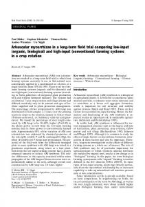
Fig. 1 shows the results of the gel filtration of the soluble matrix of Loligo sp., Spirula spirula and Sepia sp. shells. When the same shell extract was read at different wavelengths (224, 240, 256 and 280 nm), the positions of the peaks sometimes differed, indicating that several substances were included in those peaks: Moreover, the peaks were not always bell-shaped and symmetrical. In Loligo sp. and Sepia sp., molecular weights > 100 kdaltons were recorded. The profile for Spirula spirula differs from those of the other genera:
E
c
The following shells were used: Loli9o sp. (probably Atlantic), Spirula spirula (New Caledonia) and Sepia sp. (New Caledonia). The different layers of the mineralized shells were not isolated for SpiruIa spiruIa and Sepia sp., since some layers were too thin. Although the LoIigo sp. pen is not calcified, all samples were prepared by the same procedure. BSA (bovine serum albumin, Eurobio) and ribonuclease A (Sigma) were used for calibration. BSA has several pIs: 4.9, 5.2, 5.4, 5.5 and 5.6; ribonuclease A has two main pIs: 8.9 and 9.3. The samples were washed first with sodium hypochlorite to remove contaminating superficial amino acids and peptides, then with distilled, deionized water. They were crushed, decalcified with acetic acid (pH 4), and centrifuged at 3000 • for 45 rain. The supernatant containing the soluble matrix (SM) was filtered against distilled water using a Filtron membrane with a molecular weight cut-off at 3 kdaltons, and was then lyophilized. Gel filtration chromatography [TSK (Tosohaas) G2000 SW (silice water) XL] using citrate buffer (pH 4.7) and eluted at 15 mlh i
OJ
>100 kdaltons
0
=o c~ o
'
Spirula
! 20 rain
, ,2o,..4 32.50 rain
elution time
Fig. 1 Loligo sp., Spirula spirula, Sepia sp. High-performance liquid chromatography of soluble extracts of cephalopod shells. Spirula spirula and Sepia sp. molecular weights are not well separated. (Not to scale)
527
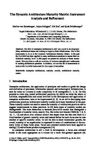
there was no soluble high-molecular material, the profile was initially horizontal, changing to a low regular slope. Peaks were faint and only low-molecular weights were present (10 kdaltons). Previous chromatographs of the soluble matrix of Sepia sp. (Superosel2 and 6) revealed two main peaks: a small peak at ~ 220 kdaltons, and a main peak at --~22 kdaltons (Dauphin and Kervadec 1994). The Sepia sp. HPLC profile shows a stronger, smooth slope, peaking at ~ 19 and ~ 13.5 kdaltons. These faint peaks were more distinct when first- and second-order derivatives of the profile were used. The peaks of the Loligo sp. profile were more distinct, and indicated molecular weights of ~ 29.5, 16 and 12 kdaltons, respectively. The results of the IEF of the soluble matrix of LoIigo sp., Spirula spiruIa and Sepia sp. shells are shown in Fig. 2. In Lotigo sp., the most prominent heavilystained band was seen at basic pIs between 7 and 9. The highest basic band was broad and composed of four minor bands. The band at pI 7.8 is unique to Loligo sp. The acidic band (5.7 to 6.3) was composed of two faint bands. In Spirula spirula and Sepia sp., the main bands were located on the acidic side only. Several bands were scattered between pI 4 and 6 in the Spirula spiruta
BSA
Ribonuclease A
soluble organic matrix. In occurred at pI 100 kdaltons) recorded for this species, but not for Spiruta spirula Fig. 1). This idea is supported by the fact that the acidic amino acids of the insoluble part of non-mineralized horny plate of Sepia sp. are less abundant than in the spongy, aragonitic, ventral part (Wada 1966; Drozdova and al. 1971). Even if it is difficult to correlate the molecular weights and pI of the soluble matrices of the studied species, comparison of chromatograms and electrophoretic profiles of the two mineralized shells (Spirula spirula and Sepia sp.) are more similar than those of the Loligo sp. pen: molecular weights and pIs are better separated in the soluble matrix of Loligo sp. than in the matrices of Spirula spirula and Sepia sp. Only the amino-acid compositions of the insoluble matrices of some coleoid shells are available in the
529
Literature. Thus a comparison between theoretical pI (calculated from the amino-acid contents) and the observed pl is not possible for the soluble matrices. It is well known that in molluscan shells the acidic amino acid content of soluble matrix is higher than that of insoluble matrix (Gr6goire et al. 1955; Hare 1963; Degens et al. 1967; Weiner 1979; Watabe et al. 1993). However, the acidity of the insoluble matrices of various molluscan shells is heterogeneous (Wada 1966). Total acidic amino acid contents are higher in the mineralized part of a shell. For example, the aspartic acid content in the insoluble part of the Loligo sp. pen is 7 % (Ghiselin et al. 1967; Hunt and Nixon 1981), and in the insoluble part of the horny plate of Sepia sp. it is 6 % (Drozdova and al. 1971). Glutamic acid contents are ~ 4 and 8%, respectively, in the same shells. The aspartic acid of the ventral part of Sepia sp. is ~ 17%, and glutamic acid content is 7% (Wada 1966; Drozdora and al. 1971). The basic pI of the soluble matrix of the Loligo sp. pen and the acidic pI of the soluble matrices of the mineralized shells of Spirula spirula and Sepia sp. support the view that the acidity of the organic matrix is related to mineralization processes.
References Bandel K, Boletzky S yon (1979) A comparative study of the structure, development and morphological relationships of chambered cephalopod shells. Veliger 21:313-354 Barskov IS (1973) Microstructure of the skeletal layers of Sepia and Spirula compared with the shell layers of other molluscs. Paleont J Wash 3:285-294 Boggild OB (1930) The shell structure of the mollusks. Kgl Danske Vidensk Selsk Skr (Naturvidensk og math) 9/2:231-326 Crenshaw MA (1972) The soluble matrix from Mercenaria mercenaria shell. ForschBer Biomineralis Akad Wiss Lit, Mainz (Biomineralization Res Rep) 6:6-11 Crenshaw MA, Ristedt H (1976) The histochemical localization of reactive groups in septal nacre from Nautilus pompilius L. In: Watabe N, Wilbur KM (eds) The mechanisms of mineralization in the invertebrates and plants. University of South Carolina Press, Columbia, pp 355-367 Dauphin Y (1976) Microstructure de coquilles de c6phalopodes. I. Spirula spirula L. (Dibranchiata, Decapoda). Bull Mus natn Hist nat, Paris, 36 s~r. (sect Sciences Terre) 54 (382): 197-238 Dauphin Y (1981) Microstructure des coquilles de c6phalopodes. II. La seiche (Mollusca, Coleoida). Palaeontographica (Abt A) 176: 35 51 Dauphin Y, Keller JP (1982) Mise en 6vidence d'un type structural coquillier sp6cifique des c6phalopodes dibranchiaux. C r hebd S6anc Acad Sci, Paris II 294:409-412 Dauphin Y, Kervadec GW (1994) Comparaison des diagen+ses subies par les phases min6rale et prot6ique soluble des tests de mollusques cbphalopodes colfioides. Palaeontographica (Abt A) 232:85-98 Dauphin Y, Marin F (1995) The compositional analysis of recent cephalopod shell carbohydrates by Fourier transform infrared spectrometry and high performance anion exchange pulsed amperometric detection. Experientia 51:278-283
Degens ET, Spencer DW, Parker RH (1967) Paleobiochemistry of molluscan shell proteins. Comp Biochem Physiol 20:533-579 Drozdova TV, Karyakin AV, Krasnova VA (1971) Chemical composition and infrared absorption spectra of the organic matrix of the shell in the squid Sepia pharaonis. Zh evolyut Biokhim Fiziol 7:350-356 [In Russ] Ghiselin MT, Degens ET, Spencer DW, Parker PH (1967) A phylogenetic survey of molluscan matrix proteins. Breviora 262:1-35 Gr6goire C (1972) Structure of the molluscan shell. In: Florkin M, Scheer BT (eds) Chemical zoology. Vol. 7. Academic Press, New York, pp 45-102 Gr6goire C, Duch/tteau GH, Florkin M (1955) La trame protidique des nacres et des perles. Annls Inst oc~anogr, Monaco 31:1-8 Hare PE (1963) Amino acids in the proteins from aragonite and calcite in the shells of Mytilus californianus. Science, Wash 139: 216-217 Hopkins B, Boletzky S yon (1994) The fine morphology of the shell sac in the squid genus Loligo (Mollusca: Cephalopoda): features of a modified conchiferan program. Veliger 37:344 357 Hunt S, Nixon M (1981) A comparative study of protein composition in the chitin-protein complexes of the beak, pen, sucker-disc, radula and oesophageal cuticle of cephalopods. Comp Biochem Physiol 68B: 535-546 Krampitz G, Drolshagen H, H/iusle J, Hof-Irmscher K (1983) Organic matrices of mollusc shells. In: Westbroek P, de Jong EW (eds) Biomineralization and biological metal accumulation. D. Reidel, pp 231-247 Krampitz G, Engels J, Cazaux C (1976) Biochemical studies on water-soluble proteins and related components of gastropod shells. In: Watabe N, Wilbur KM (eds) The mechanisms of mineralization in the invertebrates and plants. University of South Carolina Press, Columbia, pp 55-173 Peters W (1972) Occurrence of chitin in Mollusca. Comp Biochem Physiol 41B: 541-550 Samata T, Sanguansri P, Cazaux C, Harem M, Engels, J, Krampitz G (1980) Biochemical studies on components of mollusc shells. In: Omori M, Watabe N (eds) The mechanisms of biomineralization in animals and plants. Tokai University Press, Tokyo, pp 37-47 Sillero A, Ribeir0 JM (1989) Isoelectric points of protein: theoretical determination. Analyt Biochem 179:319-325 Simkiss K (1965) The organic matrix of the oyster shell. Comp Biochem Physiol 16:427-435 Wada K (1966) Studies on the mineralization of the calcified tissue in molluscs. XII. Specific patterns of non-mineralized layer conchiolin in amino acid composition. Bull Jap Soc scient Fish 32: 304-311 Watabe N, Kingsley RJ, Kawaguchi T (1993) Functions of organic matrices in some invertebrate calcifying systems. In: Kobayashi I, Mutvei H, Sahni A (eds) Structure, formation and evolution of fossil hard tissues. Tokai University Press, Tokyo, pp 3-11 Weiner S (1979) Aspartic acid-rich proteins: major components of the soluble organic matrix of mollusk shells. Calcif Tissue int 29: 163-167 Weiner S, Hood L (1975). Soluble protein of the organic matrix of mollusk shells: a potential template for shell formation. Science, Wash 190: 987-989. Weiner S, Traub W (1984) Macromolecules in mollusc shells and their functions in biomineralization. Phil Trans R Soc (Set B) 304:425-434 Wilbur KM (1964) Shell formation and regeneration. In: Wilbur KM, Younge CM (eds) Physiology of Mollusca. I. Academic Press, New York, pp 243-282 Wilbur KM, Simkiss K (1968) Calcified shells. In: Florkin M, Stotz EH (eds) Comprehensive biochemistry. Vol 26A. Elsevier, New York, pp 229 295