Apr 21, 1983 - was discussed by Hunter & Ludwig (1962), but a re- examination (Browne & Kent, 1975) with ethyl acetimidate demonstrated that an alternative.
Biochem. J. (1984) 217, 589-594 Printed in Great Britain
589
The preparation of fully N-s-acetimidylated cytochrome c Carmichael J. A. WALLACE*t and David E. HARRIS*: *Laboratory of Molecular Biophysics, Department of Zoology, University of Oxford, South Parks Road, Oxford OX] 3PS, U.K., and tDepartement de Biochimie Medicale, Centre Medical Universitaire, 9 Avenue de Champel, 1211 Geneve 4, Switzerland
(Received 21 April 1983/Accepted 4 October 1983) We have re-examined the acetimidylation and subsequent deprotection of cytochrome c by published methods in the light of recent findings on the tendency of protein acetimidylation reactions to yield side products of differing net charges. We find that the protection methods do indeed yield a mixture of products, some of which have considerably diminished biological activity. Our observations support a postulated mechanism for the generation of side products, and we have been able to identify the major factor responsible for their formation by published methods. The deprotection method appears to be free of side reactions. We describe a new procedure for acetimidylation that will produce fully N-e-acetimidylated cytochrome c. This derivative, lacking detectable side products and having good biological-activity, is useful for structure-function studies and as an intermediate in the semisynthesis of cytochrome c analogues.
The reaction of alkyl acetimidates (imidoesters) with primary amines converts them into acetimidines (Hunter & Ludwig, 1962), e.g.: NH
(G. R. Moore & C. J. A. Wallace, unpublished work). Thus high solubility and, very often, biological activity is retained by the protected protein. NH
-(CH2]4-NH2 + CH3-C-OCH3 -* -[CH2]4-NH-C-CH3 + CH30H The acetimidyl (acetimidoyl) group is an effective blocking group for lysine c-amino functions (cytochrome c has an acetylated a-amino group), exhibiting exceptional stability to acid and to mild base. The group is smoothly removable by ammonium (Hunter & Ludwig, 1972) or methylamine (DuBois et al., 1981) buffers of pH 11.3-11.5. A number of proteins, including ribonuclease A (Reynolds, 1968) and cytochrome c (Harris & Offord, 1977), have been subjected to a protection and deprotection cycle with this blocking group without apparent loss of activity. It has found particular favour in semisynthesis (Offord, 1973; Garner & Gurd, 1975; Slotboom & de Haas, 1975; Wang et al., 1977; Harris & Offord, 1977; Wallace & Offord, 1979; Wallace, 1984a,b) for these reasons and because the charge of the original lysine residue is conserved. The pK of the e-acetimidino group in free c-acetimidyl-lysine is 12.7-12.8 t Present address: Department of Chemistry, University of Indiana, Bloomington, IN 47405, U.S.A. Vol. 217
The blocked lysine residue is rendered insensitive to amine-directed agents such as the Edman reagent (Gamer & Gurd, 1975; Wallace & Offord, 1979), except where it is N-terminal, and to specific enzymes such as trypsin (Harris & Offord, 1977), clostripain (S. Yagisawa, unpublished work) and carboxypeptidase A (although, conveniently enough, sensitivity to carboxypeptidase B is retained; Wallace & Offord, 1979). The mechanism of the acetimidylation reaction was discussed by Hunter & Ludwig (1962), but a reexamination (Browne & Kent, 1975) with ethyl acetimidate demonstrated that an alternative reaction, the formation of N-alkyl imidates [compound (II), Scheme 1], was predominant at the lower end of the pH range normally used for this reaction (7-10). Browne & Kent (1975) also proposed a scheme for the production of side products in protein acetimidylation, through intramolecular cross-linking (III). In a subsequent study of the acetimidylation of myoglobin by methyl acetimidate under a wide
590
C. J. A. Wallace and D. E. Harris NH2+
1
RNH2 +CH3- C-OR' o
1
F
NH2
CH3 -C-OR' RNH-C-OR' 1 0.
H20
[
CH3
NH2 +
H+
I
1
-k RNH-C-CH3 (I) +R'OH
RNH+= f OR' (II)+NH3 CH3 '
NH2
NHR" H+ R"NH+ [RNH-COR' 1 RNH-C-CH3 (III) I~~~~~~~~~~~~~~~~~~~~~I + R'OH CtI3 Scheme 1. Scheme proposedfor theformation of N-acetimidylated amines (I) and side products (II and III) by the reaction of methyl acetimidate with primary amines (after Browne & Kent, 197S) See the text for a detailed discussion.
range of conditions Di Marchi et al. (1978) observed the formation of such side products, i.e. protein in which one or more of the lysine side chains, although modified, had no residual charge at neutral pH. They succeeded in defining conditions that would decrease the formation of such side products. When the product of a published method for the acetimidylation of cytochrome c (Wallace, 1976; Wallace & Offord, 1979; based on the method of Hunter & Ludwig, 1962) was chromatographed on the cation-exchanger SP- (sulphopropyl-)Sephadex C-25, it too was seen to contain multiple molecular forms. Conditions employed by other workers (Reynolds, 1968; used by Pettigrew et al., 1976) were also seen to lead to side-product formation. We have studied the effect of varying the reaction conditions during cytochrome c protection and have isolated the principal cause of such charge loss. This has allowed us to define conditions that abolish side-product formation altogether.
Experimental Materials Horse heart cytochrome c (type III) was obtained from Sigma Chemical Co., St. Louis, MO, U.S.A. Methyl acetimidate hydrochloride was prepared by the method of Hunter & Ludwig (1962). Other chemicals were of analytical grade.
Acetimidylation of cytochrome c Method (i). The method of Wallace (1976) and Wallace & Offord (1979) for cytochrome c is a modification of the general method of acetimidylation described by Hunter & Ludwig (1962). Methyl acetimidate hydrochloride (0.66g) was dissolved in 0.8 ml of 5 M-NaOH to give an approximately neutral solution, which was added to 100mg of cytochrome c in lOml of 0.1 M-Na2B407 solution. The reaction mixture was left at 0°C for 30min, during which time the pH rose from 8.4 to 9.0. It was then allowed to reach room temperature (24°C). The pH was adjusted to 9.5 and the mixture was left at 24°C for 1 h. Then 0.33g of reagent in 0.27 ml of 5M-NaOH was added and the pH re-adjusted to 9.5. After 1 h at 240C this latter procedure was repeated. Finally 0.66g of reagent in 0.6ml of 5M-NaOH was added and the mixture was left at 4°C overnight. Protected protein was separated from unwanted compounds by repeated dialysis against 0.1 M-Na2B407. Method (ii). This is based on the method of Di Marchi et al. (1978) for myoglobin. A 100mg portion of the protein was dissolved in 3.5 ml of 0.1 Msodium borate buffer, pH9.5. To this was added 100mg of reagent dissolved in 0.12ml of 5MNaOH. The pH of the reaction mixture was re-adjusted and maintained by addition of 0.1 M-NaOH for 40min. These basic conditions were varied in a number of ways (see the Results and discussion section). The protein was freed of excess of reagent 1984
591
Acetimidylated cytochrome c: preparation
and other products by gel filtration on a column (30 cm x 2.2 cm) of Sephadex G-25 (fine grade) equilibrated and run in 0.1 M-Tris/HCl buffer, pH9.4. Method (iii). Cytochrome c was acetimidylated in accordance with the method of Pettigrew et al. (1976), itself based on the method of Reynolds (1968). A 40mg portion of cytochrome c was dissolved in 5 ml of 0.1 M-sodium borate buffer, pH 10. Three additions, of 325mg each, of methyl acetimidate hydrochloride were made at 20min intervals, and the pH was adjusted to 10 each time with 6M-NaOH. The product was gel-filtered on the Sephadex G-25 column described above. Method (iv). A 100mg portion of cytochrome c was dissolved in 3.5ml of 0.1 M-Na2B407; 0.1 MNaOH was added to adjust the pH to 10.5. A 100mg portion of methyl acetimidate hydrochloride was dissolved rapidly in sufficient SMNaOH to make the pH of the solution greater than 10.5. The quantity of NaOH solution depends on the amount of adsorbed HCI that the methyl acetimidate contains, but preparations with a m.p. of 93-95°C will normally require 0.18-0.20 ml. The reagent solution was then added at once to the cytochrome c solution, the pH lowered, if necessary, to 10.5 with conc. HCI (to decrease the rate of hydrolysis of the reagent), and the mixture left for 40min at room temperature before gel filtration as described above. Removal of acetimidyl groups Deprotection was effected by the method of Hunter & Ludwig (1972) through exposure of the protein to a buffer prepared by adding conc. NH3 solution (sp.gr. 0.880) to acetic acid to a pH of 11.3.
were performed in the manner described by Harris & Offord (1977).
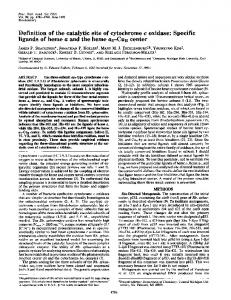
Redox potentials Potentials were determined by the method of mixtures (Davenport & Hill, 1952). Results and discussion Most of the acetimidylation methods tested gave rise to multiple molecular forms. Up to five peaks were seen on cation-exchange chromatography (Fig. 1), though their relative proportions varied (Table 1). The last peak to be eluted from the column (peak V in Fig. 1), and thus the most positively charged fraction, appears when the molarity of the eluent was about 0. 17M. The c-acetimidyllysine content of the protein was determined by the method of Milman (1974). It proved to be between 98 and 102% of the expected lysine content. The other peaks I, II, III and IV are therefore presumed to correspond to materials containing 4, 3, 2 or 1 less positive charge(s) respectively than the correctly modified material. Table 1 records the proportions of these fractions found under the various conditions employed in methods (i) and (ii). The results obtained by Di Marchi et al. (1978) for myoglobin showed clear trends towards lower proportions of the less positively charged materials with increasing pH, and decreasing temperature, of the reaction mixture. No such conclusion could be drawn from our results. Variation of the concentration of protein or of reagent revealed no consistent trends either. Browne & Kent (1975) had demonstrated that at pH 10 the product of reaction is the desired N-alkyl acetimidate [compound (I), Scheme 1], whereas at
Ion-exchange chromatography
Chromatography on SP-Sephadex C-25 was used as a method of detection of side products and of comparison of the products of the various acetimidylation methods employed. Solutions, of products were diluted with 1.5vol. of water after gel filtration on Sephadex G-25 or dialysis and applied to a column of SP-Sephadex C-25 cation-exchanger, previously equilibrated with a 10-fold dilution of a stock buffer prepared by adjusting the pH of 0.4M K2HPO4 to 7.2 with conc. H3PO4. The column was then developed with a linear gradient formed from the diluted buffer and the stock buffer. Protein fractions were collected and quantified spectrophotometrically. Biological assays Assays of the ability of ion-exchange fractions of acetimidylated cytochrome c to restore succinate oxidation in cytochrome c-depleted mitochondria Vol. 217
.;00.3
I
I
0
,-I1
X,,
10
20
Iv l
III I
30
-
v
-
I VI
40
50
60
I
I
70
Tube no.
Fig. 1. Elution profile of a product of method (ii) of acetimidylation on native horse heart cytochrome c, on ionexchange chromatography For full experimental details see the text.
592
C. J. A. Wallace and D. E. Harris
Table 1. Relative production of the five ion-exchange fractionsfrom acetimidylation methods (i) and (ii) with various reaction conditions See the Experimental section for details of the procedures and of the ion-exchange separation method. Reaction conditions
Method (i) (ii)
Mass of reagent
Temp.
(mg)
(OC)
2500 100 100 200 500 500 500 100 100 100 500 500
24 24 4 4 4 4 24 14 4 24 24 4
Concn. of protein pH 10.0 9.5 9.5 9.5 9.5 9.5 9.5 10.5 9.5 10.0 10.0 10.0
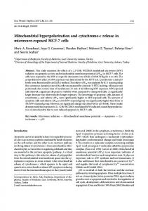
pH 8 the first product is an N-alkyl imidate. This can then either re-form the amine or give the acetimidate (in approx. 2:1 ratio). They pointed out that the imidate is susceptible to attack by other amines to give compound (III) of Scheme 1. We therefore hypothesized that the momentary decrease in the pH on addition of a reagent solution at or near neutral pH (methods i and ii) was the major cause of the problem. If the N-alkyl imidate formation is so rapid as to be largely complete within the few seconds between addition of reagent and re-adjustmentof pH, then an opportunity would be presented for amine attack on the imidate, where pairs of lysine residues exist in proximity, to give compound (III) of Scheme 1. We designed method (iv) for the acetimidylation of cytochrome c to avoid this possibility. The product of the method shows a single symmetrical peak in the ion-exchange chromatography system described (Fig. 2, trace A). To confirm the correctness of our supposition, we employed method (iii), in which the reagent is added as a solid, thus causing a very sharp fall in pH, which is immediately re-adjusted. The elution profile on ion-exchange chromatography is as trace B of Fig. 2, where approximately half of the product is not fully charged. It seems likely that the mechanism proposed by Browne & Kent (1975) is operative in the cytochrome c case. Horse heart cytochrome c has one -Lys-Lys-Lys- sequence and three -Lys-Lys- sequences. If we allow that cross-linking between residues distant in the three-dimensional structure is unlikely, this would allow a maximum charge loss of 4, and thus five different ion-exchange fractions, which is what we observe. What is slightly
(mg/ml)
Recovery in each peak (%) ,
I 0 0 2 1 0 3 2 3 0 2 9 3
8 30 30 30 30 30 30 30 10 30 10 30
II 2 2 9 7 0 14 9 12
0 3 27
16
III 8 6
IV 24 28
26 22 1 26 20 26 5 16 32 33
37 38 10 37 40 37 22 43 17 29
V 66 64 26 30 89 22 28 22 73 36 5 18
0.4
0.3 e
0.2
0.2 v. 0
s1 0.1
0
100
200
300
Elution volume(ml)
Fig. 2. Comparison of the properties on ion-exchange chromatography of the products ofmethod (iv) (trace A) and method (iii) (trace B) of acetimidylation of cytochrome c For full experimental details see the text.
surprising is the extent to which the formation of less positively charged materials occurs in some of the examples in Table 1. The implication is that imidate formation proceeds very rapidly and that its attack by amine is favoured over hydrolysis or ammonolysis. The three fractions collected from the ion-exchange chromatography of the product of method (iii) (Fig. 2, trace B) showed marked differences from one another in the biological assay system, fraction IV exhibiting approx. 60%, and fraction III 30%, of the specific activity of fraction V. Thus those published methods that are liable, like method (i), or certain, like method (iii), to yield 1984
Acetimidylated cytochrome c: preparation side reactions will exhibit a decreased activity for the unpurified product. Conclusions drawn from studies of such preparations might thus be misleading. Despite the biological activity differences, the redox potential determined for fraction IV was almost identical, at about 240mV (Table 2), with those determined for fraction V and the method (iv) product. This lack of further change in redox potential implies that the internal structure, the haem environment, is unperturbed (Myer et al., 1979) by the loss of surface charge (all 19 lysine residues of horse heart cytochrome c are external; Mandel et al., 1977). The decrease in biological activity is thus likely to be a direct consequence of effects on the cytochrome's ability to bind to oxidase and reductase, as with other forms of lysine modification (Capaldi et al., 1982). It is noteworthy that, of the four lysine tracts susceptible to cross-linking by the mechanism proposed, three (7-8, 72-73 and 86-88) have been assigned such a functional role (Capaldi et al., 1982). The charge loss incurred by this side reaction is irreversible, and full lysine content by amino acid analysis is not restored on deprotection (Reynolds, 1968; Di Marchi et al., 1978). DuBois et al. (1981) have ascribed the loss of charge in the Reynolds (1968) preparation and in the preparations discussed by Ludwig & Hunter (1967) to the deprotection step, rather than to the protection step as we do here, and conclude that the deprotection conditions are too severe. This interpretation was investigated by deprotection of material from peaks V (fully N-e-acetimidylated cytochrome c) and IV (material lacking one positive charge) from ion-exchange purifications of the products of acetimidination methods (i) and (ii). The method used was that of Hunter & Ludwig (1972). Before treatment, peak IV was
Table 2. Redox potentials determined for cytochrome c (horse heart) and derivatives The assay method employed was that of Davenport & Hill (1952), with a phosphate buffer of pH 7.0 as described. Literature values for the native protein range from 254 to 260mV. Redox potential Cytochrome c or derivative (mV) Horse cytochrome c 258 Amidino-cytochrome c 236 Acetimidyl-cytochrome c (method iv) 242 240 Acetimidyl-cytochrome c (method i, peak V) 237 Acetimidyl-cytochrome c (method i, peak IV) Deprotected peak V (cytochrome c) 258 Deprotected peak IV 240
Vol. 217
593
eluted at a molarity of 0. 1 SM, and peak V at 0. 17M. These materials were still eluted as single symmetrical peaks after treatment, but now at 0.12 and 0.15M respectively. The latter value corresponds to the molarity of elution of native cytochrome c (D. E. Harris & C. J. A. Wallace, unpublished work). It is thus evident that such a method ofdeprotection does not induce heterogeneity in the protein product; however, the heterogeneity obtained on reaction with methyl acetimidate is retained. The products of deprotection were tested for biological activity and their oxidation-reduction potentials were measured. Peak V material after deprotection had a redox potential restored to 258mV, identical with that determined for native cytochrome c. Peak IV material retained a redox potential of about 240mV on deprotection. Bioassays showed that it had only about 40% of the specific activity of the deprotected peak V material. The method of DuBois et al. (1981) for removal of acetimidyl groups (3.44M-methylamine, buffered with HCI to pH 11.5, for 4h at 25°C) was tried on acetimidyl-cytochrome c. A product of khakibrown colour lacking biological activity was obtained. Conclusions (1) When applied to cytochrome c, some published methods for acetimidylation are certain, and others are likely, to result in the formation of side products irreversibly lacking one or more positive charges. The side products have decreased biological activity and thus render the amino-protected protein unsuitable for semisynthetic procedures without subsequent purification and consequent loss of material. (2) Our studies on the generation and properties of these side products support the mechanism proposed by Browne & Kent (1975) for their formation at intermediate pH. We have shown that the side reactions are so rapid that adjustment to alkaline pH after mixing protein and reagent is ineffective in preventing them, and that this is the source of difficulty in existing published methods. (3) Acetimidyl-cytochrome c is cleanly deprotected by the method of Hunter & Ludwig (1972) to give a product with full biological activity and normal redox potential. The side reactions attributed by DuBois et al. (1981) to these deprotection conditions are more likely to have occurred during the protection step. The alternative deprotection conditions proposed by DuBois et al. (1981) are not suitable for acetimidyl-cytochrome c. (4) We suggest a procedure for cytochrome c acetimidylation that gives no detectable side products.
594 We are grateful to Professor R. E. Offord for his help and encouragement. We thank the Medical Research Council of Great Britain and the Fonds National Suisse de la Recherche Scientifique for financial support, and Mme. Monique Rychner and Mme. Barbara Battistolo for technical assistance.
References Browne, D. T. & Kent, S. B. H. (1975) Biochem. Biophys. Res. Commun. 67, 126-132 Capaldi, R. A., Darley-Usmar, V., Fuller, S. & Millett, F. (1982) FEBS Lett. 138, 1-7 Davenport, H. E. & Hill, R. (1952) Proc. R. Soc. London Ser. B 139, 327-347 Di Marchi, R. D., Garner, W. H., Wang, C. C., Hanania, G. I. H. & Gurd, F. R. N. (1978) Biochemistry 17, 2822-2829 DuBois, G. C., Robinson, E. A., Inman, J. K., Perham, R. N. & Appella, E. (1981) Biochem. J. 199, 335-340 Garner, W. M. & Gurd, F. R. N. (1975) Biochem. Biophys. Res. Commun. 63, 262-268 Harris, D. E. & Offord, R. E. (1977) Biochem. J. 161, 2125 Hunter, M. J. & Ludwig, M. L. (1962) J. Am. Chem. Soc. 84, 3491-3494
C. J. A. Wallace and D. E. Harris Hunter, M. J. & Ludwig, M. L. (1972) Methods Enzymol. 27, 585-596 Ludwig, M. L. & Hunter, M. J. (1967) Methods Enzymol. 11, 595-604 Mandel, N., Mandel, G., Trus, B. L., Rosenberg, J., Carlson, G. & Dickerson, R. E. (1977) J. Biol. Chem. 252, 4619-4636 Milman, J. D. (1974) D.Phil. Thesis, University of Oxford Myer, Y. P., Saturno, A. F., Verma, B. C. & Pande, A. (1979) J. Biol. Chem. 254, 11202-11207 Offord, R. E. (1973) in Peptides 1972 (Hanson, H. & Jakubke, H.-D., eds.), pp. 52-56, North-Holland, Amsterdam Pettigrew, G. W., Aviram, I. & Schejter, A. (1976) Biochem. Biophys. Res. Commun. 68, 807-813 Reynolds, J. H. (1968) Biochemistry 7, 3131-3135 Slotboom, A. J. & de Haas, G. H. (1975) Biochemistry 14, 5394-5399 Wallace, C. J. A. (1976) D.Phil. Thesis, University of Oxford Wallace, C. J. A. (1984a) Biochem. J. 217, 595-599 Wallace, C. J. A. (1984b) Biochem. J. 217, 601-604 Wallace, C. J. A. & Offord, R. E. (1979) Biochem. J. 179, 169-182 Wang, C. C., Gamer, W. H. & Gurd, F. R. N. (1977) Fed. Proc. Fed. Am. Soc. Exp. Biol. 36, 890
1984