IJC International Journal of Cancer
The receptor tyrosine kinase Axl in cancer: Biological functions and therapeutic implications Juliano D. Paccez1,2, Matjaz Vogelsang1,3, M. Iqbal Parker1,2 and Luiz F. Zerbini1,2 1
International Centre for Genetic Engineering and Biotechnology (ICGEB), Cape Town, South Africa Division of Medical Biochemistry, University of Cape Town, Cape Town, South Africa 3 Laboratory for Molecular Biology and Nanobiotechnology, National Institute of Chemistry, Ljubljana, Slovenia
Mini Review
2
The receptor tyrosine kinase Axl has been implicated in the malignancy of different types of cancer. Emerging evidence of Axl upregulation in numerous cancers, as well as reports demonstrating that its inhibition blocks tumor formation in animal models, highlight the importance of Axl as a new potential therapeutic target. Furthermore, recent data demonstrate that Axl plays a pivotal role in resistance to chemotherapeutic regimens. In this review we discuss the functions of Axl and its regulation and role in cancer development, resistance to therapy, and its importance as a potential drug target, focusing on acute myeloid leukemia, breast, prostate and non-small cell lung cancers.
Tyrosine Kinases Tyrosine kinases (TKs), proteins that represent a major portion of all oncoproteins, are essential mediators of the signal transduction process and play essential roles in cell differentiation, migration, proliferation, apoptosis and metabolism. TKs are a class of enzymes which selectively catalyze phosphorylation of selected tyrosine residues in target proteins using ATP. This post-translational modification is a critical component for normal cellular communication and homeostasis and it is associated with several steps of development and progression of cancers.1,2 TKs’ activity usually contributes to sensitivity to programmed cell death and prevents uncontrolled proliferation. Deregulation of TKs together with modification of their signaling cascades can eventually lead to malignant transformation via multiple cellular processes. TKs consist of two major groups, the receptor tyrosine kinases (RTK) and the non-receptor tyrosine kinases (NRTK). NRTK are cytoplasmic proteins, with significant structural variability which possess kinase and protein-protein interacting domains (such as SH2, SH3 and the PH domains).3 Importantly, more than 90 tyrosine kinases have been
Key words: receptor tyrosine kinases, cancer, Axl, cancer therapy, drug resistance Grant sponsor: International Centre for Genetic Engineering and Biotechnology Post-Doctoral Fellowship DOI: 10.1002/ijc.28246 History: Received 12 Feb 2013; Accepted 24 Apr 2013; Online 3 May 2013 Correspondence to: Luiz F Zerbini, International Centre for Genetic Engineering and Biotechnology (ICGEB), Wernher and Beit Building (South), UCT Campus, Anzio Road, Observatory 7925, Cape Town, South Africa, Tel.: 127-21-6507627, Fax: 127-21-650-7717, E-mail:
[email protected]
C 2013 UICC Int. J. Cancer: 134, 1024–1033 (2014) V
described since the completion of the Human Genome Project, and around two-thirds of them are RTKs.4 RTKs are transmembranic proteins and are activated by ligand binding to their extracellular domain. Ligands are extracellular signal molecules that induce receptor dimerization and activation. It is important to highlight that different ligands utilize different strategies by which they accomplish stable dimeric conformation and consequent receptor activation.3 The intracellular ATP binding domain catalyzes receptor autophosphorylation and exhibits the highest level of conservation between the RTKs. The ATP binding site is also a docking site for the specific binding of cytoplasmic signaling proteins containing protein tyrosine binding (PTB) and Src homology-2 (SH2) domains. This event leads to the activation of a cascade of biochemical signals, leading to the activation/repression of genes and thus resulting in the biological response (Fig. 1). In this review we will focus on the receptor tyrosine kinase, Axl, and its signaling pathway and role in cancer development, especially in acute myeloid leukemia, breast, prostate and non-small cell lung cancer studies. Furthermore, we will discuss the role of Axl in chemotherapy resistance and the progress in the field for development of a potential therapeutic agent to target this RTK.
The Receptor Tyrosine Kinase Axl The Axl subfamily of mammalian RTKs, also known as TAM family, consists of Axl, Tyro3 (or Sky) and Mer. Axl is a tyrosine phosphorylated 140 kDa protein which has transforming abilities and is ubiquitously expressed in cell lines of epithelial, mesenchymal, and hematopoietic origins, as well as in non-transformed cells.5–8 It was originally isolated from patients with chronic myelogenous leukemia (CML) and chronic myeloproliferative disorder.9
Figure 1. Schematic representation of receptor tyrosine kinase activation. Receptors are activated by ligand binding to their extracellular domain. The intracellular ATP binding domain catalyzes receptor auto-phosphorylation. This phosphorylation leads to activation of target proteins resulting in activation of intracellular biochemical signals, which activate/repress target genes allowing a biological response to signals.
The extracellular domain of Axl receptor family members is composed of a combination of two N-terminal immunoglobulin (Ig)-like domains and two fibronectin Type III (FNIII) repeats (Fig. 2). The domains similar to those present in adhesion molecules of the cadherin and immunoglobulin family, as well as receptor tyrosine phosphatases. TAM receptor tyrosine kinases are known to play a role in cell adhesion as well as in intracellular signaling. In fact, McCloskey et al. (1997) observed that Axl mediates adhesion of 32D myeloid cell line,10 whereas Lee et al. (1999), demonstrated that increased expression of the Axl receptor induces transformation of NIH 3T3 cells in highly tumorigenic cells in nude mice.11 Furthermore, Axl together with two closely related receptor tyrosine kinases, Tyro 3 and Mer, has been postulated to play an essential immunoregulatory role, as mutant mice lacking expression of these receptors develop a severe lymphoproliferative disorder accompanied by broad-spectrum autoimmunity.12–14 Axl activation and regulation
The ligand for Axl is the vitamin K-dependent protein Gas-6 (Growth Arrest-Specific gene 6). Interaction between Axl and Gas6 was reported by Mark et al. (1996). Gas6 is composed of an N-terminal region containing multiple gamma-carboxyglutamic acid residues (Gla), which are responsible for the specific interaction with the negatively charged phospholipid membrane, and binding of Gas-6 to the receptor.15,16 In addition, Gas6 has a loop region (present after the Gla region), followed by four epidermal growth factor (EGF)-like repeats and a globular sex hormone binding globulin (SHBG)-like region, comprising a pair of laminin G-like (LG) C 2013 UICC Int. J. Cancer: 134, 1024–1033 (2014) V
1025
domains.17 The general structure of Gas 6 is demonstrated in Figure 2. Axl activation has been linked to signaling cascades and is closely related to progression and development of tumors, such as the phosphatidylinositol 3-OH kinase (PI3K) pathway, including its downstream targets S6K and Akt, MAP kinases, STAT and NF-jB signal transductions pathways.18–20 The anti-apoptotic effect of Axl signaling was confirmed by studies using fibroblasts from Axl knock-out mice.21 Mouse embryonic fibroblasts (MEF) from wild-type and Axl knockdown animals were prepared and used to compare apoptosis induced by serum starvation. Interestingly, cells derived from Axl knockdown mice demonstrated more sensitivity to undergo apoptosis than MEF derived from wild-type mice after serum deprivation, indicating a pivotal role for this receptor in the induction of apoptosis.21 The AXL gene is located on chromosome 19, encoded by 20 exons, and distributed over a region of 44 kb. Schulz et al. (1993) described different isoforms of AXL mRNA generated by alternative splicing of exon 10.22 The AXL 5’upstream region is GC rich and lacks TATA and CAAT boxes. The characterization of the promoter region was first described by Mudduluru and Allgayer (2008).23 Using cloning and 5’ deletions they demonstrated that a minimal GC-rich region (2556/17) is sufficient for AXL promoter activity. Moreover, binding sites for various transcription factors within the –556 to –182 bp core region upstream of the AXL translational start codon were identified, including five binding sites for Sp a-e, one for MZF1, and one for AP1.23–25 Schematic representation of the Axl promoter is described in Figure 3. Further analysis demonstrated that the two sites (Sp-a and Spb), positioned close to the translation start site, are indispensable for promoter activity. Interestingly, Sayan et al. (2012) demonstrated a close relationship between Fra-1 and Axl, since the tyrosine kinase was directly upregulated by Fra-1 in bladder cancer. The authors showed that Fra-1 is highly expressed in 80% of invasive forms of carcinoma of the bladder expressing the Axl protein, compared to superficial bladder cancer (80% vs. 42%). Furthermore, Fra-1 expression affects cell morphology, motility and proliferation. These findings indicate that Axl is a potential therapeutic target in cancers expressing high Fra1 levels.26 Another important characteristic of the Axl promoter, evaluated by the same group, is the control of promoter activation by CpG methylation. CpG methylation plays a critical role in gene expression and consequently in the control of the cell cycle, as well as tumor development and differentiation.27–29 In eukaryotes, gene expression is controlled in different ways and DNA methylation represents one of the most common epigenetic modifications that cells use to repress gene expression. Mudduluru and Allgayer (2008) described 19 CpG sites within the region 2669/297 as putative methylation sites of the Axl promoter region.23 Methylation of these CpG sites
Mini Review
Paccez et al.
The role of Axl in cancer
Mini Review
1026
Figure 2. Structure of the receptor tyrosine kinase Axl and its ligand Gas6. Axl is composed of two Ig-domains in the amino-terminal region and two FNIII repeats in the carboxy-terminal. Gas6 is composed of an N-terminal region containing multiple Gla, followed by a loop region, four EGF-like repeats and a pair of LG-domains.
Figure 3. Axl promoter representation with transcription start sites.
within Sp transcription factor binding motifs and their flanking regions, can affect the binding and the transactivation potential of Sp1 and Sp3.30 By using cell lines with either low (WiDr) or high (RKO) expression of Axl, Mudduluru and Allgayer were able to demonstrate that the CpG sites within Sp family binding motifs, or within adjacent CG rich regions, were partially methylated in cell lines with low Axl expression, when compared to cells showing high expression, where no methylation was observed. Treatment with a demethylating agent-with 5-Aza-20 -deoxycytidine (5-aza-dC)-activated Axl gene expression in the low Axl-expressing WiDr cell line.31,32 Recent discoveries in gene transcription and protein translation have revealed the importance of different molecules, which were previously considered “junk” DNA, in the regulation of essential biological processes. Amongst them we can cite micro RNAs (miRNAs)—short-length RNAs—that have been shown to play a role in mRNA stability and translation. Moreover, these molecules have been described as being responsible for the control of many biological functions, including cellular differentiation and apoptosis.33,34 Cho et al. (2007) and Tili et al. (2007) demonstrated that miRNAs can also have a dual role, acting both as oncogenes or tumor
suppressors. The twofold role of miRNAs promises to have a fundamental impact on cancer initiation and progression.35,36 Recent reports have demonstrated the epigenetic regulation of miRNAs in cancer; as shown for miR-9-1, miR-107, miR-127, miR-193a, miR-137, miR-342, miR-203, miR-34b/c and miR-1.37,38 The role of miRNA in Axl expression was first reported by Mudduluru et al. (2009).39 Using luciferase reporter assay with wild-type and deleted specific miRNA, they have demonstrated that miR-34a and miR-199a/b target the 3’-UTR of Axl. It is noteworthy that this group demonstrated an inverse correlation between Axl protein and miR34a expression in a panel of non-small cell lung cancer (NSCLC), colorectal cancer (CRC) and breast cancer (BRC) cell lines. Additionally, they demonstrated that miR-34a, miR-199a/b can inhibit the expression and functions of Axl such as migration, invasion and metastasis of cancer cells.39,40 The mechanism of how these three microRNAs affect Axl expression was evaluated by analyzing the methylation status of each of them. Analysis demonstrated that mRNA is methylated (i.e. repressed) in tumor cells where expression of Axl is upregulated. In addition, treatment with 5-aza-dC induces constitutive expression of miR-34a, miR-199a, and miR-199b and inhibition of Axl protein expression, corroborating the C 2013 UICC Int. J. Cancer: 134, 1024–1033 (2014) V
1027
Paccez et al.
Axl upregulation/ implications
Axl and drugresistant cancer
Regulation of AXL transcription
Axl as a target in cancer treatment
Breast cancer
[45,52–55]
–
[39,56]
[57–59]
Leukemia
[6,7,60–62]
[63,64]
[24]
–
Lung cancer
[50,51]
[65]
[39,66]
[58,67–69]
Prostate cancer
[47,70–73]
–
–
[70]
Cancers of GI tract (incl. esophagus, stomach and colon)
[46,74]
[75,76]
[25,39]
[77]
Ovarian cancer
[48]
[78]
–
[49,79]
Melanoma
[80,81]
–
–
[82]
Pancreatic cancer
[83]
–
–
[84]
Bladder cancer
[26]
–
[85]
–
Hepatocellular carcinoma
–
–
[86]
[87]
Renal cell carcinoma
[88–90]
–
–
–
Thyroid cancer
[91–93]
–
–
–
Brain cancer
[40]
–
–
–
Kaposi sarcoma
[94]
–
–
–
Glioma
[95]
–
–
–
Ocular cancer
[96]
–
–
–
Osteosarcoma
[97]
–
–
–
Uterine cancer
[98,99]
–
–
–
Cervical cancer
–
–
[25]
–
Astrocytoma cell
–
–
–
[100]
References are represented in brackets.
notion that miR-34 and miR-199a/b function as tumor suppressors.39–43
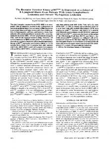
The Role of Axl in Cancer RTKs, including Axl, have been implicated in the pathophysiology of many cancers. Axl was first described in CML patients and its overexpression in fibroblasts revealed Axl’s transforming activity.7,9,44 Following its original identification, upregulation of Axl has been reported in a variety of cancers including breast,45 gastric,46 prostate,47 ovarian,48,49 lung50,51 and other (Table 1). Overexpression of Axl was shown to correlate with poorer prognosis,52,60 as well as increased invasiveness53 and xenograft growth of human cancers,40,57,101 further indicating that Axl has strong oncogenic potential. Activation of the tyrosine kinase domain of Axl in cancer cell lines was shown to be mediated through ligand Gas6 binding or overexpression of Axl7,91,102 and seems to be linked to several downstream signal transduction pathways.41,70 Figure 4 shows a summary of signal transduction pathways involved in Axl activation. Axl was further shown to be a downstream effector of the epithelial-to-mesenchymal transition (EMT) program, which is an enhancer of invasive mobility of malignant cells.52,103,104 C 2013 UICC Int. J. Cancer: 134, 1024–1033 (2014) V
Recent studies reported that Axl is overexpressed and activated in several drug-resistant cancer cell lines, suggesting that Axl may play a role in chemotherapy-resistant cancers. Increased Axl levels have been linked with Imatinib-resistant gastrointestinal stromal tumors, Nilotinib-resistant chronic myeloid leukemia cells, BMS-754087-resistant Rhabdomyosarcoma, Lapatinib-resistant HER-2 positive breast tumor cells and resistance to cisplatin in ovarian and esophageal adenocarcinoma (EAC).54,63–65,75,78,105 Although Axl is consistently associated with resistance to chemotherapy in cancer cells, the underlying pathways of Axl upregulation in this context remain unknown.106 The emerging role of Axl in cancer holds potential for cancer treatment and therefore represents an important direction for future research. In the next sections we will discuss in detail recent advances in the understanding of Axl signaling in four types of cancer: leukemia, breast, prostate and non-small lung cancer. Axl in myeloid leukemias
The Axl receptor was initially isolated from two patients with CML and its transforming activity was observed when cDNA was cloned and overexpressed in NIH 3T3 fibroblasts and
Mini Review
Table 1. Studies reporting on Axl in cancer.
1028
Mini Review
The role of Axl in cancer
Figure 4. Schematic representation of Axl signaling pathway. Activation of Axl leads to activation of the PI3K pathway, including S6K, Akt and MAP kinases, leading to tumor progression. Antiapoptotic effect of Axl is associated with activation of STAT and NF-jB signal transduction pathways. The Axl/Akt/NF-jB pathways act in order to generate the maximal antiapoptotic effect in cancer cells. This event is also induced by IL-6 secretion and activation of JAK kinases which leads to activation of STAT3.
murine myeloid precursor 32D cells.7,9,44 Recently, several studies reported upregulation of Axl in Imatinib (IM) and Nilotinib-resistant CML cell lines and patients.64,106–108 Dufies et al. (2011)106 went on to present evidence that the kinase domain of Axl is necessary for IM-sensitive cells to confer resistance to IM-treatment. In addition, it has been shown that Axl overexpression is regulated at the transcriptional level by the activator protein 1 (AP1) transcription factor and through protein kinase C (PKC) and ERK1/2 pathways in IM-resistant and phorbol-12-myristate-13-acetate (PMA)-stimulated leukemia cells.7,24,44,106 Axl was also found to be overexpressed in acute myeloid leukemia (AML) patients, where levels of its expression correlated with Bcl2 (an apoptosis regulator) and CD34 (an adhesion molecule with a role in early hematopoiesis) expression levels. Moreover, patients with AML that had high mRNA levels of AXL were found to have worse progressionfree and overall survival.60 Hong et al. (2008) observed that, similar to CML, upregulation of Axl in AML patients became refractory to doxorubicin/cytoarabine multi-agent therapy, confirming the notion that Axl expression is associated with drug resistance.63 The same study showed that expression of the endogenous Axl could be induced by chemotherapeutic drugs (doxorubicin, VP16 and cisplatin) in a dose-dependent manner in AML U937 cells.63 This induction was dependent on the methylation status of CCWGG site of the AXL
promoter, as only the CCWGG-unmethylated AXL promoter was responsive to chemotherapeutic drugs in the luciferase reporter assay.63 While overexpression of Axl by itself was insufficient to confer drug resistance, addition of exogenous Gas6 in U937 cells ectopically expressing Axl induced phosphorylation of Axl and activation of Akt and ERK1/2 survival pathways, and increased resistance to chemotherapy.63 Axl in breast cancer
Deregulation of Axl at the mRNA and protein level was found in some breast cancer cell lines, indicating that Axl is overexpressed in a subgroup of breast cancers.109 In a study by Berclaz et al. (2001) immunohistochemical (IHC) staining for Axl and estrogen receptor ER expression was performed on a panel of 23 normal and 111 malignant breast cancer samples.45 High correlation between Axl and ER expression in malignant breast tissue was observed, accompanied by a significant correlation between Axl expression and the tumor stage.45 Moreover, a large study by Gjerdrum et al. (2010) comprising 190 breast cancers showed that high expression of Axl is a negative prognostic indicator for survival in breast cancer patients.52 Holland et al. (2005) provided functional evidence that Axl signaling may contribute to human tumor growth by showing that RNAi-mediated inhibition of Axl expression reduced the growth of MDAMB-231 human breast carcinoma cells in a xenograft C 2013 UICC Int. J. Cancer: 134, 1024–1033 (2014) V
model.101 Additionally, Axl knockdown in highly metastatic breast cancer cell lines (MDA-MB-231) indicated that Axl is required for mesenchymal-like invasiveness of metastatic breast carcinoma cells, but has no effect on cell proliferation.52,53 It has also recently been shown that decreased AXL mRNA and protein levels correlate with cell migration potential and decreased phospho-Akt protein expression level in MDA-MB-231 cells, without having any significant concomitant effect on the viability or induction of apoptosis in these cell lines.56 Results of the Gjerdrum et al. study (2010) recognized the role of Axl as an Epithelial-to-mesenchymal transition (EMT)-induced downstream effector, that is required for breast cancer metastasis and progression.52 Vuoriluoto et al. (2011) recently found that the intermediate filament protein vimentin functionally contributes to EMT activation and is also required for Axl upregulation, which contributes to lung extravasation of breast cancer cells in mice.53 Regarding regulation of Axl expression in breast cancer, Mackiewicz et al. (2011)56 identified miR-34a as a key regulator of AXL expression in two independent triple-negative breast cancer cell lines (MDA-MB-231 and Hs578T). A target site for miR-34a was also found within the 3’UTR of AXL.39,56
Axl and metastatic potential of prostate cancer
The expression of Axl was found to be higher in the metastatic prostate carcinoma cell line DU145 than in the less aggressive prostate carcinoma cell line PC-3 or in normal prostate cells.47,71 Moreover, Sainaghi et al. (2005) demonstrated that Axl/Gas6 interaction induces mitogenic activity in DU145 and PC-3 cell lines, which is not mediated by inhibition of apoptosis and is proportional to Axl expression.71 Further studies demonstrated that PI3K/AKT and MEK pathways are involved in Axl/Gas6-induced proliferation of DU145 cells, however, only MEK phosphorylation was shown to be essential for growth signaling.71 However, we have recently shown that mRNA and protein expression levels of Axl are significantly higher in both androgen-insensitive prostate carcinoma cell lines, PC-3, CL1 and DU145, compared to androgen-sensitive LNCaP, CW22 and CW19 cells.70 We demonstrated that AXL is upregulated in 50% of human prostate tumors. Furthermore, we show that blockage of Axl expression inhibits proliferation, invasion and migration of prostate cancer cell lines, as well as reduces tumor formation in a xenograft mice model. We further showed that inhibition of Axl leads to inactivation of the NF-jB pathway through inhibition of Akt and IKKa activity, blocking the IL-6-STAT-3 signaling pathway.70 A suggested link between Axl and the IL-6-Stat3 signaling pathway is also described in Figure 4. These findings have an impact on prostate cancer biology and corroborate the notion that Axl may be employed as a therapeutic target for treatment of advanced prostate cancer. C 2013 UICC Int. J. Cancer: 134, 1024–1033 (2014) V
In contrast to our findings, Shiozawa et al. (2010) suggested that Gas6 inhibits proliferation of prostate carcinoma cell lines DU145 and PC-3 while protecting them from apoptotic cell death.72 The reason for this discrepancy is not yet clear, however, according to Shiozawa et al. (2010), their findings are based on the interesting concept that metastasis of prostate cancer cells to the bone marrow is similar to the “homing” behavior of hematopoietic stem cells (HSCs) to the marrow, where they reside in an area that is defined as the stem cell “niche”. Similarly, prostate cancer cells, shed from the primary tumor and disseminating to the bone marrow, are thought to use the stem cell niche for homing to the bone marrow where they lie dormant for long periods before they can be activated to form metastases.110–112 This notion is supported by previous reports where 10% of prostate cancer patients have bone metastases, and almost all patients who die of prostate cancer have skeletal involvement.113
The role of Axl in non-small cell lung cancer (NSCLC): A key player in resistance against chemotherapeutics
As previously mentioned, overexpression of Axl has been related to increased resistance of cancer cells to chemotherapeutic drugs in numerous types of cancer. Axl has been shown to be overexpressed in NSCLC50,51 and recently two independent studies reinforced the notion that Axl is important for NSCLC. Both articles demonstrated that Axl plays a key role in acquiring resistance to chemotherapy. In an impressive report by Zhang et al. (2012),67 the authors demonstrated increased activation of Axl and evidence for epithelial-to-mesenchymal transition (EMT) in the epidermal growth factor receptor (EGFR)-mutant lung cancer models with acquired resistance to erlotinib (a tyrosine kinase inhibitor used in the treatment of lung cancer) in vitro and in vivo. Inhibition of Axl leads to restoration of sensitivity to erlotinib, indicating that Axl may represent a promising therapeutic target whose inhibition could prevent or overcome acquired resistance to drugs.67 Using cells with blocked vimentin (EMT biomarker), they demonstrated that Axl expression is significantly reduced, proving that Axl-mediated resistance occurs during EMT. In addition they showed that erlotinib-resistant cells exhibit higher rates of migration and adhesion; however, blockage of vimentin or Axl leads to reduction in these characteristics. Evaluation of expression of Axl in patients before treatment with an EGFR inhibitor (erlotinib or gefitinib) and after, when resistance to the inhibitor had developed, further demonstrated that upregulation of Axl is critical for the development of resistance to both erlotinib and gefitinib.67 In an independent report, Byers et al. (2012) developed and validated a 76-gene EMT signature using gene expression profiles from 54 NSCLC cell lines and patients treated in a clinical study using three different arrays.68 The signature was used in predicting resistance to inhibitors (EGFR and PI3K/Akt) and identified Axl as a pivotal mediator of
Mini Review
1029
Paccez et al.
Mini Review
1030
resistance to EGFR inhibitor associated with mesenchymal phenotype. They interrogated their signature in two groups of NSCLC cell lines; epithelial and mesenchymal. Interestingly, the signature strongly correlates with the mesenchymal group but not with the epithelial one. The authors identified upregulation of Axl in the mesenchymal group, but not in the epithelial group. Additionally, they observed that inhibition of Axl in mesenchymal cells was able to reverse the acquired resistance to the drug treatments.68 Taken together, these findings highlight two other features related to Axl and cancer: (i) the association with the EMT phenotype and (ii) the role in resistance to chemotherapeutic drugs.
Axl as a Potential Target for Cancer Therapy As previously discussed, aberrant activation of more than half of RTKs, as well as many cytoplasmic TKs, has been implicated in a whole variety of cancer types. Genetic and possibly epigenetic deregulations are the main mechanisms leading to aberrant activation of RTKs in human cancers. TKs represent a major class of proto-oncogenes involved in tumor growth, progression and metastasis of cancer cells. For this reason, this class of proteins is being actively studied as a target for therapeutic intervention. In fact, tyrosine kinase inhibitors, such as Imatinib mesylate (Gleevec), Herceptin, Tarceva and Iressa have shown efficacy in clinical trials.114 In this context, the development of specific inhibitors for Axl would improve the treatment of different types of cancer in which Axl is known to play an important role in malignancy. Recent studies, using RNAi and therapeutic agents, validate the therapeutic value of Axl inhibition. Ye et al. (2010)58 developed a monoclonal antibody (YW327.6S2) against Axl, which recognizes the receptor with high affinity. The YW327.6S2 antibody has been shown to diminish xenograft tumor growth and potentiates the effect of anti-VEGF treatment. Moreover, treatment with YW327.6S2 also enhances the effect of erlotinib and chemotherapy in reducing NSCLC tumor growth, as well as the reduction of breast cancer metastasis to distant organs.58 Development of small molecule Axl inhibitors
After characterization of Axl as an important mediator in the progression of cancer, the search for specific inhibitors intensified. The identification of 3-quinolinecarbonitrile compounds was achieved using the Nested Chemical Library technology— a hit and lead finding method for rational drug design of kinase inhibitors, developed by Keri et al.115 This class of compounds demonstrated potent inhibitory activity against Axl as well as strong inhibition of motility and invasiveness, especially in breast cancer cells.59 After analysis of around 300 compounds, they identified that NA80x1 directly inhibits Axl phosphorylation in a dose-dependent manner and has previously been reported to display inhibitory activity against the Src kinase.116,117 It is of note that the same approach also identified SKI-606, a Src/Abl inhibitor as a potent Axl inhibitor.
The role of Axl in cancer
The compound was shown to inhibit Axl in doses 20 times lower than NA80x1. The authors suggest that both small molecules (SKI-606 and NA80x1) might be potential therapeutic agents for blocking breast cancer growth and metastasis. However, SKI-606 and NA80x1 are not specific for Axl inhibition, and show effects in the micro-molar range. In 2010, Holland et al.57 described the development of a new small molecule that inhibits Axl in the nanomolar range. The molecule named R428 ((1-(6,7-dihydro-5H-benzo[6,7]cyclohepta[1,2-c] pyridazin-3-yl)-N3-((7-pyrrolidin-1-yl)-6,7,8, 9-tetrahydro-5Hbenzo[7]annulene-2-yl)-1H-1,2,4-triazole-3,5diamine) inhibits Axl activity and blocks Axl-dependent signaling events, such as Akt phosphorylation, cancer cell invasion, and pro-inflammatory cytokine production. A dosedependent reduction of EMT transcriptional regulator Snail and an inhibition of angiogenesis and tumor formation was also achieved. Of significance is the fact that extension in survival was observed in vivo using a mouse model. Animals receiving treatment with R428 have median survival rates of over 80 days in breast (MDA-MB-231) and lung (4T1) cancer models, while non-treated animals’ survival rate was 52 days. The compound also showed synergistic effects in mice treated with cisplatin, leading to suppression of liver metastasis.57 Mollard et al. (2011), using an in silico approach—the scaffold-based flexile docking method (described by Abagyan et al. (1994)),118 developed a series of compounds with Axl inhibitory properties119 and identified 2,4,5-trisubstitued pyrimidines as potent Axl inhibitors with activity in the nanomolar range.119 Moreover, Cerchia et al. (2012)120 used the concept of RNA Aptamers to develop GL21.T, a new specific Axl inhibitor. Aptamers are molecules (generally stable DNA or RNA) that bind targets with high affinity and specificity. This Axl inhibitor compound was able to bind the ectodomain regions of Tyro3 (Kd 5 43 nmol/L) and Axl (Kd 5 13 nmol/L).120 Using a xenograft fluorescent model, the authors further demonstrated that (i) aptamer specifically accumulates in the tumor region and (ii) mice treated with GL21.T developed 68.2% smaller tumors than non-treated controls.120 The description of molecules with different structures (antibodies, compounds and nucleic acid aptamers) reveal the versatility that can be applied in the development of Axl inhibitors and may represent an innovative approach in drug development.
Concluding Remarks The receptor tyrosine kinase Axl has been shown to play important roles in tumorigenesis. Upregulation of Axl has been related to poor diagnosis in different types of cancer and its inhibition has been proven to be an important mechanism in inhibiting cancer progression. In this context, the development of therapies involving the inhibition of Axl expression as well as its downstream effectors may represent a novel approach in the fight against cancer. Future work employing high throughput screening strategies with natural and synthetic libraries to C 2013 UICC Int. J. Cancer: 134, 1024–1033 (2014) V
1031
Paccez et al.
identify and develop small molecule drugs with potential to selectively inhibit Axl expression and its pathways in cancer cells is clearly a worthy field for exploration.
Acknowledgements J.D.P. and M.V. are supported by an International Centre for Genetic Engineering and Biotechnology post-doctoral fellowship.
1.
2. 3.
4.
5.
6.
7.
8.
9.
10.
11.
12.
13.
14.
15.
16.
Hunter AJ, Ottoson N, Boerth N, et al. Cutting edge: a novel function for the SLAP-130/FYB adapter protein in beta 1 integrin signaling and T lymphocyte migration. J Immunol 2000;164:1143–7. Schlessinger J. Cell signaling by receptor tyrosine kinases. Cell 2000;103:211–25. Schenk PW, Snaar-Jagalska BE. Signal perception and transduction: the role of protein kinases. Biochim Biophys Acta 1999;1449:1–24. Robinson DR, Wu YM, Lin SF. The protein tyrosine kinase family of the human genome. Oncogene 2000;19:5548–57. Graham DK, Bowman GW, Dawson TL, et al. Cloning and developmental expression analysis of the murine c-mer tyrosine kinase. Oncogene 1995;10:2349–59. Neubauer A, Fiebeler A, Graham DK, et al. Expression of axl, a transforming receptor tyrosine kinase, in normal and malignant hematopoiesis. Blood 1994;84:1931–41. O’Bryan JP, Frye RA, Cogswell PC, et al. axl, a transforming gene isolated from primary human myeloid leukemia cells, encodes a novel receptor tyrosine kinase. Mol Cell Biol 1991;11:5016–31. Angelillo-Scherrer A, de Frutos P, Aparicio C, et al. Deficiency or inhibition of Gas6 causes platelet dysfunction and protects mice against thrombosis. Nat Med 2001;7:215–21. Janssen JW, Schulz AS, Steenvoorden AC, et al. A novel putative tyrosine kinase receptor with oncogenic potential. Oncogene 1991;6:2113–20. McCloskey P, Fridell YW, Attar E, et al. GAS6 mediates adhesion of cells expressing the receptor tyrosine kinase Axl. J Biol Chem 1997;272:23285–91. Lee WP, Liao Y, Robinson D, et al. Axl-gas6 interaction counteracts E1A-mediated cell growth suppression and proapoptotic activity. Mol Cell Biol 1999;19:8075–82. Angelillo-Scherrer A, Burnier L, Flores N, et al. Role of Gas6 receptors in platelet signaling during thrombus stabilization and implications for antithrombotic therapy. J Clin Invest 2005;115:237–46. Braunger J, Schleithoff L, Schulz AS, et al. Intracellular signaling of the Ufo/Axl receptor tyrosine kinase is mediated mainly by a multisubstrate docking-site. Oncogene 1997;14:2619– 31. Gebhardt F, Burger H, Brandt B. Modulation of EGFR gene transcription by secondary structures, a polymorphic repetitive sequence and mutations–a link between genetics and epigenetics. Histol Histopathol 2000;15:929–36. Tanabe K, Nagata K, Ohashi K, et al. Roles of gamma-carboxylation and a sex hormonebinding globulin-like domain in receptorbinding and in biological activities of Gas6. FEBS Lett 1997;408:306–10. Mark MR, Chen J, Hammonds RG, et al. Characterization of Gas6, a member of the superfamily of G domain-containing proteins, as a ligand for Rse and Axl. J Biol Chem 1996;271:9785–9.
17.
18.
19.
20.
21.
22.
23.
24.
25.
26.
27.
28.
29.
30.
31.
C 2013 UICC Int. J. Cancer: 134, 1024–1033 (2014) V
Manfioletti G, Brancolini C, Avanzi G, et al. The protein encoded by a growth arrest-specific gene (gas6) is a new member of the vitamin Kdependent proteins related to protein S, a negative coregulator in the blood coagulation cascade. Mol Cell Biol 1993;13:4976–85. Goruppi S, Ruaro E, Schneider C. Gas6, the ligand of Axl tyrosine kinase receptor, has mitogenic and survival activities for serum starved NIH3T3 fibroblasts. Oncogene 1996;12:471–80. Goruppi S, Ruaro E, Varnum B, et al. Requirement of phosphatidylinositol 3-kinasedependent pathway and Src for Gas6-Axl mitogenic and survival activities in NIH 3T3 fibroblasts. Mol Cell Biol 1997;17:4442–53. Goruppi S, Yamane H, Marcandalli P, et al. The product of a gas6 splice variant allows the release of the domain responsible for Axl tyrosine kinase receptor activation. FEBS Lett 1997;415:59–63. Bellosta P, Zhang Q, Goff SP, et al. Signaling through the ARK tyrosine kinase receptor protects from apoptosis in the absence of growth stimulation. Oncogene 1997;15:2387–97. Schulz AS, Schleithoff L, Faust M, et al. The genomic structure of the human UFO receptor. Oncogene 1993;8:509–13. Mudduluru G, Allgayer H. The human receptor tyrosine kinase Axl gene–promoter characterization and regulation of constitutive expression by Sp1, Sp3 and CpG methylation. Biosci Rep 2008;28:161–76. Mudduluru G, Leupold JH, Stroebel P, et al. PMA up-regulates the transcription of Axl by AP-1 transcription factor binding to TRE sequences via the MAPK cascade in leukaemia cells. Biol Cell 2010;103:21–33. Mudduluru G, Vajkoczy P, Allgayer H. Myeloid zinc finger 1 induces migration, invasion, and in vivo metastasis through Axl gene expression in solid cancer. Mol Cancer Res 2010;8:159–69. Sayan AE, Stanford R, Vickery R, et al. Fra-1 controls motility of bladder cancer cells via transcriptional upregulation of the receptor tyrosine kinase AXL. Oncogene 2012;31:1493– 503. Bartolomei MS, Webber AL, Brunkow ME, et al. Epigenetic mechanisms underlying the imprinting of the mouse H19 gene. Genes Dev 1993;7:1663–73. Carlone DL, Lee JH, Young SR, et al. Reduced genomic cytosine methylation and defective cellular differentiation in embryonic stem cells lacking CpG binding protein. Mol Cell Biol 2005;25:4881–91. Carlone DL, Skalnik DG. CpG binding protein is crucial for early embryonic development. Mol Cell Biol 2001;21:7601–6. Zhu WG, Srinivasan K, Dai Z, et al. Methylation of adjacent CpG sites affects Sp1/ Sp3 binding and activity in the p21(Cip1) promoter. Mol Cell Biol 2003;23:4056–65. Ammanamanchi S, Brattain MG. Sp3 is a transcriptional repressor of transforming growth
32.
33.
34.
35. 36.
37.
38.
39.
40.
41.
42.
43.
44.
45.
46.
47.
48.
factor-beta receptors. J Biol Chem 2001;276:3348–52. Ammanamanchi S, Freeman JW, Brattain MG. Acetylated sp3 is a transcriptional activator. J Biol Chem 2003;278:35775–80. Zhao Y, Srivastava D. A developmental view of microRNA function. Trends Biochem Sci 2007;32:189–97. Erson AE, Petty EM. MicroRNAs in development and disease. Clin Genet 2008;74:296–306. Cho WC. OncomiRs: the discovery and progress of microRNAs in cancers. Mol Cancer 2007;6:60. Tili E, Michaille JJ, Gandhi V, et al. miRNAs and their potential for use against cancer and other diseases. Future Oncol 2007;3:521–37. Lujambio A, Esteller M. How epigenetics can explain human metastasis: a new role for microRNAs. Cell Cycle 2009;8:377–82. Valeri N, Croce CM, Fabbri M. Pathogenetic and clinical relevance of microRNAs in colorectal cancer. Cancer Genomics Proteomics 2009;6:195–204. Mudduluru G, Ceppi P, Kumarswamy R, et al. Regulation of Axl receptor tyrosine kinase expression by miR-34a and miR-199a/b in solid cancer. Oncogene 2011;30:2888–99. Vajkoczy P, Knyazev P, Kunkel A, et al. Dominant-negative inhibition of the Axl receptor tyrosine kinase suppresses brain tumor cell growth and invasion and prolongs survival. Proc Natl Acad Sci USA 2006;103:5799–804. Hafizi S, Dahlback B. Signalling and functional diversity within the Axl subfamily of receptor tyrosine kinases. Cytokine Growth Factor Rev 2006;17:295–304. Bommer GT, Gerin I, Feng Y, et al. p53mediated activation of miRNA34 candidate tumor-suppressor genes. Curr Biol 2007;17:1298–307. Welch C, Chen Y, Stallings RL. MicroRNA-34a functions as a potential tumor suppressor by inducing apoptosis in neuroblastoma cells. Oncogene 2007;26:5017–22. McCloskey P, Pierce J, Koski RA, et al. Activation of the Axl receptor tyrosine kinase induces mitogenesis and transformation in 32D cells. Cell Growth Differ 1994;5:1105–17. Berclaz G, Altermatt HJ, Rohrbach V, et al. Estrogen dependent expression of the receptor tyrosine kinase axl in normal and malignant human breast. Ann Oncol 2001;12:819–24. Wu CW, Li AF, Chi CW, et al. Clinical significance of AXL kinase family in gastric cancer. Anticancer Res 2002;22: 1071–8. Jacob AN, Kalapurakal J, Davidson WR, et al. A receptor tyrosine kinase, UFO/Axl, and other genes isolated by a modified differential display PCR are overexpressed in metastatic prostatic carcinoma cell line DU145. Cancer Detect Prev 1999;23:325–32. Sun W, Fujimoto J, Tamaya T. Coexpression of Gas6/Axl in human ovarian cancers. Oncology 2004;66:450–7.
Mini Review
References
1032
49.
50.
51.
Mini Review
52.
53.
54.
55.
56.
57.
58.
59.
60.
61.
62.
63.
64.
Rankin EB, Fuh KC, Taylor TE, et al. AXL is an essential factor and therapeutic target for metastatic ovarian cancer. Cancer Res 2010;70:7570–9. Shieh YS, Lai CY, Kao YR, et al. Expression of axl in lung adenocarcinoma and correlation with tumor progression. Neoplasia 2005;7:1058– 64. Wimmel A, Glitz D, Kraus A, et al. Axl receptor tyrosine kinase expression in human lung cancer cell lines correlates with cellular adhesion. Eur J Cancer 2001;37:2264–74. Gjerdrum C, Tiron C, Hoiby T, et al. Axl is an essential epithelial-to-mesenchymal transitioninduced regulator of breast cancer metastasis and patient survival. Proc Natl Acad Sci USA 2010;107:1124–9. Vuoriluoto K, Haugen H, Kiviluoto S, et al. Vimentin regulates EMT induction by Slug and oncogenic H-Ras and migration by governing Axl expression in breast cancer. Oncogene 2011;30:1436–48. Liu L, Greger J, Shi H, et al. Novel mechanism of lapatinib resistance in HER2-positive breast tumor cells: activation of AXL. Cancer Res 2009;69:6871–8. Johnson CE, Gorringe KL, Thompson ER, et al. Identification of copy number alterations associated with the progression of DCIS to invasive ductal carcinoma. Breast Cancer Res Treat 2012;133:889–98. Mackiewicz M, Huppi K, Pitt JJ, et al. Identification of the receptor tyrosine kinase AXL in breast cancer as a target for the human miR-34a microRNA. Breast Cancer Res Treat 2011;130:663–79. Holland SJ, Pan A, Franci C, et al. R428, a selective small molecule inhibitor of Axl kinase, blocks tumor spread and prolongs survival in models of metastatic breast cancer. Cancer Res 2010;70:1544–54. Ye X, Li Y, Stawicki S, et al. An anti-Axl monoclonal antibody attenuates xenograft tumor growth and enhances the effect of multiple anticancer therapies. Oncogene 2010;29:5254–64. Zhang YX, Knyazev PG, Cheburkin YV, et al. AXL is a potential target for therapeutic intervention in breast cancer progression. Cancer Res 2008;68:1905–15. Rochlitz C, Lohri A, Bacchi M, et al. Axl expression is associated with adverse prognosis and with expression of Bcl-2 and CD34 in de novo acute myeloid leukemia (AML): results from a multicenter trial of the Swiss Group for Clinical Cancer Research (SAKK). Leukemia 1999;13:1352–8. Challier C, Uphoff CC, Janssen JW, et al. Differential expression of the ufo/axl oncogene in human leukemia-lymphoma cell lines. Leukemia 1996;10:781–7. Neubauer A, O’Bryan JP, Fiebeler A, et al. Axl, a novel receptor tyrosine kinase isolated from chronic myelogenous leukemia. Semin Hematol 1993;30:34. Hong CC, Lay JD, Huang JS, et al. Receptor tyrosine kinase AXL is induced by chemotherapy drugs and overexpression of AXL confers drug resistance in acute myeloid leukemia. Cancer Lett 2008;268: 314–24. Gioia R, Leroy C, Drullion C, et al. Quantitative phosphoproteomics revealed interplay between Syk and Lyn in the resistance to nilotinib in
The role of Axl in cancer
65.
66.
67.
68.
69.
70.
71.
72.
73.
74.
75.
76.
77.
78.
79.
80.
81.
chronic myeloid leukemia cells. Blood 2011;118:2211–21. Lay JD, Hong CC, Huang JS, et al. Sulfasalazine suppresses drug resistance and invasiveness of lung adenocarcinoma cells expressing AXL. Cancer Res 2007;67:3878–87. Lee JH, Voortman J, Dingemans AM, et al. MicroRNA expression and clinical outcome of small cell lung cancer. PLoS One 2011;6:e21300. Zhang Z, Lee JC, Lin L, et al. Activation of the AXL kinase causes resistance to EGFR-targeted therapy in lung cancer. Nat Genet 2012;44:852– 60. Byers LA, Diao L, Wang J, et al. An epithelialmesenchymal transition (EMT) gene signature predicts resistance to EGFR and PI3K inhibitors and identifies Axl as a therapeutic target for overcoming EGFR inhibitor resistance. Clin Cancer Res 2013;19:279–90. Yakes FM, Chen J, Tan J, et al. Cabozantinib (XL184), a novel MET and VEGFR2 inhibitor, simultaneously suppresses metastasis, angiogenesis, and tumor growth. Mol Cancer Ther 2011;10:2298–308. Paccez JD, Vasques GJ, Correa RG, et al. The receptor tyrosine kinase Axl is an essential regulator of prostate cancer proliferation and tumor growth and represents a new therapeutic target. Oncogene 2013;32:689–98. Sainaghi PP, Castello L, Bergamasco L, et al. Gas6 induces proliferation in prostate carcinoma cell lines expressing the Axl receptor. J Cell Physiol 2005;204:36–44. Shiozawa Y, Pedersen EA, Patel LR, et al. GAS6/AXL axis regulates prostate cancer invasion, proliferation, and survival in the bone marrow niche. Neoplasia 2010;12:116–27. Jansen FH, van Rijswijk A, Teubel W, et al. Profiling of antibody production against xenograft-released proteins by protein microarrays discovers prostate cancer markers. J Proteome Res 2012;11:728–35. Sawabu T, Seno H, Kawashima T, et al. Growth arrest-specific gene 6 and Axl signaling enhances gastric cancer cell survival via Akt pathway. Mol Carcinog 2007;46:155–64. Mahadevan D, Cooke L, Riley C, et al. A novel tyrosine kinase switch is a mechanism of imatinib resistance in gastrointestinal stromal tumors. Oncogene 2007;26:3909–19. Hong J, Peng D, Chen Z, et al. ABL regulation by AXL promotes cisplatin resistance in esophageal cancer. Cancer Res 2013;73: 331–40. Hector A, Montgomery EA, Karikari C, et al. The Axl receptor tyrosine kinase is an adverse prognostic factor and a therapeutic target in esophageal adenocarcinoma. Cancer Biol Ther 2010;10:1009–18. Macleod K, Mullen P, Sewell J, et al. Altered ErbB receptor signaling and gene expression in cisplatin-resistant ovarian cancer. Cancer Res 2005;65:6789–800. Jiao Y, Ou W, Meng F, et al. Targeting HSP90 in ovarian cancers with multiple receptor tyrosine kinase coactivation. Mol Cancer 2011;10:125. Tworkoski K, Singhal G, Szpakowski S, et al. Phosphoproteomic screen identifies potential therapeutic targets in melanoma. Mol Cancer Res 2011;9:801–12. Green J, Ikram M, Vyas J, et al. Overexpression of the Axl tyrosine kinase receptor in cutaneous
82.
83.
84.
85.
86.
87.
88.
89.
90.
91.
92.
93.
94.
95.
96.
97.
98.
SCC-derived cell lines and tumours. Br J Cancer 2006;94:1446–51. Sensi M, Catani M, Castellano G, et al. Human cutaneous melanomas lacking MITF and melanocyte differentiation antigens express a functional Axl receptor kinase. J Invest Dermatol 2011;131:2448–57. Song X, Wang H, Logsdon CD, et al. Overexpression of receptor tyrosine kinase Axl promotes tumor cell invasion and survival in pancreatic ductal adenocarcinoma. Cancer 2011;117:734–43. Koorstra JB, Karikari CA, Feldmann G, et al. The Axl receptor tyrosine kinase confers an adverse prognostic influence in pancreatic cancer and represents a new therapeutic target. Cancer Biol Ther 2009;8:618–26. Yeh CY, Shin SM, Yeh HH, et al. Transcriptional activation of the Axl and PDGFR-alpha by c-Met through a ras- and Srcindependent mechanism in human bladder cancer. BMC Cancer 2011;11:139. He L, Zhang J, Jiang L, et al. Differential expression of Axl in hepatocellular carcinoma and correlation with tumor lymphatic metastasis. Mol Carcinog 2010;49:882–91. Xu MZ, Chan SW, Liu AM, et al. AXL receptor kinase is a mediator of YAP-dependent oncogenic functions in hepatocellular carcinoma. Oncogene 2011;30:1229–40. Gustafsson A, Bostrom AK, Ljungberg B, et al. Gas6 and the receptor tyrosine kinase Axl in clear cell renal cell carcinoma. PLoS One 2009;4:e7575. Gustafsson A, Martuszewska D, Johansson M, et al. Differential expression of Axl and Gas6 in renal cell carcinoma reflecting tumor advancement and survival. Clin Cancer Res 2009;15:4742–9. Chung BI, Malkowicz SB, Nguyen TB, et al. Expression of the proto-oncogene Axl in renal cell carcinoma. DNA Cell Biol 2003;22:533–40. Avilla E, Guarino V, Visciano C, et al. Activation of TYRO3/AXL tyrosine kinase receptors in thyroid cancer. Cancer Res 2011;71:1792–804. Ito M, Nakashima M, Nakayama T, et al. Expression of receptor-type tyrosine kinase, Axl, and its ligand, Gas6, in pediatric thyroid carcinomas around chernobyl. Thyroid 2002;12:971–5. Ito T, Ito M, Naito S, et al. Expression of the Axl receptor tyrosine kinase in human thyroid carcinoma. Thyroid 1999;9:563–7. Liu R, Gong M, Li X, et al. Induction, regulation, and biologic function of Axl receptor tyrosine kinase in Kaposi sarcoma. Blood 2010;116:297–305. Hutterer M, Knyazev P, Abate A, et al. Axl and growth arrest-specific gene 6 are frequently overexpressed in human gliomas and predict poor prognosis in patients with glioblastoma multiforme. Clin Cancer Res 2008;14:130–8. van Ginkel PR, Gee RL, Shearer RL, et al. Expression of the receptor tyrosine kinase Axl promotes ocular melanoma cell survival. Cancer Res 2004;64:128–34. Nakano T, Tani M, Ishibashi Y, et al. Biological properties and gene expression associated with metastatic potential of human osteosarcoma. Clin Exp Metastasis 2003;20:665–74. Sun WS, Fujimoto J, Tamaya T. Coexpression of growth arrest-specific gene 6 and receptor
C 2013 UICC Int. J. Cancer: 134, 1024–1033 (2014) V
99.
100.
101.
102.
103.
104.
105.
tyrosine kinases Axl and Sky in human uterine endometrial cancers. Ann Oncol 2003;14:898– 906. Sun WS, Fujimoto J, Tamaya T. Clinical implications of coexpression of growth arrestspecific gene 6 and receptor tyrosine kinases Axl and Sky in human uterine leiomyoma. Mol Hum Reprod 2003;9:701–7. Keating AK, Kim GK, Jones AE, et al. Inhibition of Mer and Axl receptor tyrosine kinases in astrocytoma cells leads to increased apoptosis and improved chemosensitivity. Mol Cancer Ther 2010;9:1298–307. Holland SJ, Powell MJ, Franci C, et al. Multiple roles for the receptor tyrosine kinase axl in tumor formation. Cancer Res 2005;65:9294–303. Varnum BC, Young C, Elliott G, et al. Axl receptor tyrosine kinase stimulated by the vitamin K-dependent protein encoded by growth-arrest-specific gene 6. Nature 1995;373:623–6. Thiery JP. Epithelial-mesenchymal transitions in tumour progression. Nat Rev Cancer 2002;2:442–54. Polyak K, Weinberg RA. Transitions between epithelial and mesenchymal states: acquisition of malignant and stem cell traits. Nat Rev Cancer 2009;9:265–73. Huang F, Hurlburt W, Greer A, et al. Differential mechanisms of acquired resistance to insulin-like growth factor-i receptor antibody therapy or to a small-molecule inhibitor, BMS-
106.
107.
108.
109.
110.
111.
112.
C 2013 UICC Int. J. Cancer: 134, 1024–1033 (2014) V
754807, in a human rhabdomyosarcoma model. Cancer Res 2010;70:7221–31. Dufies M, Jacquel A, Belhacene N, et al. Mechanisms of AXL overexpression and function in Imatinib-resistant chronic myeloid leukemia cells. Oncotarget 2011;2:874–85. Gamas P, Marchetti S, Puissant A, et al. Inhibition of imatinib-mediated apoptosis by the caspase-cleaved form of the tyrosine kinase Lyn in chronic myelogenous leukemia cells. Leukemia 2009;23:1500–6. Grosso S, Puissant A, Dufies M, et al. Gene expression profiling of imatinib and PD166326resistant CML cell lines identifies Fyn as a gene associated with resistance to BCR-ABL inhibitors. Mol Cancer Ther 2009;8:1924–33. Meric F, Lee WP, Sahin A, et al. Expression profile of tyrosine kinases in breast cancer. Clin Cancer Res 2002;8:361–7. Morgan TM, Lange PH, Porter MP, et al. Disseminated tumor cells in prostate cancer patients after radical prostatectomy and without evidence of disease predicts biochemical recurrence. Clin Cancer Res 2009;15:677–83. Nash KT, Phadke PA, Navenot JM, et al. Requirement of KISS1 secretion for multiple organ metastasis suppression and maintenance of tumor dormancy. J Natl Cancer Inst 2007;99:309–21. Pantel K, Alix-Panabieres C, Riethdorf S. Cancer micrometastases. Nat Rev Clin Oncol 2009;6:339–51.
113. Landis SH, Murray T, Bolden S, et al. Cancer statistics, 1999. CA Cancer J Clin 1999;49:8–31. 114. Gschwind A, Fischer OM, Ullrich A. The discovery of receptor tyrosine kinases: targets for cancer therapy. Nat Rev Cancer 2004;4:361– 70. 115. Keri G, Szekelyhidi Z, Banhegyi P, et al. Drug discovery in the kinase inhibitory field using the Nested Chemical Library technology. Assay Drug Dev Technol 2005;3: 543–51. 116. Boschelli DH, Barrios Sosa AC, Golas JM, et al. Inhibition of Src kinase activity by 7-ethynyl-4phenylamino-3-quinolinecarbonitriles: identification of SKS-927. Bioorg Med Chem Lett 2007;17:1358–61. 117. Boschelli DH, Wang YD, Ye F, et al. Synthesis and Src kinase inhibitory activity of a series of 4-phenylamino-3-quinolinecarbonitriles. J Med Chem 2001;44:822–33. 118. Abagyan R, Totrov M, Kuznetsov D. ICM—a new method for protein modeling and design: applications to docking and structure prediction from the distorted native conformation. J Comput Chem 1994;15:488–506. 119. Mollard A, Warner SL, Call LT, et al. Design, synthesis and biological evaluation of a series of novel Axl kinase inhibitors. ACS Med Chem Lett 2011;2:907–12. 120. Cerchia L, Esposito CL, Camorani S, et al. Targeting Axl with an high-affinity inhibitory aptamer. Mol Ther 2012;20:2291–303.
Mini Review
1033
Paccez et al.