Available online at www.sciencedirect.com
ScienceDirect Procedia Technology 22 (2016) 1106 – 1112
9th International Conference Interdisciplinarity in Engineering, INTER-ENG 2015, 8-9 October 2015, Tirgu-Mures, Romania
The relationship between cortisol and the hippocampal volume in depressed patients – a MRI pilot study Theodor Moicaa, Adrian Gligorb, Sorina Moicab,* a b
First Clinic of Psychiatry Tirgu-Mures, 38 Gh. Marinescu street, Tirgu-Mures 540088, Romania ”Petru Maior” University of Tirgu-Mures, 1 Nicolae Iorga street, Tirgu-Mures 540139, Romania
Abstract Stress is responsible for increased cortisol levels in depression, affecting different areas of the brain, especially the prefrontal cortex and the hippocampus. Our paper is analysing the interrelation between cortisol – as a marker of neuroendocrine response to stress and the hippocampal volume subfields – as markers of neurogenesis and neuroplasticity theory. The main goal of the research was focused to investigate and to prove the relation between the increased level of cortisol and the reduced volume of hippocampus in depression. We measured the morning serum cortisol level in a fasting state. For the volume of the hippocampus all patients underwent a MRI of the brain examination. The investigations have shown that both the right and the left hippocampal volumes are reduced in MDD patients. Higher levels of cortisol are associated with lower hippocampal subfield volumes, especially on the right. © by Elsevier Ltd. This is an open access article under the CC BY-NC-ND license © 2016 2016Published The Authors. Published by Elsevier Ltd. (http://creativecommons.org/licenses/by-nc-nd/4.0/). Peer-review under responsibility of the “Petru Maior” University of Tirgu-Mures, Faculty of Engineering. Peer-review under responsibility of the “Petru Maior” University of Tirgu Mures, Faculty of Engineering Keywords: cortisol; depression; hippocampus; applied statistics.
1. Introduction Mental Health Organization considers that depression is the disease of our epoch and around 5% of the world population suffers from this disease. It seems that females around 35-40 years are more predisposed to depression than males, where the incidence is higher after 55 years old [1]. Major Depressive Disorder (MDD) is a chronic
* Corresponding author. Tel.: +40-729-980-619; fax: +40+265-233-112. E-mail address:
[email protected]
2212-0173 © 2016 Published by Elsevier Ltd. This is an open access article under the CC BY-NC-ND license (http://creativecommons.org/licenses/by-nc-nd/4.0/). Peer-review under responsibility of the “Petru Maior” University of Tirgu Mures, Faculty of Engineering doi:10.1016/j.protcy.2016.01.156
Theodor Moica et al. / Procedia Technology 22 (2016) 1106 – 1112
1107
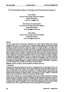
disease that affects about 15% of the population in USA [2]. The estimated prevalence in Europe is the same in some epidemiological trials [3] or even higher [4]. In a report published in 2011, authors showed that every year over a third of the total European Union population suffers from mental disorders and the true size and burden of these disorders is likely to be considerably larger in the future [5]. Since monoamine theories fail to fully explain the mechanism of depressive disease and the complex mechanism of action of antidepressants, in the last years new theories have been proposed. These theories are involving the role of Brain Derived Neurotrophic Factor (BDNF) and the glutamatergic system; the revaluation role of cortisol and the activation of hypothalamic-pituitary-adrenal axis (HPAA); the modifications of the hippocampal volume as a marker of the theory of neuroplasticity and neuronal regeneration. Hypersecretion of cortisol is an important factor in the pathophysiology of depression [6] determining atrophical modifications in the hippocampus, a limbic structure involved in cognition, emotions, affect and memory processes [7,8]. The degree of atrophy of the hippocampus can be assessed by measuring its volume, taking into consideration that antidepressive therapy induces neurogenesis processes at this level. When a stressing factor acts on the body and the glucocorticoid cascade is activated, the hippocampus exerts a negative feed-back on hypersecretion of the cortisol. Since currently there are just a few studies that measured both the level of cortisol and the volume of the hippocampus in depressed patients, we have realized a study regarding the assessment of correlation between the increased level of cortisol and the reduced volume of the hippocampus in depression. 2. Materials and Methods The purpose of this research was to study the relationship between cortisol and the hippocampal volume in the context of Recurrent Major Depressive Disorder (R-MDD). We conducted a study, on a number of 7 patients hospitalized in the First Clinic of Psychiatry Tirgu-Mures, diagnosed with recurrent MDD. Major Depressive Episode. All clinical investigations have been conducted in accordance with the guidelines in The Declaration of Helsinki and have been formally approved by The Ethics Committee of the Institution. All patients gave their consent to participate in the study by signing an informed consent and within 7 days from the time of admission, morning serum cortisol level was measured. Blood samples were collected from all patients in the morning, in a fasting state, at 08:00 AM, about 30-40 minutes after awaking and analyzed in Bioclinica Laboratory, Tirgu-Mures. For measuring the volume of the hippocampus all patients underwent a magnetic resonance imaging (MRI) of the brain and the imaging data were analyzed with FreeSurfer Version 5.3.0 (FreeSurfer; The General Hospital Corporation, Boston MA, USA) on Linux workstation for obtaining numerical measures. All of the subjects were scanned with a 1.5-T MRI scanner and had the same parameters of acquisition: 1.3 mm thickness; no gap; 160 slices; scanning time 10 min 13 s; repetition time/echo time 10/4.3 ms; number of signal averages 1; matrix 256×256; field of view 22×22 cm; and flip angle 8° [9]. The statistical analysis was performed with Statistical Package for Social Sciences (SPSS) Version 20. FreeSurfer is a computer software tool which provides computational algorithms for the cortical and subcortical volumes and surfaces determination, including the hippocampus. It is assigning a neuroanatomical label to each voxel in a MRI volume based on probabilistic information. Freesurfer morphometric procedures have been demonstrated to show good test-retest reliability across scanner manufacturers and across field strengths [10,11]. The technical details related with FreeSurfer were described in previous publications [12,13]. 3. Results The mean age of the entire group was Mage = 39.60 with a Standard deviation of 3.89. The mean number of Major Depressive Episodes (MDE) was M = 2.7 and the mean duration of the disease were 3.7 years. Mean volume for the right hippocampus was MRH = 3083.20, SDRH = 636.59, SERH = 240.60 (see Figure 1).
1108
Theodor Moica et al. / Procedia Technology 22 (2016) 1106 – 1112
Fig. 1: Histogram of right hippocampus volume
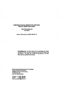
Fig. 2: Histogram of left hippocampus volume
We applied two normality tests (see Table 1) and our data regarding the volume of right hippocampus passed the normality tests, so we can say that there is a normal, parametric distribution of the data (p>0.05, H0 is accepted, meaning that there is no difference between our group and the general population). Table 1: Tests of Normality for the volumes of right hippocampus
Kolmogorov-Smirnov Statistic Right_Hippocampus
df
Shapiro-Wilk Sig.
.205
7
Statistic .200
df
Sig.
.913
7
.414
Mean volume for the left hippocampus was MLH = 2739.26, SDLH = 503.64, SELH = 190.35 (see Figure 2). Our data regarding the volume of left hippocampus also passed the normality tests (see Table 2), so we can say that there is a normal, parametric distribution of the data (p>0.05, H0 is accepted). Table 2: Tests of Normality for the volumes of Left Hippocampus
Kolmogorov-Smirnov Statistic Left_Hippocampus
.197
df
Shapiro-Wilk Sig.
7
Statistic .200
.914
df
Sig. 7
.423
The Coefficient of Variation (CV) for a single variable aims to describe the dispersion of the variable in a way that does not depend on the variable's measurement unit. The variability of left and right hippocampus was very close, almost equal (CVRH = 20.64 and CVLH = 18.386).
Theodor Moica et al. / Procedia Technology 22 (2016) 1106 – 1112
1109
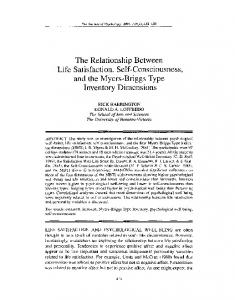
The total volume of the hippocampus and the subfield volumes were measured automatically with FreeSurfer v.5.3.0 (see Figure 3) on a Linux workstation.
Fig. 3. Subfield segmentation of hippocampus with FreeSurfer;
As it is shown in Table 3, the values obtained for one sample t test when comparing the volumes of the right hippocampus with the normal values were: t = -3.884, p-value = .008, CI = 95% (-1523.24 to -345.75). Table 3: One sample t test for right hippocampus
Test Value = 4017.7
Right_Hippocampus
t
Df
Sig. (2-tailed)
-3.884
6
.008
Mean Difference 95% Confidence Interval of the Difference Lower Upper -934.50 -1523.24 -345.75
The values obtained for one sample t test when comparing the volumes of the left hippocampus with the normal values (see Table 4) were: t = -5.587, p-value = .001, CI = 95% (-1529.42 to -597.84). Table 4: One sample t test for left hippocampus
Test Value = 3802.9
Left_Hippocampus
t
df
Sig. (2-tailed)
-5.587
6
.001
Mean Difference 95% Confidence Interval of the Difference Lower Upper -1063.63 -1529.42 -597.84
1110
Theodor Moica et al. / Procedia Technology 22 (2016) 1106 – 1112
High levels of cortisol were found in 3 of 7 patients, the morning biological value of the reference being in the range from 4.3 to 22.4 μg/dl. As it is shown in Table 5, the Pearson Correlation between the level of serum cortisol and the subfields of hippocampus volume were: r cortisol- Right_CA1= -0.596; r cortisol- Right_CA2_3= -0.616; r cortisol- Left_CA1= -0.038 and r cortisol- Left_ CA2_3= -0.295. Table 5: Correlations between cortisol level and volumes of hippocampus subfields
Cortisol Right_CA1 Right_CA2_3 Left_CA1 Left_CA2_3 Cortisol Pearson Correlation 1 -.596 -.616 -.038 -.295 Right_CA1 Pearson Correlation 1 .962 .577 .797 Right_CA2_3 Pearson Correlation 1 .516 .774 Left_CA1 Pearson Correlation 1 .926 Left_CA2_3 Pearson Correlation 1 Our results indicate the existence of a negative correlation with moderate association between the level of cortisol and CA1 (-0.596) and CA2-3 (-0.616) subfields of the right hippocampus and a weak association with the left CA2-3 (-0.295) subfield of the hippocampus. A very small correlation was revealed between the level of cortisol and CA1 subfield of the left hippocampus (-0.038). A high positive correlation was found between Right CA1 and Right CA2-3 subfield volumes (0.962) and also between Left CA1 and Left CA2-3 subfield volumes (0.926). 4. Discussion Whether it favorites the installation of the disease or act as a trigger, stress is almost always involved in the development of depressive disorders, causing an activation of hypothalamic-pituitary-adrenal axis (HPAA) and an increase of cortisol, which causes important apoptotic phenomena in the brain [14]. In addition to the increased level of glutamate, hypercortisolemia is involved in neuroplasticity processes and apoptosis by decreasing the number of dendritic spines and synapses, reducing the number of glial cells and by causing dendritic atrophy. Certain areas of the brain, like hippocampus and prefrontal cortex, are more prone to neurotoxicity. In the hippocampus are located numerous glucocorticoid and mineralocorticoid receptors and in depression there is an imbalance between these receptors, with an increase of the glucocorticoid receptor density at this level [15,16]. In a meta-analysis of 12 studies, the volume of the hippocampus has been shown to be consistently and significantly decreased in patients with MDD compared with controls, the degree of this reduction being directly proportional to the number and duration of untreated depressive episodes. Even after they entered in remission, recurrent MDD patients continued to have significantly less hippocampal volumes compared with healthy control group [16]. A meta-analysis performed by Videbech on a number of 351 patients and 279 healthy subjects showed an average reduction of hippocampal volume of 8% in the left hemisphere and 10% in the right hemisphere in depressed patients relative to comparison subjects. The number of depressive episodes seems to correlate with the reduction of hippocampal volume especially on the right side [17]. Tae and all have published in 2008 a comparative study between the manual and the automated measures (Free Surfer and IBASPM) of the hippocampal volume. Since we have used the same parameters of acquisition on MRI, we compared our data of hippocampal volumes for depressed patients with the mean for the control group from this study (4017.7 mm3 for the right and 3802.9 mm3 for the left hippocampus) [9]. Our results were consistent with the previous reports and the values obtained for “one sample t test” showed that the total volumes of the hippocampus of our depressed patients were significantly lower both on the right and the left side. The new generation of antidepressants has the ability to exert a neuroprotective effect and can induce neuroplasticity. In an animal model study, published in 2011, Marinescu et al. have demonstrated the neuroprotective effect of agomelatine in the hippocampus and prefrontal cortex, against the aggression of the increased level of cortisol, which is an important marker of depression [20].
Theodor Moica et al. / Procedia Technology 22 (2016) 1106 – 1112
1111
Although we cannot state with certainty whether it is a trigger or a secondary factor for depression, about 50% of patients with MDD has a hypersecretion of cortisol, but this percentage depends a lot on the analyzed population [21,22]. In our research, a high level of morning serum cortisol was observed in 3 of the 7 analyzed patients (33.3%). This small percentage might be explained by the fact that all patients included in the study had a long period of depression and the endocrine response to stress is altered in time, during the long term evolution of depressive illness. The hippocampus can be divided into several subfields with distinctive histological characteristics: the subiculum, the four cornu ammonis sectors (CA1–4) and the dentate gyrus. These subfields are functionally interconnected but they are responsible for different things: CA1 seems to be involved in temporal pattern association and intermediate term memory while CA3 is responsible for short-term memory, spatial pattern association and detection of novelty [23]. In the last years, preclinical and post mortem studies have showed that the analysis of different subfields of the hippocampus on MRI it is recommended, because the response to stress and the neuroplasticity in response to antidepressive treatment is differentiated in these subfields. There is preclinical evidence that hippocampal neuroplasticity is localized especially in cornu ammonis (CA1-4) and dentate gyrus, the same hippocampal subfields where volume reduction (measured on MRI) is more pregnant in case of MDD patients [24]. Previous studies have shown that in the first psychotic episode there is a correlation between cortisol and hippocampus, meaning that smaller hippocampal volume can partly be explained by stress-related processes in the brain, as measured by cortisol hyper-secretion [25]. Regarding the depressed patients, we found just a few data in the literature about the relationship between increased levels of cortisol and the degree of the hippocampal atrophy. In an article published in 1993, Axelson and all found a relationship between hippocampal volume and 11 p.m. cortisol concentration, but no differences in hippocampal volume were observed between patients and control subjects. This study measured the entire amygdala-hippocampal complex and not only the hippocampal volume and did not take into consideration the subfields measurements of the hippocampus [26]. In a recent study, cortisol awakening response (CAR) in MDD patients was proved to be negatively correlated with total hippocampal volume and also with CA1 subfield volume (27). In our research, the Pearson Correlation between the level of serum cortisol and the subfields of hippocampus volume were: r cortisol- Right_CA1= -0.596; r cortisol- Right_CA2_3= -0.616; r cortisol- Left_CA1= -0.038 and r cortisol- Left_ CA2_3= -0.295. Our results indicate the existence of a negative correlation with moderate association between the level of cortisol and CA1 (-0.596) and CA2-3 (-0.616) subfields of the right hippocampus and a weak association with the left CA2-3 (-0.295) subfield of the hippocampus. A very small correlation was revealed between the level of cortisol and CA1 subfield of the left hippocampus (-0.038). This means that higher levels of cortisol are associated with lower hippocampal subfield volumes. Since these are the sections of the hippocampus where neurogenesis takes place, long term follow up interventional studies are required to better understand the interrelation between cortisol - as a marker of neuroendocrine response and the hippocampal volume subfields in depression - as a marker of neurogenesis and neuroplasticity Because the correlations obtained is mild to moderate type, we can say that in the case of depressed patients, there are other factors involved in the relationship between the body's endocrine response to stress and the neuroanatomical modification of the hippocampal volume, as a part of the neuroplasticity of the brain. To our knowledge, this is the first pilot study in Romania that is assessing the hippocampal subfields volumes using FreeSurfer and is evaluating the correlation of these volumes with the levels of cortisol in depressed patients. 5. Conclusions The current researches show that both the right and the left hippocampal volumes are reduced in MDD patients. Higher levels of cortisol are associated with lower hippocampal subfield volumes, especially on the right. Since these are the sections of the hippocampus where neurogenesis takes place, long term follow up interventional studies are required to better understand the interrelation between cortisol - as a marker of neuroendocrine response and the hippocampal volume subfields in depression - as a marker of neurogenesis and neuroplasticity.
1112
Theodor Moica et al. / Procedia Technology 22 (2016) 1106 – 1112
Acknowledgments This paper was published under the frame of European Social Found, Human Resources Development Operational Programme 2007-2013, project no. POSDRU/159/1.5/S/136893. References [1] Stiemerling D. „10 abordări psihoterapeutice ale depresiei” (10 psychotherapeutic approaches of depression). Bucharest: Editura Trei; 2006. [2] Kessler R, et a. Prevalence, Severity, and Comorbidity of 12-Month DSM-IV Disorders in the National Comorbidity Survey Replication. JAMA. 2003; 289: 3095-3105. [3] Jacobi F, et al. Estimating the prevalence of mental and somatic disorders in the community: aims and methods of the German National Health Interview and Examination Survey. Int. J. Method. Psych. 2002; 11: 1-18. [4] Rorsman B, et a. A prospective study of first-incidence depression. Br. J. Psychiatry. 1990; 156: 336-342. [5] Wittchen H, Jacobi F, Rehm J, Gustavsson A, Svensson M, Jönsson B, et al. The size and burden of mental disorders and other disorders of the brain in Europe 2010. Eur Neuropsychopharmacol. 2011; 21(9): 655-679. [6] Monnier J, Hobfoll SE, Dunahoo CL, HuIizer MR, Johnson R. There’s more than rugged individualism in coping. Part 2: Construct validity and further model testing. Anxiety, Stress, Coping: An International Journal. 1998; 11(3):247-272. [7] Sadock BJ, Sadock VA. Kaplan and Sadock’s Synopsis of Psychiatry. In Lippincott Williams &Wilkins; 2007; Philadelphia. p. 530. [8] Squire LR, Stark CE, E CR. The medial temporal lobe. Annu Rev Neurosci. 2004; 27: 279-306. [9] Tae WS, Kim SS, Lee KU, Nam EC, Kim KW. Validation of hippocampal volumes measured using a manual method and two automated methods (FreeSurfer and IBASPM) in chronic major depressive disorder. Neuroradiology. 2008; 50(7):569-81. [10] Reuter M, Schmansky N, Rosas HD, Fischl B. Within-Subject Template Estimation for Unbiased Longitudinal Image Analysis. Neuroimage. 2012. [11] Han X, Jovicich J, Salat D, van der Kouwe A, Quinn B, Czanner S, et al. Reliability of MRI-derived measurements of human cerebral cortical thickness: the effects of field strength, scanner upgrade and manufacturer. Neuroimage. 2006; 32:180-194. [12] Jovicich J, Czanner S, Greve D, Haley E, van der Kouwe A, Gollub R, et al. Reliability in multi-site structural MRI studies: effects of gradient non-linearity correction on phantom and human data. Neuroimage. 2006 April; 30(2):436-43. [13] Segonne F, Pacheco J, Fischl B. Geometrically accurate topology-correction of cortical surfaces using nonseparating loops. IEEE Trans Med Imaging. 2007; 26:518-529. [14] Walhovd KB, Fjell AM, Reinvang I, Lundervold A, Dale AM, Eilertsen DE, et al. Effects of age on volumes of cortex, white matter and subcortical structures. Neurobiol Aging. 2005 October; 26(9):1261-70. [15] Talău G, Duică L, Nicoară D, Talău RD, Sântu A, Băhnean I. InterrelaĠii hipocamp – axa hipotalamo-hipofizo-corticosprarenaliană în tulburarea depresivă. Romanian Journal of Psychofarmacology. 2005; 5(1-2):45-50. [16] Maletic V, Robinson M, Oakes T, Iyengar S, Ball SG, Russell J. Neurobiology of depression: an integrated view of key Findings. Int J Clin Pract. 2007; 61 (12):2030–2040. [17] Videbech P, Ravnkilde B. Hippocampal volume and depression: a meta-analysis of MRI studies. Am J Psychiatry. 2004 November; 161(11):1957-66. [18] McKinnon M, Yucel K, Nazarov A, MacQueen G. A meta-analysis examining clinical predictors of hippocampal volume in patients with major depressive disorder. J Psychiatry Neurosci. 2009 January; 34(1):41-54. [19] Moldovan L. Innovative method of peer assisted learning by technology and assessment of practical skills. Procedia Technology 2014;12:667-674. [20] Marinescu D, Mogoanta L, Udristoiu T, Udristoiu I, Pirici D. The neuroprotective potentially of agomelatine - animal model study. European Psychiatry. 2011; 26(1):1256. [21] Cowen PJ. Cortisol, serotonin and depression: all stressed out? BJ Psych. 2002; 180:99-100. [22] Mihăilescu A, Năstase S, Matei V, Greabu M, Totan A. Investigation of emotional distress and salivary cortisol in young healthy subjects in the period of acute stress. Revista Medicală Română. 2011; LVIII (1):45-51. [23] Kesner R, Lee I, Gilbert P. A behavioral assessment of hippocampal function based on a subregional analysis. Rev Neurosci. 2004; 15:333– 51. [24] Malykhin N, Coupland N. Hippocampal neuroplasticity in major depressive disorder. Neuroscience. 2015 April; 15: p. S0306-4522. [25] Pariante CM, Navari S, Aas M, D'Albenzio A, Di Forti M, et al. Higher cortisol levels are associated with smaller left hippocampal volume in first-episode psychosis. Schizophr Res. 2010 June; 119(1-3):75-8. [26] Axelson D, Doraiswamy P, McDonald W, Boyko O, Tupler L, Patterson L, et al. Hypercortisolemia and hippocampal changes in depression. Psychiatry Research. 1993; 47(2): 163-173. [27] Wolkowitz O, Rowen J, Mason S, Mellon S, Reus V, Epel E, Mueller S. Cortisol awakening response and cortisol/DHEA ratio associations with hippocampal volume in MDD. European Journal of Psychotraumatology. 2012; 3(0): p. 1.