In the presence of central vascular shunts,. 1 From the Symposium Control of Arterial Blood. Gases: Cardiovascular and Ventilatory Perspectives presented at ...
AMER. ZOOL., 37:12-22 (1997)
The Role of Cardiac Shunts in the Regulation of Arterial Blood Gases1 TOBIAS WANG*, EGLE H. KROSNIUNASI , AND JAMES W. HICKS| *Institute of Biology, University of Odense, DK-5230 Odense M, Denmark ^Department of Ecology and Evolutionary Biology, University of California at Irvine, Irvine CA 92717, USA
The pulmonary and systemic circulations are not completely separated in reptiles and amphibians, so oxygen-rich blood returning from the lungs can mix with oxygen-poor blood returning from the systemic circuit (cardiac shunts). In these animals, the arterial blood gas composition is determined by both lung ventilation and the cardiac shunt. Therefore, changes in cardiac shunting patterns may participate actively in the regulation of arterial blood gases. In turtles the cardiac shunt pattern changes independently of ventilation and the cardiac R-L shunt (pulmonary bypass of systemic venous blood) is reduced under circumstances where the demands on efficient gas exchange are high (hypoxia, hypoxemia or exercise). We propose, therefore, that the size of cardiac shunts is regulated independently of ventilation and hypothesize that there exist at least two groups of peripheral chemoreceptors with different reflex roles. SYNOPSIS.
THE ANATOMICAL BASIS FOR CARDIAC SHUNTS IN NON-CROCODILIAN REPTILES
In all non-crocodilian reptiles (and amphibians), central vascular shunts result from the incomplete anatomical separation of the pulmonary and systemic circuits within the ventricle of the heart. These shunts are consequently referred to as cardiac shunts. The cardiac shunts can be de-
1 From the Symposium Control of Arterial Blood Gases: Cardiovascular and Ventilatory Perspectives presented at the Annual Meeting of the Society for Integrative and Comparative Biology, 26-30 December 1995, at Washington, D.C.
12
Downloaded from icb.oxfordjournals.org by guest on July 16, 2011
arterial systemic blood is a mixture of systemic venous blood and blood returning from the lungs. Arterial blood gas composition therefore is determined by both lung gases and the degree of admixture. This is in contrast to mammals and birds, where arterial blood gases closely resemble those within the lung and can be regulated exclusively by means of ventilation. The main focus of this chapter is to discuss the possible role of cardiac shunts on arterial blood gas regulation. Following a brief review of the anatomical basis for cardiac shunts and their effects on blood gases, we will discuss the possible neural regulations of blood flows. Although most of this chapter is based on our own recent data on turtles, we will, albeit selectively, draw comparisons to previous studies on other reptiles and amphibians.
INTRODUCTION
The pulmonary and systemic circulations of reptiles, amphibians and many airbreathing fish are not completely separated, and blood flows to the lungs and body can be altered independently (e.g., Johansen and Burggren, 1980). As a result, systemic venous blood returning from the body can bypass the pulmonary circulation (right-to-left shunt), whereas blood returning from the lungs can recirculate into the pulmonary circulation (left-to-right shunt). Although the functional significance of this cardiovascular design remains largely unknown (c/., Burggren, 1987; Hicks and Wang, 1996), numerous studies attest that the blood flows change in a predictable fashion. In particular, large increases in pulmonary blood flow during ventilation have been characterized for many different species (e.g., Johansen et al., 1970; Shelton, 1970; Shelton and Burggren, 1976; West et al., 1992; Wang and Hicks, 1996a). The underlying control of these changes is not well understood. In the presence of central vascular shunts,
13
CARDIAC SHUNTS
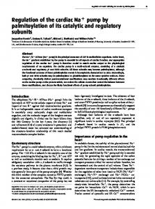
Pressure Shunts
"Ill Washout Shunts
II Systole
FIG. 1. The basic features of the pressure and washout hypotheses for Right-to-Left cardiac shunts in reptiles (RA,, right atrium; LA,, left atrium; CP, cavum pulmonale; CA, cavum arteriosum; CV, cavum venosum; LAO, left aortic arch; RAO, right aortic arch; PA, pulmonary artery). See text for further explanation. Modified from Hicks and Malvin (1995).
fined as right-to-left cardiac shunt (R-L shunt) and left-to-right cardiac shunt (L-R shunt), where R-L shunt refers to systemic blood that bypasses the lungs and L-R shunt refers to pulmonary venous blood that reenters the pulmonary circulation. Two dominating hypotheses {pressure shunt and washout shunt) have been advanced to explain the mechanisms of cardiac shunts in non-crocodilian reptiles. These hypotheses differ regarding the intraventricular flow patterns during systole (Heisler and Glass, 1985; Hicks and Malvin, 1995; Fig. 1). The ventricle is divided into two main chambers (the cavum pulmonale and a larger dorso-lateral chamber, the cavum dorsale) by a septum-like structure called the muscular ridge. In most species, the cavum dorsale is further subdivided into the cavum arteriosum and the cavum venosum. The pulmonary artery emerges from the cavum pulmonale,
whereas the two aortic arches arise from the cavum venosum. During diastole, O2 poor blood from the right atrium enters the cavum venosum and flows to the cavum pulmonale, while O2 rich blood enters the cavum arteriosum directly from the left atrium. During systole, blood is ejected into the systemic arteries from the cavum venosum and into the pulmonary arteries from the cavum pulmonale. The washout hypothesis proposes that the muscular ridge effectively separates the cavum pulmonale and the cavum venosum at the onset of systole and that the R-L shunt results from the O 2 poor blood residing in the cavum venosum at the end of diastole. Similarly, the O2 rich blood remaining in the cavum venosum following systole is washed into the cavum pulmonale during diastole account for the L-R shunt. The pressure shunt hypothesis proposes that the muscular ridge does not separate the cavum pulmonale and the
Downloaded from icb.oxfordjournals.org by guest on July 16, 2011
* Deoxy
14
T. WANG ET AL.
primary site of the resistance appears to be at a narrow region of the ventricular outflow tract of the pulmonary artery (Burggren, 19776; Smith and Maclntyre, 1979; Lillywhite and Donald, 1994). In addition, smooth muscle within the lung may regulate Rpul in snakes (Lillywhite and Donald, 1994), but the contribution at this site appears to be small in lizards and turtles. In all reptiles studied, the regulation of pulmonary resistance and, thus, pulmonary blood flow (Qpul) is controlled by the vagus nerve. Electrical stimulation of the efferent vagus or intravenous infusions of acetylcholine increase Rpul and reduce heart rate, and these changes are abolished following administration of atropine (Burggren, 1977a; Comeau and Hicks, 1994; Hicks and Comeau, 1994). The central vascular blood flows are also controlled adrenergically. In turtles, intravenous injection of epinephrine elicits a tachycardia, a reduction in Rpu, and a L-R shunt (Comeau and Hicks, 1994). Electrical stimulation of vagal afferents results in similar cardiovascular changes that are blocked by administration of bretylium (Comeau and Hicks, 1994), suggesting that the cardiovascular changes, often associated with brief periods of ventilation, may have an adrenergic component. Finally, non-adrenergicnon-cholinergic factors seem to be involved in regulating systemic and pulmonary vascular resistances and may influence cardiac shunt patterns (Lillywhite and Donald, 1994). THE IMPACT OF CARDIAC RIGHT-TO-LEFT SHUNT ON ARTERIAL OXYGEN LEVELS
In the presence of cardiac shunts, the blood gases of systemic arterial and pulmonary arterial blood are different from the venous blood entering the heart from the pulmonary and systemic circulations. Specifically, L-R shunt elevates the O2 content of pulmonary arterial blood relative to systemic venous blood, and R-L shunt reduces the O 2 content of the systemic arterial blood relative to that entering the heart from the lung. With the exception of alligators and crocodiles, R-L and L-R shunt normally occur simultaneously (Hicks, 1994). Nevertheless, because L-R shunt only influences
Downloaded from icb.oxfordjournals.org by guest on July 16, 2011
cavum venosum during systole. According to this view, blood will flow between these cava if differences exist in the outflow resistances. The washout shunt hypothesis and the pressure shunt hypothesis are not mutually exclusive. Rather, the extent to which one or the other mechanism can account for the shunt depends on when the muscular ridge separates the cavum pulmonale and the cavum venosum during systole. The respective contribution of these two types of shunts probably varies greatly among species. In the turtle Trachemys scripta, both pressure and washout shunts contribute, but because the muscular ridge is poorly developed, most of the shunt is due to pressure shunting (Hicks et al., 1996). In contrast, the muscular ridge is well developed in Varanus and the cavum pulmonale and the cavum venosum are separated early in systole. Most of the shunt in Varanus can accordingly be ascribed to washout shunt (Heisler et al., 1983; Heisler and Glass, 1985). To date, the mechanism of shunting remains largely undescribed for other reptilian genera. Regulation of cardiac shunts presumably differs according to the underlying mechanism. In animals with a prominent washout shunt, the degree of shunting is largely changed through changes in end-diastolic and end-systolic volumes of the ventricular chambers. Whether these volumes indeed are under active control remains to be experimentally verified. In Varanus niloticus the size of cardiac shunt does not change during hypoxia, hypercapnia or prolonged diving (Millard and Johansen, 1974) suggesting that the washout shunt is not actively regulated. In animals with a dominating pressure shunt, the net direction and magnitude of the shunt flow is affected by the resistance of the pulmonary circuit relative to that of the systemic circulation, and an active regulation of pulmonary arterial resistance is well documented (Johansen and Burggren, 1980; Hicks, 1994). In turtles and lizards, contraction or dilation of smooth muscle in the extrinsic pulmonary artery alters pulmonary arterial resistance (RpUl) (Burggren, 1977a; Milsom et al., 1977; Berger, 1973), whereas in snakes the
CARDIAC SHUNTS
15
the blood gas composition of pulmonary arterial blood, only R-L shunt needs to be considered to evaluate the impact of cardiac shunts on systemic arterial blood gases. The reduction in O 2 content caused by R-L shunt takes place in the equivalent of a closed system. Arterial PO2 therefore becomes a dependent variable determined by the resulting arterial HbO2 saturation (O2 content relative to blood O2 carrying capacity) and blood O2 affinity (Wood, 1984). The impact of R-L shunt on arterial O2 content ([O2]a) can be quantified as the weighted mean of the O2 content of pulmonary venous and systemic venous blood ([O2]pv and [O2]sv, respectively):
tive to blood oxygen affinity (i.e., P o of the blood leaving the lung is positioned at the flat portion of the oxygen dissociation curve), because increased ventilation, in that case, only causes a small increases in O 2 content of blood leaving the lung. Therefore, the cardiorespiratory response that secures O2 delivery seemingly depends on the conditions of O2 loading in the lungs. For example, both increased ventilation and reduced R-L shunt improve O2 delivery during hypoxia, but during anemia (reduced O 2 carrying capacity of the blood) a reduction in R-L shunt is more effective than ventilatory changes in maintaining O2 delivery (Wang and Hicks, 1996ft).
[OJ« = (QPu. X [O2]pv + QR_L X [O2]sv)/(Qpul + QR_L),
RECEPTORS FEED-BACK REGULATING BLOOD FLOWS AND CARDIAC SHUNT
(1)
The cardiovascular system is regulated, in part, by a number of chemo- and mechanoreceptors. In mammals, the majority of afferent input from these receptors projects to the nucleus of the tractus solitarius of the medulla, but the final integration of receptor feedback involves multiple brain centers with interaction of afferent input from different receptor groups (Daly, 1983; Dampney, 1994; Marshall, 1994). The anatomical structures and the physiological basis responsible for the integration of afferent receptor feed-back have not been investigated in reptiles or amphibians. Trigeminal, nasal and upper airway receptors Stimulation of trigeminal receptors of the face and receptors in the upper airways evokes bradycardia and systemic vasoconstriction in most mammals and birds (Butler and Jones, 1982), but the role of these receptors is not well known in reptiles or amphibians. During forced dives in snakes, bradycardia only develops if the nose is wet (Johansen, 1959; Murdaugh and Jackson, 1962), and immersion may be important for the development of bradycardia in alligators (Andersen, 1962). Nevertheless, in turtles the bradycardia and the increase in R-L shunt associated with breath hold develops regardless of whether or not the face is immersed (Burggren, 1975; T. Wang unpublished). However, these observations do not
Downloaded from icb.oxfordjournals.org by guest on July 16, 2011
where Qpul is pulmonary blood flow and QR-L denotes the R-L shunt blood flow (Qpui + QR-L = Qsys)- Thus, in the presence of R-L shunt, arterial P o is a composite variable depending on any factor that influences O2 content of either pulmonary or systemic venous blood or the O2 affinity of the blood (Wood, 1984; Wang and Hicks, 1996ft). Arterial blood gases in reptiles therefore can change independently of lung PO2. For example, if systemic oxygen extraction increases (e.g., during increased metabolic rate) at a constant cardiac R-L shunt, arterial POz will decrease even if ventilation is matched to metabolic rate. In this case lung P o would not change, and the reduction in arterial PO2 could not be predicted from measurements of ventilation or endtidal gas composition. This is never the case in healthy mammals. The effects of R-L shunt on arterial PO2 can be quantified theoretically using the two-compartment model (e.g., Wood, 1984). Recently, we used this model to evaluate the impact of increasing ventilation or eliminating R-L shunt on arterial P o (Wang and Hicks, 1996ft). Based on published values for ventilation, oxygen uptake, and degree of R-L shunt, we found that changes in the R-L shunt are as important as realistic changes in ventilation in determining arterial P o in turtles (Wang and Hicks, 1996ft). The relative effect of R-L shunt is largest when lung P o is high rela-
16
T. WANG ET AL.
exclude the possibility that these receptors participate in the regulation of QpuJ and/or cardiac shunts. The CO2 sensitive chemoreceptors located in the upper airways and/or lungs of most amphibians and reptiles are important for ventilatory control, but their effect on the cardiovascular system has not been investigated.
Effects of altering blood Po2 and O2 content: Possible roles of peripheral chemoreceptors Because many reptiles and amphibians display pronounced cardiorespiratory interactions (tachycardia, increased Q pu | and a reduction in R-L shunt during breathing), distinguishing the direct effects of a given stimulus on the cardiovascular performance from a secondary effect arising from changes in ventilation is difficult. For example, during hypoxia, an increase in mean Qpui is to be expected, simply because the animal spends more time breathing. Indeed, in three species of turtles (Testudo pardalis, Pelomedusa subrufa and Chelonia mydas), mean Q pu | increases during hypoxia, but the attained flows resemble those occurring during ventilation (Burggren et al., 1977; West et al., 1992). It may, therefore, be more informative to describe the cardiovascular parameters during a defined period of either ventilation or breath hold. Figure 3 shows Qpul and Q sys during breath hold (longer than 180 sec) or a ventilatory period (2—4 breaths) at four levels of inspired oxygen fractions (FjO2) in six turtles. At any given F ; o 2 , Qpu) is higher during ventilation than during breath hold, which reflects the cardiorespiratory interaction. However, the blood flows during breath holds or ventilatory periods were not affected by hypoxia per se. Therefore, although mean blood
Downloaded from icb.oxfordjournals.org by guest on July 16, 2011
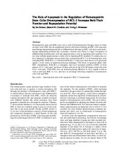
Pulmonary stretch receptors (PSR) As in other air-breathing vertebrates, reptiles and amphibians possess pulmonary stretch receptors (PSR) that convey information regarding lung pressure and volume to the central nervous system (Milsom, 1997). Stimulation of PSR during breathing could provide a stimulus to increase Qpu, and heart rate, but the contribution of PSRs to the cardiorespiratory interactions is not resolved (see West and Van Vliet [1992] for a review on amphibians). In turtles, artificial tidal ventilations via a chronically implanted lung catheter elicited increases in heart rate and Qpul that closely resembled those occurring during voluntary breathing (Johansen et al., 1978). We have, however, not been able to reproduce this response in the turtle, Trachemys scripta (Wang and Hicks, unpublished). In our experiments, turtles were equipped with blood flow probes (Transonic, 2R) around the left aortic arch and the left pulmonary artery. In addition, a catheter (PE 160) was placed in each lung and connected to a T-piece allowing simultaneous inflation or deflation of both lungs and measurements of lung pressure. Three to ten days after surgery, the turtles were placed in an experimental set-up in which they could dive freely but were constrained to breathe in a small funnel connected to a pneumotachograph for measurements of ventilation. Figure 2 shows the pronounced increase in Qpul during ventilation and that withdrawal of lung gas (marked as A and C) and injection of air into the lungs (B) did not affect Qpul. In all experiments (n = 5), artificial manipulation of lung volume/pressure had absolutely no effect on blood flows or heart rate. In several instances, manipulations of lung volume initiated breathing which was associated with the normal increase in Qpul during voluntary ventilation. Johansen et al. (1978) did not measure ventilation, so the
manipulation of lung volume in their experiments may also have elicited voluntary breathing, which could explain the observed changes in blood flows. Given the conflicting results obtained in different studies, it is difficult to construct a simple model to explain the role of PSR's in the regulation of central vascular blood flows. Our experiments on turtles described above indicate that afferent input arising from PSRs alone is not sufficient to elicit cardiovascular changes. Increase in Qpul during ventilation may therefore be due to a feed-forward mechanism acting simultaneously on both the respiratory and vagal motor neurons (see also, West et al., 1992). This hypothesis does not preclude the possibility that PSRs contribute to the final expression of the cardio-respiratory interactions.
CARDIAC SHUNTS
17
Downloaded from icb.oxfordjournals.org by guest on July 16, 2011
1 minute FIG. 2. The effect of manipulating lung volume and pressure (P|ung) on pulmonary (Qpul; 2 X QLPA) and left aortic blood (QLAo) flows in a 1.3 kg turtle. At the marked events lung gas was withdrawn (A and C) or air was injected (B) through catheters placed in the lungs (T. Wang and J. W. Hicks, unpublished).
flows and heart rate were elevated during hypoxia (data not shown), no selective effect of hypoxia on the cardiovascular system is indicated. Based on the theoretical prediction that a reduction in cardiac R-L shunt is particu-
larly beneficial for gas exchange when blood O 2 carrying capacity is reduced (Wang and Hicks, 1996a), we investigated the effect of experimental reductions in haematocrit on blood flows in turtles. Figure 4 show QpU), Q sys and the net shunt flow
18
T. WANG ET AL.
VP (2-4 breaths)
NVP> 180 sec
120 1
c
pul
(ml
E
o
1^
0 140 120 100 80 60 40 20 0
0.05 0.10 0.15 0.20
0.05 0.10 0.15 0.20
FIG. 3. Pulmonary and systemic blood flows (Qpu! and Qsys) in six turtles at normoxia and during inhalation of three different hypoxic gas mixtures at 25°C. Turtles were instrumented with a flow probes around the left pulmonary artery (Q pu | = 2 X QLPA) and flow probes around the left aortic arch (LAo), the right branch of the right aortic arch (RRAo) and a probe around the right subclavian and carotid arteries (Rsub and Rca) for calculation of Q sys (Qsys = Q LAo + QR QRC»)- Each data point represents a mean value for an individual turtle during non-ventilatory periods (NVP) lasting longer than 180 second and during ventilatory periods (VP) consisting of 2-4 breaths. Note that several animals did not exhibit any long lasting non-ventilatory periods during severe hypoxia (T. Wang and J. W. Hicks, unpublished).
(Qpui ~~ Qsys) during breath holds and ventilation as a function of haematocrit. In anemic turtles, Qpu| remained high during breath holding, whereas Q sys was not affected by reductions of haematocrit. As a result, the net R-L shunt which normally prevails during breath hold is greatly reduced in turtles with low blood O 2 content and even reversed to a net L-R shunt in severely anemic turtles (Figure 4). Because reductions in blood O2 content by inhalation of CO or by nitrite (NaNO2) infusions elicit similar responses (Wang et al., 1996), un-
intentional changes in blood volume are unlikely causes for the increase in Qpul. In none of the experiments did the reduction in blood O2 content affect ventilation or the ventilatory response to hypoxia. Similarly, reductions in blood O 2 content by bleeding does not affect the hypoxic ventilatory response in toads, but elicits large increases in heart rate (Wang et al., 1994). The finding that blood O2 content appears to alter Qpu, and net shunt flows in the absence of changes in P a o 2 could be explained by the existence of an O 2 content sensitive
Downloaded from icb.oxfordjournals.org by guest on July 16, 2011
E
100 80 60 40 20
CARDIAC SHUNTS
19
NVP > 180 sec
VP (2-4 breaths)
'c
E
1 "5 CL
o "E
o
1
U( }
o o
°o
6
o
0
c o
V)
•o
60 40 'c 20 E 0 -20 -40 -60 •O -80
o
oo \ o o
o ^
o
%
u o o o
o o
°o
o
o 1
0
1
i
i
1
5 10 15 20 25 30 0 Haematocrit
1
1
1
1
0
o1
5 10 15 20 25 30 Haematocrit
FIG. 4. Pulmonary and systemic blood flows in turtles with varying haematocrit during normoxia at 25°C (see Figure 3 for calculation of blood flows). Each data point represents a mean value for an individual turtle during non-ventilatory periods lasting (NVP) longer than 180 second and during ventilatory periods (VP) consisting of 2^4 breaths. Haematocrit was artificially reduced by bleeding and blood volume was maintained constant by reinfusion of plasma and dextran. Linear regression are included when the correlation between haematocrit and blood flow was a significant (P s 0.05) (T. Wang and J. W. Hicks, unpublished).
Downloaded from icb.oxfordjournals.org by guest on July 16, 2011
o o
i
20
T. WANG ET AL.
Exercise 30 -
Rest I air
30
submerged
20 Rest in air
Submerged rest
_L
a>
L-R shunt
10 0
R-L shunt
-10 - -20 - -30
chemoreceptor that selectively influences the cardiovascular system. In mammals, the aortic bodies respond to changes in arterial O 2 content and predominantly influence the cardiovascular system {e.g., Lahiri et al., 1981), while ventilatory responses are controlled by the Po2 sensitive carotid bodies. The aortic bodies are believed to be sensitive to O 2 content because they are supplied with a low perfusion relative to O 2 uptake. This could also be the case in reptiles and amphibians, but O2 content sensitive receptors have not been identified in these animals so far. In fact, the O 2 sensitivity of the chemoreceptors in the carotid labyrinth in toads is not affected by blood O 2 content, indicating that Po2 rather than O 2 content exerts the primary stimulus for these receptors (Van Vliet and West, 1992). Alternatively, the cardiovascular response to reductions in arterial O2 content can be explained by the existence of Po 2 sensitive chemoreceptors in the venous circulation. Venous O 2 levels (O 2 content and Po2) are determined by systemic O 2 delivery (Q sys * arterial O 2 content) relative to metabolic rate. Therefore, if Qsys and metabolic rate remain constant, a reduction of arterial O 2 content will reduce venous PO2 and O 2 content. The existence of chemoreceptors within the venous circulation has not been identified in any vertebrate group. However, Ishii et al. (1985) described O 2 sensitive chemorecep-
tors on the pulmonary artery in turtles. The blood in this vessel is perfused predominantly by venous systemic blood, although the exact composition depends on the level of L-R shunt. Perhaps O2 sensitive chemoreceptors on the pulmonary artery are responsible for cardiovascular control while the arterial chemoreceptors are responsible for ventilatory control. Clearly this hypothesis needs to be verified experimentally through direct recording of the peripheral chemoreceptors to determine the exact receptor modalities, as well as by establishing the reflex roles of the different receptor groups. CARDIAC SHUNTS DURING EXERCISE AND DIFFERENT TYPES OF BEHAVIOUR
In most animals, different types of behavior are associated with changes in central vascular blood flows. For example, during swimming and terrestrial locomotion, Qpul increases 2-3 fold over resting values in turtles (Shelton and Burggren, 1976; West et al., 1992; Krosniunas and Hicks, 1995) and similar changes have been reported in frogs (Johansen et al., 1970). Recently, Jones and Shelton (1993) showed that alligators eliminate the R-L shunt if disturbed. Recently, Krosniunas and Hicks (1995) assessed the effects of behavioral state on blood flows Trachemys scripta. In these experiments, turtles were instrumented with
Downloaded from icb.oxfordjournals.org by guest on July 16, 2011
FIG. 5. Net cardiac shunt (Qpu, - Qsys) during different types of voluntary behavior in Trachemys scripta (mean ± 1 s.e.m.; N = 7 - 12; Tb = 20 ± 2°C). All values were obtained for bouts of behavior one minute or longer in duration (E. Krosniunas and J. W. Hicks, unpublished).
21
CARDIAC SHUNTS
CONCLUSIONS
The cardiac shunt in amphibians and reptiles is actively regulated and has important implications for arterial blood gas composition. The development of a comprehensive model to explain blood gas control in these animals must encompass both ventilatory and cardiovascular control. The neural aspects of cardiovascular control is poorly understood, but cardiac shunt patterns appear to be controlled by centrally generated feedforward mechanisms and feed-back from peripheral receptors. Large interspecific differences apparently exist in the relative importance of the different receptors. In addition, the effects of some receptors are entirely unknown; for example the role of central chemoreceptors in cardiovascular control has not been studied. Based on studies to date, large net R-L cardiac shunts in amphibians and reptiles are only apparent at rest. When the demands on gas exchange are increased (hypoxia, hypoxemia, exercise) the R-L shunt is reduced and may be reversed to a net L-R shunt. This pattern seems advantageous with respect to the systemic O 2 delivery, as
elimination of R-L shunt increases arterial O2 content. ACKNOWLEDGEMENTS
We thank Drs. N. H. West, S. Reid and E. W. Taylor for providing critical comments and numerous suggestions on an early version of the manuscript. TW was supported by a NSERC international post doctoral fellowship and JWH was supported by NSF (IBN-9218936). REFERENCES Andersen, H. T. 1962. Physiological adjustments to prolonged diving in the American alligator, Alligator mississippiensis. Acta Physiol. Scand. 53:23-54. Berger, P. J. 1973. Autonomic innervation of the visceral and vascular smooth muscle of the lizard lung. Comp. Gen. Pharmacol. 4:1-10. Burggren, W. W. 1975. A quantitative analysis of ventilation tachycardia and its control in two chelonians, Chrysemys scripta and Testudo graeca. J. Exp. Biol. 63:367-380. Burggren, W. W. 1977a. The pulmonary circulation of the chelonian reptile: morphology, haemodynamics and pharmacology. J. Comp. Physiol. 116:303-323. Burggren, W. W. \911b. Circulation during intermittent lung ventilation in the garter snake Thamnophis. Can. J. Zool. 55:1720-1725. Burggren, W. W. 1987. Form and function in reptilian circulations. Amer. Zool. 27:15-19. Burggren, W. W., M. L. Glass, and K. Johansen. 1977. Pulmonary ventilation: Perfusion relationships in terrestrial and aquatic chelonian reptiles. Can. J. Zool. 55:2024-2034. Butler, P. J. and D. R. Jones. 1982. The comparative physiology of diving in vertebrates. Adv. Comp. Physiol. Biochem. 8:179-364. Comeau, S. G. and J. W. Hicks. 1994. Regulation of central vascular blood flows in the turtle. Am. J. Physiol. 267:R569-R578. Daly, M. de B. 1983. Peripheral arterial chemoreceptors and the cardiovascular system. In H. Acker and R. G. O'Regan (eds.), Physiology of the peripheral arterial chemoreceptors, pp. 325—393. Elsevier, Amsterdam. Dampney, R. A. L. 1994. Functional organization of central pathways regulating the cardiovascular system. Physiol. Rev. 74:323-364. Heisler, N., P. Neumann, and G. M. O. Maloiy. 1983. The mechanism of intracardiac shunting in the lizard Varanus exanthematicus. J. Exp. Biol. 105:1531. Heisler, N. and M. L. Glass. 1985. Mechanism and regulation of central vascular shunts in reptiles. In K. Johansen and W. W. Burggren (eds.), Cardiovascular shunts: Phylogenetic, ontogenetic and clinical aspects, pp. 334-353. Munksgaard, Copenhagen.
Downloaded from icb.oxfordjournals.org by guest on July 16, 2011
flow probes and followed for several months in an enriched laboratory setting. There the animals performed a range of spontaneous activities in either an aquatic or terrestrial environment. The behavior was divided into six categories: walking, rest in air, rest in water, active diving (movement into water from air), feeding and swimming. The latter four types of behavior were operationally defined as those in which turtles were completely submerged and therefore unable to breathe. The cardiac shunt pattern during voluntary behavior in turtles is primarily associated with activity level (Fig. 5). Activity leads to an increase in Qpu] and reduction or elimination of the net R-L cardiac shunt, both in the presence or absence of pulmonary ventilation. Longer bouts of exercise, those lasting one minute or more, resulted in the development of a net L-R shunt, irrespective of submergence (Fig. 5). These findings point to an independent feed-forward regulation of blood flows, because Qpu, increased before or quickly after the onset of exercise.
22
T. WANG ET AL.
Responses to hypoxia, hypercarbia and diving. J. Exp. Biol. 60:871-880. Milsom, W. K., B. L. Langille, and D. R. Jones. 1977. Vagal control of pulmonary vascular resistance in the turtle, Chrysemys scripta. Can. J. Zool. 55:359-367. Milsom, W. K. 1997. Cardiorespiratory stimuli: Receptor cell versus whole animal. Amer. Zool. 37:000-000. Murdaugh, H. V. and J. E. Jackson. 1962. Heart rate and blood lactic acid concentration during experimental diving in water snakes. Am. J. Physiol. 202:1163-1165. Shelton, G. 1970. The effect of lung ventilation on blood flows to the lungs and body of the amphibian, Xenopus laevis. Respir. Physiol. 9:183-196. Shelton, G. and W. Burggren. 1976. Cardiovascular dynamics of the chelonia during apnoea and lung ventilation. J. Exp. Biol. 64:323-343. Smith, D. G. and D. H. Macintyre. 1979. Autonomic innervation of the visceral and vascular smooth muscle of a snake lung (Ophidia: Colubridae). Comp. Biochem. Physiol. 62C: 187-191. Van Vliet, B. N. and N. H. West. 1992. Functional characteristics of arterial chemoreceptors in an amphibian (Bufo marinus). Respir. Physiol. 88:113-127. Wang, T, L. G. S. Branco, and M. L. Glass. 1994. Ventilatory responses to hypoxia in the toad Bufo paracnemis before and after a decrease in haemoglobin oxygen-carrying capacity. J. Exp. Biol. 186:1-8. Wang, T. and J. W. Hicks. 1996a. Cardio-respiratory synchrony in turtles. J. Exp. Biol. 199:1791-1800. Wang, T. and J. W. Hicks. 19966. The interaction of pulmonary ventilation and cardiac shunts on arterial oxygen levels. J. Exp. Biol. 199: 2121-2129. Wang, T, J. W. Hicks, and W. K. Milsom. 1996. Changes in arterial O 2 content affect cardiac shunt but not ventilation in turtles. Physiologist 39:A7.27. West, N. H., P. J. Butler, and R. M. Bevan. 1992. Pulmonary blood flow during rest and swimming in the green sea turtle, Chelonia mydas. Physiol. Zool. 65:287-310. West, N. H. and B. N. Van Vliet. 1992. Sensory mechanisms regulating the cardiovascular and respiratory systems. In M. E. Feder and W. W. Burggren (eds.), Environmental physiology of the Amphibia, pp. 151—182. Chicago University Press, Chicago, London. Wood, S. C. 1984. Cardiovascular shunts and oxygen transport in lower vertebrates. Am. J. Physiol. 247:R3-R14.
Downloaded from icb.oxfordjournals.org by guest on July 16, 2011
Hicks, J. W. 1994. Adrenergic and cholinergic regulation of intracardiac shunting. Physiol. Zool. 67:1325-1345. Hicks, J. W. and S. G. Comeau. 1994. Vagal regulation of intracardiac shunting in turtles. J. Exp. Biol. 186:109-126. Hicks, J. W. and G. M. Malvin. 1995. Mechanism of intracardiac shunting in reptiles: Pressure vs washout shunting. In N. Heisler (ed.), Comparative and environmental physiology. Mechanisms of systemic regulation: Respiration and circulation, pp. 137-157. Springer Verlag, Berlin, Heidelberg, New York. Hicks, J. W. and T. Wang. 1996. Functional role of cardiac shunts in reptiles. J. Exp. Zool. 275:204216. Hicks, J. W., A. Ishimatsu, S. Molloi, A. Erskin, and N. Heisler. 1996. The mechanism of cardiac shunting in reptiles: A new synthesis. J. Exp. Biol. 199:1435-1446. Ishii, K., K. Ishii, and T. Kusakabe. 1985. Electrophysiological aspects of the reflexogenic area in the chelonian, Geoclemmys reevesii. Respir. Physiol. 59:45-54. Johansen, K. 1959. Heart activity during experimental diving of snakes. Am. J. Physiol. 197:604-606. Johansen, K., C. Lenfant, and D. Hanson. 1970. Phylogenetic development of the pulmonary circulation. Fedn Am. Socs Exp. Biol. 29:1135-1140. Johansen, K. and W. W. Burggren. 1980. Cardiovascular function in the lower vertebrates. In G. H. Bourne (ed.) Hearts and heart-like organs, pp. 61117, Academic Press, New York. Johansen, K., W. W. Burggren, and M. L. Glass. 1977. Pulmonary stretch receptors regulate heart rate and pulmonary blood flow in the turtle, Pseudemys scripta. Comp. Biochem. Physiol. 58A:185-191. Jones, D. R. and G. Shelton. 1993. The physiology of the alligator heart: Left aortic flow patterns and right-to-left shunts. J. Exp. Biol. 176:247-269. Krosniunas, E. H. and J. W. Hicks. 1995. Intracardiac shunts during voluntary activity and rest in the turtle. Amer. Zool. 35:A63. Lahiri, S., E. Mulligan, T. Nishino, A. Mokashi, and R. O. Davies. 1981. Relative responses of aortic body and carotid body chemoreceptors to carboxyhemoglobinemia. J. Appl. Physiol. 50:580-586. Lillywhite, H. B. and J. A. Donald. 1994. Neural regulation of arterial blood pressure in snakes. Physiol. Zool. 67:1260-1283. Marshall, J. M. 1994. Peripheral chemoreceptors and cardiovascular regulation. Physiol. Rev. 74:543594. Millard, R. W. and K. Johansen. 1974. Ventricular outflow dynamics in the lizard, Varanus niloticus: