Journal-Cardiovascular Surgery 2015:3(2):30-34 doi; 10.5455/jcvs.2015322
Original Research
30
The role of positron emission tomography in the diagnosis of vascular prosthetic graft infections Sefer Usta1, Hamit Serdar Basbug2*, Ergun Haliloglu1, Savaş Karyagar3, Umit Mentese1, Orhan Veli Dogan4 Department of Cardiovascular Surgery, Ahi Evren Thoracic and Cardiovascular Surgery Training and Research Hospital, Trabzon, Turkey, Department of Cardiovascular Surgery, Kafkas University Faculty of Medicine, Kars, Turkey, 3Department of Nuclear Medicine, Kanuni Training and Research Hospital, Trabzon, Turkey, 4Department of Cardiovascular Surgery, Mustafa Kemal University, Hatay, Turkey *Correspondence Author: Department of Cardiovascular Surgery, Kafkas University Faculty of Medicine, Kars, Turkey Tel.: +90-505-2612372 Fax: +90-474-2251193 e-mail:
[email protected] Key words: Blood vessel prosthesis; infection; positron-emission tomography; multidetector computed tomography Received: 25.01.2015 Accepted: 03.02.2015 e-published: 04.03.2105 1 2
Abstract Objective: Vascular prosthetic graft infections can be fatal, especially if it involves the intra-abdominal region. Definitive treatment is the removal of the infected graft and administration of appropriate antibiotic therapy. Removal of the prosthetic graft causes the problem of blood supply leading the extremity to ischemia. For this reason, it is important to decide whether the infected graft would be removed. We aimed to evaluate the role and efficiency of positron emission tomography (PET) for the diagnosis of vascular prosthetic graft infections. Materials and Methods: Between May 2013 and January 2015, eight patients were diagnosed with graft infection. They were assessed with PET. All the patients were male, and the average age was 52 ± 7. PET-computed tomography was performed following 4 weeks of antibiotherapy to avoid misinterpretation of the inflammatory response and fluid accumulation as an infection. Localization and extension of graft infections were visualized in the images. Results: Fluorodeoxyglucose (FDG) uptake by the vascular prosthesis was accepted as infection, and these grafts were removed. Grafts with no FDG uptake compatible with infection were considered as inflammation, and medical treatment was applied. All patients were discharged without complication. Conclusion: PET scan facilitates the diagnosis of infection in suspected grafts and prevents unnecessary surgical intervention. It is also accepted as a guide for diagnosis and treatment of graft infection.
Introduction
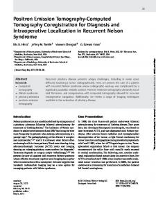
Positron emission tomography-computed tomography (PET-CT) is called as the “positron emitters.” It is a type of scintigraphic imaging modality performed using certain radiochemical compounds (radiopharmaceuticals) [1-3]. The examination depends on the various reactions of living organisms in the tissues against different radiopharmaceuticals [4]. In this wise, the functional and metabolic status of a targeted tissue can be investigated [5]. Fluorodeoxyglucose (FDG) highly accumulates and concentrates inside the tissues with increased metabolic activity like neoplastic activity or focal infection and inflammation (Figure 1). FDG accumulation during the infection mainly depends on the active macrophage infiltration due to the bacteria in the environment [6-8]. For this reason, PET-CT is a useful method for detection and
The role of positron emission tomography in the diagnosis of vascular prosthetic graft infections
follow-up of an infection and inflammation foci that is also superior to any other radiological scans. It is mostly used in the determination of localized infection as prosthetic infections and cases of fever with unknown origin [9]. During the last 45 years, artificial grafts have been successfully used in vascular surgery. Graft infection is a severe and life-threatening complication, particularly in aortic revascularization [7]. The incidence of graft infections has been reported as 1-6% in the literature [10]. As the other surgical infections, mortality and morbidity are high. High mortality and morbidity mostly depend on the complications of sepsis, anastomotic leakage or graft-enteric fistula. Clinical presentation is usually non-specific [8]. Mortality rate of aortic graft infections is 40-75%, and it is 20-50% at the level of the distal lower extremity. However, in femoropopliteal grafts,
Usta et al.
Journal-Cardiovascular Surgery 2015:3(2):30-34 doi; 10.5455/jcvs.2015322
Original Research
31
and debridement of the infected tissue is the primary concern. Any involvement in suture lines compels the removal of prosthetic graft, irrigation with local antiseptics and the use of systemic antibiotics. Infected graft may be coated with a pedicle omental flap or omentum if it is not removed. PET-CT is the most efficient radiological examination in graft infections as it evaluates both the metabolic and structural condition of the tissues at the same time. In this article, we performed diagnostic PET-CT imaging in suspected cases of vascular prosthetic graft infections and investigate its convenience in therapeutic use. a
b
Figure 1. The whole body display of positron emission tomography-computed tomography. Left femoropopliteal (a) and left iliofemoral (b) graft infection, compatible with an increased focal uptake of fluorodeoxyglucose the mortality rate has been reported as 10%. Regarding all vascular prosthetic infections, the amputation rate is between 10% and 50% [10]. Graft infections within 3 months following the operation are called as early infections. Similarly, graft infections developing later than 3 months post-operatively are called late infections. The most delayed graft infection reported in the literature is an aortic graft that was seen after 156 months post-operatively. In early infections, Staphylococcus aureus, and in the late infections, Staphylococcus epidermidis are the most common responsible microorganisms [9]. Presently, successful therapeutic results are obtained in most patients, with improved chemotherapeutic agents and modern diagnostic procedures. Fundamental principle regarding the management of the graft infections is an appropriate antibiotic therapy and total removal of the graft depending on culture antibiogram. Prosthetic graft infections usually accompany the symptoms and signs of systemic disease [6]. CT is very helpful in diagnosis. Fluid collection and the inflammatory changes around the graft are clearly demonstrated with CT. Collection of air bubbles in the liquid and the images of airfluid level are significant during the first 8 weeks after the operation [8]. In addition to CT, magnetic resonance imaging can also be used to distinguish early hematoma, fluid collection, and the inflammatory changes during the early onset of the infection. In addition, ındium-111 and gallium-67 labeled leukocytes scintigraphy and the cultures of removed graft and the surrounding tissues are also useful for the diagnosis [9]. In graft infections, the principles of treatment were defined in 1984, by Hargrove and Edmunds. Emergency reoperation
The role of positron emission tomography in the diagnosis of vascular prosthetic graft infections
Materials and Methods Between May 2013 and January 2015, eight patients who had been previously operated for peripheral arterial disease were included the study. Patients re-admitted to our clinic with complaints of fever, chills and local discharge at the incision site. Previous operations include three aortobifemoral bypass, two iliofemoral bypasses, two axillobifemoral bypasses and one femoropopliteal bypass (Table 1). All the patients were male. One patient had diabetes mellitus. Patients admitted 6.4 ± 0.4 months after admission. Laboratory blood tests, blood and effusion culture, and antibiograms were performed in all cases on admission. All patients were consulted to the infectious disease department. A broad-spectrum parenteral antibiotic (vancomycin) was administered empirically until the culture and antibiogram results were obtained S. aureus(in three patients), S. epidermidis (in two patients), Pseudomonas aureginosa (in two patients) and Escherichia coli (in one patient) were isolated in the culture (Table 1). After 4 weeks of medical treatment, PET-CT scans of all patients revealed the graft infection. PET-CT scans performed 4 weeks after the administration of antibiotherapy to avoid misinterpretation of an acute inflammatory fluid accumulation as an infection. Thus, graft status can be observed more clearly after 4 weeks of antibiotherapy in the PET-CT. In all patients, a CT scan was performed to investigate the possible presence of infection before the PET-CT scan. In PET-CT images, focally increased FDG uptake at the site of vascular graft or heterogeneous pattern of FDG uptake was considered as the presence of an infection. The infected grafts with FDG uptake in PET-CT scan were removed in three (aortobifemoral bypass) patients (Figure 2). The infected grafts of two patients (one femoropopliteal bypass and one iliofemoral bypass patient) were removed. Medical treatment was continued without removing the graft in three patients (one iliofemoral, two axillobifemoral bypass) revealing less FDG uptake in PET-CT. These three patients with unremoved infected graft were maintained
Usta et al.
Journal-Cardiovascular Surgery 2015:3(2):30-34 doi; 10.5455/jcvs.2015322
32
Original Research
Table 1: Surgical and microbiological parameters Primary operation
Operation‑ınfection ınterval
Microorganism in culture
Antibiotic
Secondary surgery/treatment
Secondary surgery material
Femoropopliteal bypass
8 months
Escherichia coli
Vancomycin/5 weeks
Femoropopliteal bypass
Saphenous vein
Aortobifemoral bypass
12 months
Staphylococcus epidermidis
Vancomycin/6 weeks
Aortobifemoral bypass
Biologic Y‑graft
Aortobifemoral bypass
10 months
Staphylococcus aureus
Vancomycin/6 weeks
Aortobifemoral bypass
Biologic Y‑graft
Aortobifemoral bypass
9 months
Staphylococcus aureus
Vancomycin/6 weeks
Axillobifemoral bypass
PTFE graft
Iliofemoral bypass
16 months
Staphylococcus epidermidis
Vancomycin/7 weeks
Aortobifemoral bypass
PTFE graft
Iliofemoral bypass
3 months
Pseudomonas auroginosa
Vancomycin/6 weeks
Medical treatment
‑
Axillobifemoral bypass
4 months
Pseudomonas auroginosa
Vancomycin/6 weeks
Medical treatment
‑
Axillobifemoral bypass
8 months
Staphylococcus aureus
Vancomycin/6 weeks
Medical treatment
‑
Results The average age of the patients was 52 ± 7. Regarding the three of the patients with aortobifemoral bypass, all infected grafts were open except an obstructed right iliac limb in a single patient. Four of the patients had clinical symptoms of fever and leukocytosis. In all patients, antibiotic therapy was started soon after admission according to the consultation of infectious diseases department. Patients were continued antibiotherapy for 4 weeks. PET-CT scans were performed after 4 weeks of antibiotherapy in all patients. All PET-CT scans revealed vascular prosthetic graft infection.
Figure 2. Images of positron emission tomography-computed tomography. Aortobifemoral graft infection, compatible with increased focal fluorodeoxyglucose uptake in the right iliac leg antibiotherapy for three more weeks despite the observation of infection according to PET-CT. Their clinical condition was better compared to the others in terms of clinical symptoms and laboratory findings. Thus, graft excision was not initially considered. However, one of the two axillobifemoral bypass patients were readmitted after 5 weeks following discharge with high values of C-reactive protein (CRP) and minor symptoms of infection. The recurrence of the graft infection was treated by suppression with the antibiotic therapy. Patient was discharged with total relief of symptoms. The first 3-months follow-up revealed no additional pathology.
The role of positron emission tomography in the diagnosis of vascular prosthetic graft infections
In three patients with axillofemoral bypasses, Y-grafts were all removed. Revascularization was reconstituted soon after the removal with new biological Y-grafts in two patients and an axillobifemoral polytetrafluoroethylene (PTFE) graft in the other patient. These three patients were administered antibiotics until discharge for an average 10.1 ± 1.4 days following the operation. Scheduled follow-up manifestations revealed no pathologic findings. Other five patients (two iliofemoral, one femora-popliteal and two axillobifemoral bypasses) with vascular graft infection were treated with appropriate antibiotherapy for 6 weeks. Three of them (one iliofemoral bypass and two axillofemoral bypass) remained without removing the grafts (Table 1). The infected grafts were not removed as the PET-CT revealed less FDG uptake compared to the aortobifemoral bypass patients (Figure 3). These three patients were discharged with total regression of symptoms. Others with the iliofemoral bypass and femoropopliteal bypass were operated, and the infected
Usta et al.
Journal-Cardiovascular Surgery 2015:3(2):30-34 doi; 10.5455/jcvs.2015322
Figure 3. The whole body display of positron emission tomography-computed tomography. Axillofemoral graft infection. Increased fluorodeoxyglucose uptake compatible with a heterogeneous pattern graft was removed. For revascularization, aortobifemoral bypass with PTFE and a new femoropopliteal bypass with saphenous vein was constituted. Scheduled follow-up manifestations also showed no evidence of infection. In two patients with an axillobifemoral bypass, the graft excision was initially considered according to the PET-CT, but medical treatment was then decided as they presented no clinical signs of infection. However, one of the patients admitted again after 5 weeks following discharge with the signs of infection. The patient had fever, discharge, and elevated CRP values. In Doppler ultrasound, the graft was patent. Appropriate antibiotic (vancomycin) was started. However, the symptoms continued despite the medical treatment. Infected graft was decided to be removed. A new biological axillobifemoral graft was constituted contralaterally after removal of the old infected prosthesis. After 2 weeks of antibiotherapy, he was discharged with no clinical symptoms. Scheduled follow-up parameters also revealed no recurrence of infection. Recurrence of the vascular prosthetic graft infection after 5 weeks demonstrated that the decision for removal of the infected graft should depend more on the PET-CT investigation than the clinical symptoms. Discussion Synthetic vascular prosthesis as a graft has been used successfully for the purpose of arterial reconstruction for over 40 years. Besides significant benefits of surgical treatment with synthetic grafts, the most important complication that should not be ignored is their high vulnerability
The role of positron emission tomography in the diagnosis of vascular prosthetic graft infections
Original Research
33
to infections. Although the graft infection incidence is low (1-6%), the mortality and morbidity are high if remained untreated [1,2]. In many series, infected graft mortality was reported between 40% and 75%. Moreover, rate of extremity loss due to failed infected graft varies between 20% and 50% [3-5]. Microorganisms from Staphylococcus species (S. epidermidis and S. aureus are the most common causes of arterial graft infections. One of the most common complications of graft infection is thrombosis [6,7]. In addition, aorto-enteric or graft-enteric fistulizations and the anastomotic pseudo aneurysms are also frequently seen complications. In addition to the medical treatment of graft infections, removal of the graft, endarterectomy, and venous autograft replacement are performed. In the presence of an aneurysm, venous autograft, and the patch plasty autograft can be alternatively applied. Definite and precise treatment constitutes the removal of the infected graft. When the graft is removed, perfusion of the extremities should be restored alternatively to prevent limb loss. For this reason, extra-anatomic bypass was first developed in 1952 by Freeman and Leeds. It is used to supply blood flow to the targeted extremity, when the initial surgical site carries a risk of operation [2]. For this purpose, many methods have been applied recently. One of the most common extra-anatomic bypasses is the axillofemoral bypass. After revascularization with a synthetic graft, inflammation, and foreign-body reaction is seen as a normal healing process. For this reason, it may result in false positive investigation regarding the FDG uptake. A diffuse FDG uptake may be seen in normal inflammatory reaction around graft, which is also associated with infection and segmental or focal involvement [9]. How should it be possible to discriminate the normal and pathological FDG uptake? We found that all patients in our study had diffuse involvement. Were they normal inflammatory process or pathological infectious manifestations? In addition, it is not clear how long the inflammation goes after aortic surgery. The duration of inflammation has not been fully determined, but the process largely depends on the patient and the extent of the operation. For various non-vascular surgical procedures, this period is said to be 6-8 weeks. In our study, we have identified this time as 4 weeks. In suspicious aortic graft infections, CT is frequently used as a first choice in the diagnosis. In 1980s, the rate of CT sensitivity and specificity in the diagnosis of graft infection was detected as 100%. However, in the next few years sensitivity has decreased to 55% especially in low-grade infections [10]. In low-grade cases, false negativity is likely to be high. CT mainly confirms structural changes secondary
Usta et al.
Journal-Cardiovascular Surgery 2015:3(2):30-34 doi; 10.5455/jcvs.2015322
to infections, while the nuclear studies detect the changes in molecular biology. Despite the use of various scintigraphic techniques in the diagnosis of graft infections over the years, attention is focused on FDG-PET. As a result of infection, leukocytes granulocytes, and macrophage inflammatory cells reveal increased uptake of FDG [11]. The use of PET and CT together provides abnormal FDG uptake to be localized more accurately. For this reason, the use of PET and CT in the diagnosis of vascular graft infections is becoming more widespread [12]. In our study, treatment strategy was decided according to the results of PET-CT in seven patients except one patient. We found that to determine the outcome of graft leads surgeon to the right way in the treatment. In order to obtain accurate PET-CT imaging, inflammation and accumulation of liquid around the graft should disappear. Administration of the antibiotic treatment for 4 weeks before the PET-CT imaging not only struggles with the infection but also diminishes hematoma and fluid around the graft. Hence, it prevents incorrect interpretation of FDG uptake [13]. According to the results of PET-CT with low uptake of FDG, infection may improve with medical treatment without the need for the removal of the graft. This further prevents the second and third revascularization or extra-anatomic bypasses that are associated with high mortality and morbidity. Our symptoms have indicated that the PET-CT imaging should come in front of clinical signs in decision making. PET-CT is effective in giving the right decision for the surgeon regarding the infected prosthetic vascular graft at the appropriate time. Depending on the PET-CT in patients who require removal of the graft, the decision can be taken more clearly without losing time by the unnecessary medical treatment. It also helps for the early and correct decision that decreases the mortality and morbidity. After 4 weeks of antibiotic therapy, PET-CT gives more accurate results. Conclusion As a result, PET-CT imaging of the infected vascular prosthetic graft is a beneficial non-invasive radiological examination for the ultimate decision whether the graft would be removed or not. It is also used in the patient’s follow-up after the surgical or medical treatment.
The role of positron emission tomography in the diagnosis of vascular prosthetic graft infections
Original Research
34
References 1) Bunt TJ. Synthetic vascular graft infections. I. Graft infections. Surgery 1983;93:733-746. 2) O’Brien T, Collin J. Prosthetic vascular graft infection. Br J Surg 1992;79:1262-1267. 3) Goldstone J, Bowersox JC. Infected prosthetic arterial grafts. In: Haimovichi’s Vascular Surgery Principles and Techniques. 4th ed. Cambridge, Massachusetts: Blackwell Science Inc.; 1996. p. 725-739. 4) Crawford ES, Bomberger RA, Glaeser DH, Saleh SA, Russell WL. Aortoiliac occlusive disease: Factors influencing survival and function following reconstructive operation over a twenty-five-year period. Surgery 1981;90:1055-1067. 5) Robbs JV, Wylie EJ. Factors contributing to recurrent lower limb ischemia following bypass surgery for aortoiliac occlusive disease, and their management. Ann Surg 1981;193:346-352. 6) Goldstone J, Moore WS. Infection in vascular prostheses. Clinical manifestations and surgical management. Am J Surg 1974;128:225-233. 7) Reilly LM, Altman H, Lusby RJ, Kersh RA, Ehrenfeld WK, Stoney RJ. Late results following surgical management of vascular graft infection. J Vasc Surg 1984;1:36-44. 8) Bandyk DF, Back MR. Infection in prosthetic vascular grafts. In: Rutherford RB, editor. Vascular Surgery. 6th ed. Philadelphia, PA: Saunders; 2005. p. 875-894. 9) Seeger JM. Management of patients with prosthetic vascular graft infection. Am Surg 2000;66:166-177. 10) Fiorani P, Speziale F, Rizzo L, et al. Detection of aortic graft infection with leukocytes labeled with technetium 99m-hexametazime. J Vasc Surg 1993;17:87-95. 11) Kaim AH, Weber B, Kurrer MO, Gottschalk J, Von Schulthess GK, Buck A. Autoradiographic quantification of 18F-FDG uptake in experimental softtissue abscesses in rats. Radiology 2002;223:446-451. 12) Keidar Z, Engel A, Nitecki S, Bar Shalom R, Hoffman A, Israel O. PET/CT using 2-deoxy-2-[18F] fluoro‑D‑glucose for the evaluation of suspected infected vascular graft. Mol Imaging Biol 2003;5:23-25. 13) Fukuchi K, Ishida Y, Higashi M, et al. Detection of aortic graft infection by fluorodeoxyglucose positron emission tomography: Comparison with computed tomographic findings. J Vasc Surg 2005;42:919-925.
Usta et al.