double-stranded DNA at a replication fork; and (ii) DNA-binding proteins (not ..... boundat the fork end ofthe aggregate, in the process an amount offree energy ...
Proc. Natl. Acad. Sci. USA Vol. 78, No. 8, pp. 4796-4800, August 1981 Biochemistry
Theoretical aspects of translocation on DNA: Adenosine triphosphatases and treadmilling binding proteins (helicases/replication fork/bioenergetics/cyclic mechanisms)
TERRELL L. HILL AND TAKASHI TSUCHIYA Laboratory of Molecular Biology, National Institute of Arthritis, Metabolism and Digestive Diseases, National Institutes of Health, Bethesda, Maryland 20205
Contributed by Terrell L. Hilt, April 27, 1981
ABSTRACT The basic kinetic and bioenergetic theory is outlined for two kinds of translocation on DNA: (i) helicases that use ATP to move along single-stranded DNA or to move on and invade double-stranded DNA at a replication fork; and (ii) DNA-binding proteins (not ATPases) that form bound aggregates on singlestranded DNA and facilitate replication by steady-state treadmilling of molecules between the ends of the aggregate. The respective resemblances to myosin-actin in muscle and to steadystate treadmilling in solution of actin or of tubulin are pointed out.
ACTIN
FORCE
--
S-S-2
'
STRETCH
MYOSIN
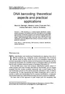
FIG. 1. Schematic actin and myosin filaments with cross-bridge (S-i, S-2) from myosin attached to actin site.
Many biological motility phenomena involve either translocation of one molecule or molecular aggregate relative to another or the assembly or disassembly of microtubules or actin filaments. Our object in this and a subsequent paper is to present some simple theoretical considerations that have relevance to these two subjects. The present paper is concerned primarily with the translocation of ATPases on single-stranded DNA (ssDNA) (1, 2), both in the absence and in the presence of a replicating fork. This is supplemented by a discussion of the facilitation of replication by some aggregated DNA-binding proteins (DBPs) (1) which can "treadmill" in a way that is similar to the behavior of aggregates of actin and tubulin in solution (3-7).
equivalent, an individual site is not symmetrical. That is, the set of sites has directionality. The particular case we shall discuss explicitly is the translocation of helicase III (11) or the rep protein (1, 2, 12) on ssDNA. The general principles of primary interest are not model dependent, but, for concreteness, we shall use a model suggested by the work of Das et al. (11) and by the presumed behavior of S-1, as shown in Fig. 1. Several generalizations will then be added in the subsections below. We assume that the basic ATPase-translocation cycle is that shown in Fig. 2. The enzyme has a "body" and an "arm"; the arm has two conformations, E1 at about 900 to the ssDNA and E2 at about 450 to the ssDNA (these details on shape and angle are completely arbitrary; all that is essential is some kind of unsymmetrical or directional inch-worm translocation mechanism associated with the ATPase activity). In states E1-T and E2-T, the body of E is very weakly bound to ssDNA (11); the conformational change (900 °-+ 450), induced by the binding of ATP, produces translocation of the body to the right (the + direction). This is also what would happen to the cross-bridge in Fig. 1 if S-2 were not attached to the myosin filament. Incidentally, the magnitude of the translocation step is much smaller here than in Fig. 1: 5 or 6 A (i.e., we assume one step is the distance between two ssDNA bases) rather than 60-80
TRANSLOCATION OF ATPASES ON DNA It is useful, for perspective, to begin with comments on the current view of the molecular basis of muscle contraction (8). Myosin cross-bridges, which are extensions from the myosin filament (Fig. 1), cyclically attach to and detach from binding sites on the actin filament. In the same cycle, ATP is bound to the S-1 portion (Fig. 1) of the cross-bridge, ATP is hydrolyzed, and its products (ADP and Pi) are released. In one step of the cycle, Pi is released (before ADP) while the cross-bridge is attached to actin (9). This is accompanied by a conformational change in S-1 that alters the angle of binding of S-1 to the actin site from 900 to 450 (Fig. 1). Because of the attachment of S-2 to the myosin filament, S-2 is stretched (10). This produces a force that tends to move the actin filament to the left relative to the myosin filament. However, movement does not result from the cyclic activity of any one cross-bridge. Rather, an asynchronous ensemble of about 10' cycling cross-bridges (single fiber; half-sarcomere) work smoothly together. Note that structural asymmetry (Fig. 1) is associated with the ATP cycle; this is essential if ATP hydrolysis is to produce a net force in a particular direction at steady state or in a transient. Translocation on DNA-Single Cycle. We turn now to the problem of the directional translocation of an ATPase on a set of equivalent one-dimensional sites. Though the sites are all
A. In states El and E2 the body is strongly bound to ssDNA (11). Consequently, the transition E2-- E1 (450 -° 900) involves the
arm but not the body. After one complete counterclockwise cycle in Fig. 2, starting from E1, one molecule of ATP has been hydrolyzed and the enzyme molecule (in state E1 again) has moved one site or step to the right. The 12 (pseudo where necessaW') first-order rate constants in the cycle (Fig. 2) are related to the operative thermodynamic force Apr = Pr - PUD- li (the free energy of ATP
hydrolysis) by (13)
fl+/fl_ -
T/RT
[1]
where H1, is the product of the six rate constants in the coun-
The publication costs of this article were defrayed in part by page charge payment. This article must therefore be hereby marked "advertisement" in accordance with 18 U. S. C. §1734 solely to indicate this fact.
Abbreviations: ssDNA, single-stranded DNA; dsDNA, double-stranded DNA; DBP, DNA-binding protein.
4796
4797
Proc. Natl. Acad. Sci. USA 78 (1981)
Biochemistry: Hill and Tsuchiya
B
)11 1
(B)
A)
E2 A
E2 'T
E2p
2~~~
FIG. 2. Counterclockwise ATPase-translocation cycle for enzyme E. T, ATP; D, ADP; P, Pi; A, arm moves; B, body moves; x, reference site. The symbols +, -, and 0 refer to direction of movement (translocation step) of body, if any.
terclockwise direction, etc. Similarly, the steady-state ATP flux JT can be expressed explicitly in terms of the 12 rate constants of the cycle in the form [2] IT = C(eAgT/RT 1), where the proportionality factor C > 0 is a rather complicated expression (13) and is omitted. The steady-state velocity v of translocation to the right, in units of the step or intersite distance, is simply v = JT in this case. As the motion occurs, ATP free energy is lost but the enzyme molecule and the ssDNA maintain a constant free energy. There is thus no free energy transduction in this system; however, the ATP hydrolysis, associated as it is with structural asymmetry (Fig. 2), is essential for the directional motion. This resembles closely the situation in steady-state treadmilling of monomers at the ends of F-actin or of microtubules in solution (3-7). Note that if Apr = 0 in Eq. 2 (by adjusting, hypothetically, the concentrations of ATP, ADP, and Pi), then JT = v = 0: there is no net motion, at equilibrium, in either direction. Of course if (hypothetically) ALe < 0, then the steady-state motion would be to the left UT = V < 0). In all cases above we are referring to net cyclic motion and flux; when Alkr # 0, cycles will be completed, occasionally, in the "wrong" direction; and when AAT= 0, cycles will be completed with equal frequency in the two directions. In summary, two features must both be present to achieve directional motion in systems of this general type: the chemical (ATP) cycle itself must be associated with structural asymmetry, and there must be an expenditure of chemical (ATP) free energy. Slippage and Resisting Force. In this subsection we generalize the above treatment in two ways. First, the obligatory B (body) and A (arm) sequence in the Fig. 2 cycle is relaxed to allow for occasional "slippage": an A transition (i.e., no body movement) is also possible at El-T :± E2 T and a B transition is possible at E2 ;± E1 (i. e., a body translocation to the left when E2- El). Both of these tend to reduce the translocation velocity v so that v < JT- The actual stoichiometry V/IT for rep protein or helicase III on ssDNA (in the absence of a fork) is presumably not known. The extended kinetic diagram is shown in Fig. 3A, in which rate constant notation is introduced for the four A and B transitions. The diagram has 16 rate constants altogether. The second generalization or complication is that, as indicated schematically in Fig. 3B, an outside force is acting against the enzyme body such that an amount of free energy AG is required in order to move the body one step to the right. In a very simplified model (see. below) this might represent translocation at a DNA fork (AG, a few kcal mold, is needed to separate one base pair of the duplex). Or, as another example, AG.could be _
X.=AG
E2 P L _ b~~~~~~~~
1L) AMT-AG
AG
Al1T
APIT
Jb
Ic
Jd
Ji
-
T
T
Jo-Ja
2
AIT +AG It
T
(C)
FIG. 3. (A) Less restrictive kinetic diagram than in Fig. 2. Notation as in Fig. 2. The two outside (curved) transitions have been added: A, arm movement despite weak binding of body; B, body movement despite strong binding of body. (B) External force associated with free energy AG restraining body movement to right. (C) Six cycles belonging to kinetic diagram in (A). Below the cycles are the cycle forces, the notation for cycle fluxes, and the consequences of counterclockwise cyclic activity (I- or -*, movement; T, ATP hydrolysis).
associated with a constant electric field (the enzyme molecule is charged). Immediate consequences ofthe existence ofAG are the relations
a+la-
=
(a/a')e-A/RT, (3-113+ (I/(1@)eAG/RT =
[3]
These follow from equilibrium constants and detailed balance at equilibrium. If slippage is included in the model but no external force, we put AG = 0 in Eqs. 3 (and below). The kinetic diagram includes six cycles (13), shown in Fig. 3C. The dominant cycle is considered to be cycle b (which is the only cycle in Fig. 2). Thus, ordinarily, a, > a and / > 8-. Each cycle K = a, b, . ... has its own force XK and net steady-state flux JK. The + cycle direction is chosen as counterclockwise in all cases. With AG > 0 and Ap > AG, all cycle forces and net fluxes are positive (JK and XK necessarily have the same sign; see Eqs. 4 and 5 below). Each force can be written in terms of rate constants (13) as (see Eq. 1)
/fi K- eX/RT. [4] Eqs. 3 are, in fact, examples for cycles a and c. The cycle fluxes K+
have the form
JK =CK (HK+
K-)
[5]
where CK > 0 is a complicated combination of all 16 rate constants in the diagram. There are straightforward rules for writing out the CK (13). The bottom row in Fig. 3C indicates the consequences of positive cyclic activity: T means ATP is hydrolyzed; the arrows show the translocation direction. Only cycle b, the main cycle, produces translocation to the right (against the direction of AG). Thus there is free energy coupling and transduction in this cycle: some of ApT (from ATP) is used to move the enzyme "uphill" (or melt the duplex, in the DNA fork example). The transduction efficiency for cycle b alone is nb =
AG/ApT.
From the bottom row in Fig. 3C, we see that the total ATP flux and the translocation velocity are given by
4798
Biochemistry: Hill and Tsuchiya
Proc.- Natl. Acad. Sci. USA 78 (1981)
IT = Jb + Jd + Je + Jf
[6]
V = -Ja + Jb - Ic - If
[7]
The overall thermodynamic efficiency is
(A)
P
I
(B)
P
P
(C)
p
p
K
If AG = 0 (slippage but no external force), Eq. 6 for JT is unchanged and.
a =1C 0, V = Jb -f, V 0. [9] =
,q
1l
P
P
P
+ lI+ +I p
p
p
L+
+-
+l
P
P
P
FIG. 4. Schematic translocating enzyme at fork, following Yarranton and Gefter (12): +, positive charge on enzyme body; P, negatively charged phosphate on DNA strand; ---, hydrogen bondeto other strand in duplex region. Note different inter-P spacing in single-stranded (left) and double-stranded (right) regions. See text for further explanation.
al. (11). On completion of this cycle (Fig. 5), one ATP has been used, one + translocation step has been taken, and one base pair has been opened up. The observed stoichiometry, however, as already mentioned, is approximately 2 ATP per base pair melted. Again, we can account for this by two kinds of "slippage" that could result in an average of about two ATPase cycles per cycle that opens a base pair. First, transition I in Fig. 5 might be used somewhat at the fork because of a reduced rate constant for the usual + or B translocation step (Fig. 4A -- Fig. 4B). This would have to be a kinetic effect, caused by the presence ofthe fork, because there is no thermodynamic reason why this rate constant should be reduced: AG, above, is not involved at this step in this model; also, the enzyme with ATP bound binds weakly to both ssDNA and dsDNA (11). If transition I is used, the enzyme remains in the Fig. 4A location: cycle d in Fig. 3C is followed. In the second kind of slippage, if state E2 P(ds) is reached in the main cycle, as described in the above paragraph, transition II might occur some fraction of the time (the enzyme remains in the Fig. 4B situation and Pi is released). The rate constant for the usual arm (A) transition, 450 -> 900 (E2 -- El), might be much reduced in Fig. 4B compared to Fig. 4C [again, not for any thermodynamic reason: AG is not involved; the free enzyme binds strongly to both ssDNA and dsDNA (11)]. If so, step III would generally follow step II (Fig. 4B -* Fig. 4A)-i.e., cycle e in Fig. 3C is used. If the main cycle (solid lines, Fig. 5) is used half of the time and cycles d and e the other half, the desired stoichiometry would result (all three cycles use ATP; only the first opens a base pair). The efficiency would be al AG/2Afr. If (as usual) DBP A ,__ E1 T
I (1° +l B
E1-_ A
B
to -t m -2
"~-E2T (ds) ii
E2(p(ds)
E2 P
E2 P (ds)
FIG. 5. Modification of Fig. 3A for the more detailed helicase model in Fig. 4.The new transition E2-P(ds)-- E2-P corresponds to Fig. 4B - Fig. 4C (opening of abasepairto lengthen single-strandedregion at expense of double-stranded region). See text for further explanation.
Biochemistry:
Hill and Tsuchiya
Proc. Natl. Acad. Sci. USA 78 (1981)
binds to and thereby stabilizes the new ssDNA, the efficiency is even smaller. Incidentally, a DBP molecule would not be bound after every base-pair-opening cycle but, at most, only after every nth cycle, where n is the number of bases covered by one DBP molecule on ssDNA. In summary: the above illustrative model is consistent with the Das et al. (11) experiments; it uses only one ATPase site and one conformational change in the enzyme; and it can explain an approximate 2:1 stoichiometry. Ifthe stoichiometry turns out to be an invariable 2:1, however, this would imply separate ATPase sites for translocation and for base pair opening, as in the work of Yarranton and Gefter (12). Translocation and Strength of Binding. Comments in the literature suggest that translocation on and strong binding to a one-dimensional system such as ssDNA is a paradoxical combination of properties. This is not necessarily the case, as is illustrated by the counterexample in Fig. 6. The rate of translocation is determined by the height of the free energy barrier to lateral motion, not by the depth of the binding free energy well. In this connection it should be kept in mind that a typical helicase spans a number ofbases on ssDNA, though it may move one base at a time. Another, more likely, possibility is illustrated by Figs. 2 and 3: there are weak and strong binding states of the enzyme in the same cycle; the enzyme usually moves when in a weak binding state (11).
TREADMILLING OF BINDING PROTEIN ON DNA The steady-state treadmilling of actin or tubulin subunits between the two nonequivalent ends of a linear aggregate in solution is now well established (3-5). Actin is an ATPase and tubulin is a GTPase; one molecule of NTP is hydrolyzed in the cyclical addition and removal of a subunit from either end of the aggregate (7, 14). A rather similar end-to-end treadmilling (ref. 1, figure 9-3, after B. Alberts) occurs with some highly cooperative DBPs, when the aggregate is bound on ssDNA (the template strand) adjacent to a replicating fork. This is illustrated schematically in Fig. 7A. However, the DBP is not an NTPase, as are actin and tubulin. The driving force for the DBP treadmilling arises, instead, from the dissimilar circumstances in the DNA itself at the two ends of the aggregate (see below). Examples of these proteins (1) are phage T4 gene 32 protein, phage M13 gene 5 protein, and Escherichia coli single-strand binding protein. A DBP molecule has a length equivalent to n base pairs, where n might be of order 10. When a new DBP molecule is bound at the fork end of the aggregate, in the process an amount offree energy nAG is required to separate or "melt" the n base pairs. At the other end of the aggregate (Fig. 7A), removal of a bound DBP molecule would be accompanied by (i) the addition of n dNMPs to the primer strand, (ii) the establishment In solution
Weak
binding
Slow
Free energy
Strong P binding Position
END 1
n&G'
nrG
_..
(B)
FIG. 7. (A) Schematic treadmillingof aggregated DBPto facilitate replication at fork [based on figure 9-3 of Kornberg (1)]. (B) Rate constant notation for adding or removing DBP molecules at two ends of bound aggregate in A.
of n paired bases with the template strand, and (iii) n translocation steps of the polymerase (assumed processive) relative to the new duplex (1). The net free energy released in these three more or less simultaneous processes is denoted by -nAG', where AG' > AG. Both AG and AG' are averaged quantities. The difference nAG' - nAG is obviously associated with the first and third processes above: the first releases free energy; the third requires some ofthis released free energy to maintain the directional motion* (as in the first section). nAG' - nAG is the thermodynamic driving force for the treadmilling shown schematically in Fig. 7A. At steady state, there is no free energy transfer from nAG' - nAG to the treadmilling DBP molecules, which have a constant free energy; the treadmilling merely facilitates the replication (1). The binding and release rate constants for the two ends of the aggregate are introduced in Fig. 7B; a and A3 are secondorder, while a' and 13' are first-order constants. The simple assumption here is that these transitions are rate determining in the overall replication process; the constants a' and 13, especially 13, are very small; a and 13' dominate. These rate contants are completely unrelated to those used in the first section (Fig. 3), even though the notation overlaps. Incidentally, because of very strong cooperativity among molecules of the aggregate (1), we are assuming that binding and release transitions occur only at the two ends of the aggregate. Let c be the local concentration of the DBP in the vicinity of the DNA duplex (there may not be many molecules to establish this "concentration"). The corresponding chemical potential is A= A + RTln c. [12] In the aggregate itself, excluding DNA free energy contributions at the ends, the DBP chemical potential is a constant, p0. In a hypothetical aggregation equilibrium at the fork end (end 1) only, now including the DNA contribution, we would have pu + RTln c(') = go + nAG, [13] where c(,) is the DBP equilibrium concentration. The presence of the positive nAG term increases the value of c that is required *
Fast
FIG. 6. Possibility of fast translocation with strong binding and translocation with weak binding.
slow
END 2
4799
In a more realistic and elaborate treatment, we would have to include the DBP treadmilling and the polymerase translocation (n steps per DBP) in a single kinetic model. In this case, the free energy released in process i alone would provide the thermodynamic driving force necessary to maintain both of these directional processes, the net effect of which is the steady-state replication of DNA (as in Fig. 7A).
4800
Biochemistry: Hill and Tsuchiya
Proc. Natl. Acad. Sci. USA 78 (1981)
in order to reach equilibrium. Because of detailed balance at equilibrium, we also have ac,(') = a', so that Eq. 13 can be written as RTln(a/a') = AO- (,uo + nAG). [14]
This equation shows how a/a' is influenced by nAG. At end 2, we have p? + RTln c(2) = o + naG', [15] 8c= 183', and
RTln(,f/p3') = AO - (pO + nAG').
[16]
In one treadmilling cycle, in which one molecule is added to end 1 and one is removed from end 2 (a clockwise cycle in Fig 7B), the thermodynamic force nAG' - nAG in the cycle is related to cycle rate constants by af3'/13a' - e(nAG'- AG)/RT [17] This follows from Eqs. 14 and 16. It should be noted, above, that c(l) is very small and c( is very large, because a' and 13 are very small. The separate rates of attachment at the two ends, at an arbitrary concentration c, are J1= ac-a' ac, J2 = Pc-a P'-I'. [18] The net or total rate of attachment is [19] J-J1 +J2 ac-a'. Steady state occurs when J = 0, that is, when c c. = (a' + P3')/(a + 13) 13'/a. [20]
Unlike cel) and c(2), c,, does not have an extreme value. The treadmilling or translocation rate, that is, the rate of adding molecules at the fork end (and the rate of removal from end 2), at steady state, is
Jm. JM(c-) = -J2(CX) = (a' - 13a')/(a + 1) 13'. [21] This is, of course, also the replication rate on the primer strand, expressed in DBP units. Note from Eq. 17 that if AG = AG', then a13' = 13a' and henceJm = 0. The aggregate does not have net movement on the template strand in this case. Treadmilling occurs, at steady state, only if AG 0 AG'. The aggregate moves to the right (Fig. 7) on the template strand if AG' > AG and Im > 0; this is the case of actual interest. There are, of course fluctuations (14), which we shall not discuss here. In the actin and tubulin systems, treadmilling occurs on the ends of an aggregate in solution because of different rate constants in the subunit NTPase cycles at the two different ends of the aggregate (4-7). Here, because DBP is not an NTPase but simply a binding protein, we would have a/a' = 13/13' and no treadmilling were it not for the DNA free energy contributions, with AG' # AG. If we regard a replicating DNA duplex and its immediate neighborhood as an effectively closed system in DBP molecules, we can see (6) that c tends naturally to approach c", as a final stable value. That is, if c > c,,, thenJ > 0 and the aggregate grows at the expense of DBP molecules in solution (thus c decreases toward c,,). On the other hand, if c < c,,, thenJ < 0 and the aggregate becomes smaller, adding DBP molecules to the solution (thus c increases toward c.). We now consider the rate of free energy dissipation in this system. At arbitrary c, the chemical potential differences be-
tween free and attached DBP molecules, including DNA contributions, are End 1: AIL,= AP + RTln c - (po + nAG) [22]
End2:
Ag2--
+ RTlnc -
(pa
+ nAG').
[23]
Eqs. 13 and 15 are special cases. The rate of free energy dissipation is then (13) T(diS/dt) = JlAAI + J2AA2 > 0. [24]
Both terms on the right are necessarily positive (this is verified in Eq. 27 below). At c = c,, using Eq. 21 and [25] A,- AA2 = nAG' - nAG, we have T(dS/dt) = Jm(nAG' - nAG) (c = c.). [26] This is the rate of free energy decrease for the "downhill," overall replication process. The DBP treadmilling is (by assumption) rate controlling, through the factorJ. (Eq. 21). Eq. 24 can also be written, using Eqs. 14 and 16, as T(diS/dt) = RT[ac - a')ln(ac/a') + (1c - 83')ln(13c/13')]. [27]
Because, as mentioned above, c naturally approaches c, in a closed system, one might expect that T(diS/dt) in Eq. 27 would be minimized with respect to c at c = c,,. However, this is easily seen not to be the case in general. For arbitrary values of c between cl) and c(, which covers practically the entire range in c, J, is positive andJ2 is negative. In this case, one can regard the total kinetic activity as made up of two components: a background of treadmilling molecules together with a surplus gain or loss of molecules at one end of the aggregate or the other. Eq. 24 can be rearranged differently, for the two possible cases, to show this explicitly: T(diS/dt) = (-J2)(nAG' - nAG) + JAAI (c. < c < c(2) [28] = Jl(nAG' - nAG) + JA/2 (C(1)