C A S E R EP O R T
pISSN: 2384-3799 eISSN: 2466-1899 Int J Thyroidol 2016 November 9(2): 200-203 https://doi.org/10.11106/ijt.2016.9.2.200
Thyroid Associated Ophthalomopathy in a Patient with Hashimoto’s Thyroiditis Ho-Jun Lee1, Yeo-Joo Kim1, Suk-Hoe Kweon2 and Ah-Jeong Ryu2 Department of Endocrinology, Soonchunhyang University College of Medicine1, Cheonan, Department of Endocrinology, Good Morning Hospital2, Pyeongtaek, Korea A 53-year-old man consulted an ophthalmologist with a chief complaint of diplopia and bilateral eyelid swelling. He was diagnosed with hypothyroidism 2 years prior at a local clinic and had been taking levothyroxine 150 mcg daily. CT scan of the orbits showed enlargement of bilateral extraocular muscles. Laboratory findings revealed hyperthyroidism due to high dose levothyroxine. Active ophthalmopathy with Hashimoto’s hypothyroidism was diagnosed and the patient was treated with steroid pulse therapy. We reported a rare case of severe ophthalmopathy with Hashimoto’s thyroiditis that needed steroid pulse therapy. Key Words: Ophthalmopathy, Hashimoto’s thyroiditis, Hypothyroidism, Hyperthyroidism, Steroid
swelling for 3 months. He was diagnosed with hypo-
Introduction
thyroidism 2 years prior at a local clinic and had been taking levothyroxine 150 mcg daily. He had no family
Thyroid associated ophthalmopathy (TAO) is an au-
history of thyroid or autoimmune diseases. He was
toimmune disorder of the extraocular muscles and
current smoker. An ophthalmological examination
surrounding orbital connective tissue which is gen-
showed mild upgaze restriction (i.e., monocular ele-
erally associated with Graves’ disease. Typical signs include upper eyelid retraction, periorbital edema,
vation deficiency) and hypotropia of the right eye (Fig.
proptosis, and impairment of eye motility. Although
of 0.01 μIU/mL (0.27-5.0 μIU/mL), free T4 level of
TAO generally occurs in patients with hyperthyroidism
1.73 ng/dL (0.93-1.7 ng/dL), antithyroglobulin (anti-Tg)
due to Graves’ disease, it may also accompany
antibody level of 581 IU/mL (0-115 IU/mL), antithyroid
Hashimoto’s thyroiditis. However, TAO in Hashimoto’s thyroiditis is uncommon. A case of severe TAO that
peroxidase antibody (anti-TPO) level of 600 IU/mL
needs steroid pulse therapy is very rare. We de-
of 13.69 IU/L (0-1 IU/L). An ultrasound examination
scribed a case of severe Hashimoto’s ophthalmopathy.
of the thyroid showed diffuse coarse heterogeneous
1). Laboratory examination results showed a TSH level
(0-34 IU/mL), and TSH receptor antibody (TRAb) level
hypoechogenicity (Fig. 2). A computed tomography
Case Report
scan of the orbits revealed both periorbital soft tissue swelling and hypertrophy and enhancement of the
A 53-year-old man consulted an ophthalmologist
superior rectus, medial rectus, and inferior rectus
with a chief complaint of diplopia and bilateral eyelid
muscles (Fig. 3). An active form of orbitopathy was di-
Received August 20, 2016 / Revised 1st October 19, 2016, 2nd October 24, 2016 / Accepted October 25, 2016 Correspondence: Yeo-Joo Kim, MD, Department of Endocrinology, Soonchunhyang University College of Medicine, 31 Soonchunhyang 6-gil, Dongnam-gu, Cheonan 31151, Korea Tel: 82-41-570-3685, Fax: 82-41-574-5762, E-mail:
[email protected] Copyright ⓒ 2016, the Korean Thyroid Association. All rights reserved. This is an open-access article distributed under the terms of the Creative Commons Attribution Non-Commercial License (http://creativecommons.org/licenses/by-nc/4.0/), which permits unrestricted non-commercial use, distribution, and reproduction in any medium, provided the original work is properly cited.
200
Thyroid Associated Ophthalomopathy in a Patient with Hashimoto’s Thyroiditis
agnosed and methylprednisolone pulse therapy was administered
(250
mg/d
intravenous
proved. His free T4 level was within the reference values.
methyl-
prednisolone for 4 succeeding days) which was fol-
Discussion
lowed by 32 mg/d oral methylprednisolone. After glucocorticoid therapy and a decrease in the
Thyroid-associated orbitopathy is a set of symp-
dose of levothyroxine, diplopia and bilateral eyelid swel-
toms caused by an autoimmune process; it is typical
ling, which were the patient’s chief complaints, im-
of Graves’ disease and rarely accompanies Hashimoto’s 1)
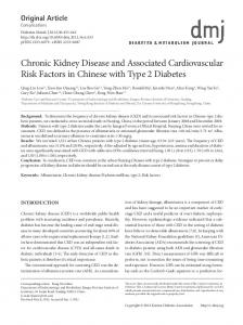
thyroiditis. In both Graves’ and Hashimoto’s diseases, anti-thyroglobulin and anti-thyroid peroxidase antibodies are detected. Therefore, transformation of Graves’ disease into Hashimoto’s thyroiditis disease and vice versa has been known to occur.2) Numerous studies have reported the prevalence of Graves’ ophthalmopathy. Similar studies about the prevalence of Hashimoto’s ophthalmopathy have been reported occasionally. Mild upper eyelid retraction is known to be predominant eye symptom in Hashimoto’s ophthalmopathy, but severe form of ophthalmopathy with Hashimoto’s thyroiditis that needs aggressive treatment was reported only a few times. Fig. 1. A 53-year-old man suffered from diplopia and bilateral eyelid swelling.
The first study about the prevalence of ophthalmopathy in Hashimoto’s thyroiditis was reported by
Fig. 2. Ultrasound examination showed no significant findings in both thyroid except diffuse goiter and inhomogeneous echogenicity.
Fig. 3. CT scan of the eyes in the patients showed enlargement of both medial rectus muscles and both inferior rectus muscles.
201 Int J Thyroidol
Ho-Jun Lee, et al
Tjiang et al.,3) who studied 91 patients recently diag-
seemed to influence the risk of any ophthalmopathy
nosed with Hashimoto’s thyroiditis and reported that the overall prevalence of any eye signs was 34%, with
subtype, namely, congestive ophthalmopathy, ocular
about one-third of patients having upper eyelid
shown to have an effect on the activity of ophthalm-
retraction. Two patients had eye muscle dysfunction
opathy: men with Hashimoto’s thyroiditis-related eye disease were 18 times more likely to develop more
and about one-third of the patients had severe in-
myopathy, or mixed disease. However, gender was
flammatory changes such as periorbital swelling, che-
active ophthalmopathy (CAS>3) than were women.
mosis, conjunctival injection, and proptosis. Kan et al.4)
Severe ophthalmopathy is rare in patients with
studied the prevalence of ophthalmopathy in 110
Hashimoto’s thyroiditis, only a few cases have been reported.6-9) Yoshihara et al.6) presented 2 cases of
Hashimoto’s thyroiditis patients as compared with healthy control subjects. Those authors reported that the prevalence of eye signs was 22.7% in patients, which was less than that reported in Tjiang’s study, and 4% in control subjects; the difference was statistically significant (p=0.002). Of all the patients, 11.8%
severe ophthalmopathy with Hashimoto’s thyroiditis. A 64-year-old woman with Hashimoto’s opthalmopathy (CAS 3) was treated with orbital irradiation (15 Gy) and orbital decompression surgery. Another patient, 44-
had upper eyelid retraction, 6.4% had proptosis, and
year-old woman with Hashimoto’s ophthalmopathy (CAS 4) was managed with orbital irradiation (15 Gy)
5.5% had ocular myopathy. Kochairi et al.5) studied the
and oral prednisolone 15 mg daily. Other studies used
risk factors which may influence the development of
steroid (intravenous or oral) as main treatment and all
ophthalmopathy in patients with Hashimoto’s thyroiditis. A retrospective cross-sectional study included 105
studies reported improvement of the orbital symptoms
patients with Hashimoto’s thyroiditis with and without ophthalmopathy and investigated 6 potential risk fac-
The exact mechanism of TAO is still unknown. However, one theory that explains the association of
tors; age, gender, smoking, vitamin D deficiency, se-
TAO with autoimmune thyroid disease is immunologic
rum TSH, and serum levels of antibodies to thyroid
cross reactivity of sensitized T lymphocytes and/or
peroxidase (TPO) and thyroglobulin (Tg). They tried to
autoantibodies against antigens common to the thyroid
find out the risk factors for i) ophthalmopathy (NOSPECS
and orbit.1,10-12) Sensitized T-cell clones trigger an in-
after the therapy.
class≥1), ii) upper eyelid retraction group (often the
flammatory process in the tissues of the orbit. As a re-
only sign in patients with Hashimoto’s thyroiditis), iii) the type of ophthalmopathy (congestive ophthalmopathy
sult, leukocytes secrete cytokines, which stimulate fi-
vs. ocular myopathy or both), iv) the activity of the eye
the swelling of orbital tissues. Kochairi et al.’s study revealed the protective effect of ageing on the devel-
disease assessed as CAS (severe ophthalmopathy was taken as a NOSPECS class≥3, more active
broblasts to secrete glycosaminoglycans, resulting in 5)
ophthalmopathy was defined as a CAS>3). They re-
opment of Hashimoto’s ophthalmopathy, and they explained that this might be due to reduced self-toler-
ported a protective effect of aging on the development
ance in young individuals and increased portion of
of ophthalmopathy in patients with Hashimoto’s thyroiditis; the risk decreased by 5.4% for each additional
regulatory T cells which maintain peripheral tolerance
year. They also reported a detrimental effect of smok-
They also mentioned a study of systemic lupus eryth-
ing, with the risk of ophthalmopathy being 5.5 times
ematosus to explain greater risk for severe auto-
greater in smokers than in non-smokers. Increased
immunity in men by a higher cumulative genetic load
serum TSH was not shown to be a risk factor for the
required
by suppressing auto-reactive T cells in old people.
for 13)
men
to
develop
the
autoimmune
presence or severity of ophthalmopathy. High serum
disease.
levels of TPO antibodies were found to be protective
possible autoantibody targets, including TRAb,14,15) the
against the development of upper eyelid retraction but
skeletal muscle calcium binding protein calsequestrin,
not ophthalmopathy. None of the tested factors
and the fibroblast cell membrane protein collagen
Several antigens have been identified as
Vol. 9, No. 2, 2016
202
Thyroid Associated Ophthalomopathy in a Patient with Hashimoto’s Thyroiditis
XIII.16-19) Evidences suggests that the TSH receptor is present in the orbit and is expressed on orbital fibroblasts. These findings support the hypothesis that TRAb is not only the cause of Graves’ disease, but is also responsible for TAO. Since majority of patients with Hashimoto’s thyroiditis test negative for TRAb, it does not explain the etiology of ophthalmopathy in Hashimoto’s thyroiditis. An alternative explanation is the specific production of an antibody against an eye muscle antigen, such as calsequestrin, flavoprotein, or G2s.20) In this case, the patient was a current smoker and male. These were thought to be risk factors for severe ophthalmopathy. CAS or NOSPECS classification was not conducted, but as we presented, we thought his clinical symptoms were at least CAS 2 or higher. Unfortunately, we did not test antibodies such as calsequestrin, flavoprotein or G2s, but the patient was reported as TRAb, TPO Ab, and TG Ab positive. The patient’s initial FT4 level was high because he was administered an overdose of synthyroid at a local clinic. After decreasing the dose of synthyroid, the FT4 level decreased to within normal limits. In conclusion, we presented a case of Hashimoto’s hypothyroidism with severe ophthalmopathy. Few cases of ophthalmopathy are reported in patients with Hashimoto’s thyroiditis, but severe form of ophthalmopathy is very rare. This case highlights that severe symptoms and functional disorders may be accompanied by ophthalmopathy in patients with Hashimoto’s thyroiditis. Thus, more intensive routine ophthalmic examinations including CAS and NOSPECS classification are needed in patients with Hashimoto’s thyroiditis and steroid pulse therapy can be considered as the treatment.
References 1) El-Kaissi S, Frauman AG, Wall JR. Thyroid-associated ophthalmopathy: a practical guide to classification, natural history and management. Intern Med J 2004;34(8):482-91. 2) Wall JR, Bernard N, Boucher A, Salvi M, Zhang ZG, Kennerdell J, et al. Pathogenesis of thyroid-associated ophthalmopathy: an autoimmune disorder of the eye muscle associated with Graves' hyperthyroidism and Hashimoto's thyroiditis. Clin Immunol Immunopathol 1993;68(1):1-8. 3) Tjiang H, Lahooti H, McCorquodale T, Parmar KR, Wall JR. Eye and eyelid abnormalities are common in patients with
203 Int J Thyroidol
Hashimoto's thyroiditis. Thyroid 2010;20(3):287-90. 4) Kan E, Kan EK, Ecemis G, Colak R. Presence of thyroidassociated ophthalmopathy in Hashimoto's thyroiditis. Int J Ophthalmol 2014;7(4):644-7. 5) Kochairi IE, Champion B, Wall JR. Risk factors for the development of ophthalmopathy in patients with Hashimoto's thyroiditis. Ophthalmology Research 2014;2(4):177-88. 6) Yoshihara A, Yoshimura Noh J, Nakachi A, Ohye H, Sato S, Sekiya K, et al. Severe thyroid-associated orbitopathy in Hashimoto's thyroiditis. Report of 2 cases. Endocr J 2011;58(5): 343-8. 7) Tateno F, Sakakibara R, Kishi M, Ogawa E. Hashimoto's ophthalmopathy. Am J Med Sci 2011;342(1):83-5. 8) Grzesiuk W, Szydlarska D, Pragacz A, Bar-Andziak E. Thyroid-associated orbitopathy in patients with Hashimoto's thyroiditis: a case report. Pol Arch Med Wewn 2008;118(5): 318-21. 9) Hiraga A, Mimura M, Kamitsukasa I. Isolated inferior rectus muscle myopathy due to Hashimoto's thyroiditis. Intern Med 2008;47(13):1283-4. 10) Jacobson DM. Dysthyroid orbitopathy. Semin Neurol 2000; 20(1):43-54. 11) Garrity JA, Bahn RS. Pathogenesis of graves ophthalmopathy: implications for prediction, prevention, and treatment. Am J Ophthalmol 2006;142(1):147-53. 12) Costa RM, Dumitrascu OM, Gordon LK. Orbital myositis: diagnosis and management. Curr Allergy Asthma Rep 2009; 9(4):316-23. 13) Hughes T, Adler A, Merrill JT, Kelly JA, Kaufman KM, Williams A, et al. Analysis of autosomal genes reveals gene-sex interactions and higher total genetic risk in men with systemic lupus erythematosus. Ann Rheum Dis 2012;71(5):694-9. 14) Bahn RS. Clinical review 157: Pathophysiology of Graves' ophthalmopathy: the cycle of disease. J Clin Endocrinol Metab 2003;88(5):1939-46. 15) Starkey KJ, Janezic A, Jones G, Jordan N, Baker G, Ludgate M. Adipose thyrotrophin receptor expression is elevated in Graves' and thyroid eye diseases ex vivo and indicates adipogenesis in progress in vivo. J Mol Endocrinol 2003;30(3):369-80. 16) Gopinath B, Ma G, Wall JR. Eye signs and serum eye muscle and collagen XIII antibodies in patients with transient and progressive thyroiditis. Thyroid 2007;17(11):1123-9. 17) Gopinath B, Musselman R, Adams CL, Tani J, Beard N, Wall JR. Study of serum antibodies against three eye muscle antigens and the connective tissue antigen collagen XIII in patients with Graves' disease with and without ophthalmopathy: correlation with clinical features. Thyroid 2006;16(10):967-74. 18) Lahooti H, Parmar KR, Wall JR. Pathogenesis of thyroidassociated ophthalmopathy: does autoimmunity against calsequestrin and collagen XIII play a role? Clin Ophthalmol 2010; 4:417-25. 19) Wall JR, Lahooti H. Pathogenesis of thyroid eye disease--does autoimmunity against the TSH receptor explain all cases? Endokrynol Pol 2010;61(2):222-7. 20) Miller A, Arthurs B, Boucher A, Liberman A, Bernard N, Rodien P, et al. Significance of antibodies reactive with a 64 kDa eye muscle membrane antigen in patients with thyroid autoimmunity. Thyroid 1992;2(3):197-202.