reproduction sur papier ou sur format .... Primer pairs used in this study. 7. ..... characterized by primer extension and S1 nuclease mapping analysis (Kim et al.,.
MOLECULAR CHARACTERIZATION OF THE HIPPURATE HYDROLASE GENE IN HIPPURATE HYDROLASE-NEGATIVE CAMPYLOBACTER JEJUNI ISOLATES
Tin Htwe Thin
A thesis submitted in conforrnity with the requirements
for the degree of Mater of Science, Graduate Department of Microbiology, in the University of Toronto
O Copyright by Tin Htwe Thin 1997
395 Wellington Street Ottawa ON K1A ON4 Canada
395, rue Wellington Ottawa ON K I A ON4 Canada Your fi/e Vofre réiérence
Our file NoIr8 réltirence
The author has granted a nonexclusive licence alîowing the National Library of Canada to reproduce, loan, distribute or sel1 copies of this thesis in microforni, paper or electronic formats.
L'auteur a accordé une licence non exclusive permettant à la Bibliothèque nationale du Canada de reproduire, prêter, distribuer ou vendre des copies de cette thèse sous la fome de microfiche/film, de reproduction sur papier ou sur format électronique.
The author retains ownership of the copyright in this thesis. Neither the thesis nor substantial extracts fi-om it may be printed or otherwise reproduced without the author's permission.
L'auteur conserve la propriété du droit d'auteur qui protège cette thèse. Ni la thèse ni des extraits substantiels de celle-ci ne doivent être imprimés ou autrement reproduits sans son autorisation.
Molecular Characterization of the Hippurate Hydrolase Gene in Hippurate Hydrolase-Negative Campylobacterjejuni Isolates Degree of Mater of Science 1997 Tin Htwe Thin Department of Microbiology University of Toronto
CampyZobacter jejuni is human entericbacterial pathogen. The present study
examined the hippurate hydrolase gene from five different hippurate hydrolasenegative C . jejuni dinical isolates. Radiolabeled single strand confomational polymorphic (SSCP) assay detected the polymorphism in F7 and F8 regions of the hippurate hydrolase gene from five negative isolates compared with the respective regions of C. jejuni TGH9011 hipO. The promoter region and the remainder coding region revealed an identical PCR-SSCP patterns compared with the respective regions of C. jejuni TGH9011 in five negative isolates. The potential mutated regions were cloned and sequenced to determine the rnolecular nature of the mutations. Most of the mutations detected were silent in nature and they do not affect for arnino acid alteration, except for valine (Val) at residue 250 altered to alanine (Ala). The Val-250 change was consistently present in al1 five negative isolates. Val-250 residue may play an important role in the hippurate hydrolase function.
Acknowledgments
My deepest thanks go to my major professor, Dr. Voo Loong (Ricky) Chan for giving me the opportunity to participate in Campylobacter jejuni genetic studies. My appreciation is also given to my comrnitee members Dr. A. Bognar and Dr. M. Kregden for their advice and editorial assistance. 1owe my sincere gratitude to my parents U Thin Tu and Daw Tin Tin Win and
my only sister, Tin 00Thin for their permanent support through my career. 1would like to extend rny apprteciation to my colleagues and fiiends, especialiy
to Helena Louie, for their help and encouragement.
1 aioie 01 conrenrs
Abstract Acknowledgement Table of contents List of figures List of tables List of abbreviations Introduction General features of the genus Campylobacter Phylogeny of Campylobacter Clinical features of Campylobacterjejuni nfect ion Virulence factors of Campylobacter jejuni Adhesins Campylobacter toxins Epidemiology of Campylobacterjejuni infection Genome size of Campylobacterjejuni Genes cloned from Campylobacterjejuni Housekeeping genes Virulence gene Antibiotics resistance gene Hippurate hydrolase of Campylobacter jejuni TGH90 1 1 Hmologous proteins belong to M40 hydrolase farnily Hippurate hydrolase-negative Campylobacter jejuni Purpose of this study Materials and Methods Bacterial strains Genomic DNA extraction OIigonucleotides Polymerase chah reaction Single strand conformational polymorphic(SSCP) assay and mutation detection enhancement (non denaturing) gel electrophoresis Cloning of the arnplified fragments Ligation of the DNA fragments Preparation of competent cells and transformation with
Kecombinant plasmid DNA preparation Preparation of the squencing grade plasmid DNA DNA sequencing and polyacrylarnide gel electrophoresis
Results Detection of the hippurate hydrolase sequence heterogeneity in the hippurate hydrolase-negative C.jejuni clinical isolates Single strand conformational polymorphic analysis of naturally occuring mutant hippurate hydrolase genes Cloning of SSCP polymorphic regions and DNA sequencing Sequence comparison Predicted hydropathy profiles
Discussion
Future studies References
Figure Biochemical reaction of hippurate hydrolase Nucleotide sequence of C. jejuni TGH9011 hippurate hydrolase Physical map of C.jejuni TGH9011 Strategy for single Strand conformational polymorphic (SSCP) assay based on polyrnerase chain reaction PCR-SSCP analysis of F7 and F8 regions of the hippurate hydrolase gene of five different hippurate hydrolase-negative C. jejuni isolates PCR-SSCP analysis of F 1,F2, and F3 regions of five different hippurate hydrolase-negative C.jejuni isolates Deduced amino acid sequence alignment of the mutated region of hippurate hydrolase-positive C .jejuni TGH90 11 and hippurate hydrolase-negative strain
D835 Alignment of C.jejuni hippurate hydrolase and related proteins Comparison of the hydrophobicity profiles for arnidohydrolases of Campylobacter, Bacillus, Pseudomonas, and Synechocystis Predicted hydropathy (hydrophobicity and flexibility) profiles of C. jejuni hippurate hydrolase Comparison of the predicted secondary structure and hydrophobicity profiles of C.jejuni hippurate hydrolase and its modified sequence
Lists of tables Table 1. 2. 3. 4. 5. 6.
7. 8.
Classification of the genus Campylobacter and its relatives Codon usage and molecular percentage of G+C content of C. jejuni TGH9011 hippurate hydrolase. Homologous proteins of C.jejuni hippurate hydrolase Bacterial strains used in this study OIigonucleotides used in this study Primer pairs used in this study Summary of PCR analysis in the hippurate hydrolase gene of hippurate hydrolase-negative C.jejuni isolates Nucleotide substitutions between nucleotide 473 and 912 of the hipO gene of C .jejuni hippurate hydrolase-negative isolates
a
P
+ %
ala A ~ P Arg ATP bp 'C C.
CHC13 Ci CO2 CIAP CJT Ch1 CLDT
cm CNW CP*
CT D DNA dATP dCTP ddNTP dGTP dNTP ddTTP DMSO Dnase DTT EDTA EtBr EtOH fig g
gly GTE H2 H20
H2S
alpha beta positive negative percent alanine ampicillin arginine adenosine triphosphate base pair degree CeIsius Campylobacter chloroform curie carbon dioxide calf intestinal alkaline phosphatase CampyIobucterjejuni toxin chlorarnphenicol cytolethal distension toxin centimeter catalase negative/weak counts per minute cholera toxin daltons deoxyribonucleic acid 5'-deoxyadenosine triphosphate 5'-deoxycytosine triphosphate 2',3'-dideoxynucleotide triphosphates 5'-deoxyguanine triphosphate 2'-deoxynucleotide triphophates 5'-deoxythymine triphosphate dimethylsulphoxide deoxyribonuclease dithiothreitol ethylenediarninotetraacetic acid ethidium brornide ethanol figure gram glycine glucose, tris, EDTA buffer hydrogen water hydrogen sulfide
H 1P hr 1AA ile IPTG K
kb kDa
Km L LB 1bs.lsq.in pCi Pg P1 M Mb mg Mg MgCl2 MH min ml mM mol%G+C MOPS M r
mFWA N N-BzNa NaCl NaOH ng nM nt OD PAGE PCR PEG PH
PI pmole PFGE phe
nlppurare nyaroiase hour Indole-3-acetic acid-arnino acid isoleucine isopropyl-P-D-thiogalactoside potassium kilobase kilodalton Michaelis constant liter Luria Bertani broth pounds per square inch microcurie microgram microliter molar megabase milligrarn magnesium magnesium chioride MuelIer Hinton minute milliliter millimolar mol percentage guanidine plus cytidine 3-[N-Morpholino]propane-sulfonicacid relative molecular mass messenger ribonucleic acid normal fi-benzoylsodium sodium chloride sodium hydroxide nanometer nanomolar nucleotide optical density polyacrylamide gel electrophoresis polmerase chain reaction polyethylene glycol power of hydrogen isoelectric pH picomole pulsed- field gel elctrophoresis phenylalanine resistance
KNA
RNase rRNA
SDS SSC
SPP* subspp. TBE TE TEMED tet TLC Tris RNA TS
val W
ri bonucleic acid ribonuclease ribosomal ribonucleic acid revoiution per minute sensitive sodium dodecyl sulphate sodium chloride/sodium citrate solution species species(plura1) subspecies Tris boric acid EDTA buffer Tris-EDTA N,N,N',N1-tetramethylenediamine tetracycline thin-layer chromatography tris(hydroxymethy1) benzidine transfer ribonucleic acid type strain unit microcurie ultraviolet volt valine watt weight per volume times [multiplication] times [concentration strength] 5-bromo-4-chloro-3-indoyw-D-galactopyranoside
General features of the prenus Campvlobacier
Members
of
the
farnily
Campylobacteraceae
are
classified
as
chemoorganotrophs. They do not ferment or oxidize carbohydrates. They appear as curved in spira-, V-, or comma-shaped or occasionally straight rods that are 0.2 to 0.9 pm wide and 0.5 to 5 pm long. They rnay occur in short or occasionally long chains. CoIonies are usually nonpigmented and are weakIy or non-hemolytic. The cells are Gram-negative and non-sporeforming.
Motility occurs as a result of single or
occasionally multiple unsheathed flagella at one or both ends of the bacterial cells. Campylobacters are usually microaerophilic with a respiratory metabolism. However. some strains may grow under aerobic or anaerobic conditions.
They use
menaquinones as the sole respiratory quinones. A variety of organic acids including amino acids are used as carbon sources. Oxidation of tricarboxylic acid intermediates and the dearnination of amino acids are the main routes through which cellular energy is derived (Hoffman and Goodman, 1982). Optimum temperature for growth ranges
from 30' to 42'C. Biochemical tests give negative reactions in the methyl red and the Voges-Proskauer tests and there is no production of indole. Gelatin is not liquified. Al1 species have oxidase activity.
Most species c m reduce nitrate and do not
hydrolyze hippurate (Smibert, 1984; Penner, 1988; Penner, 1991; Ursing et al., 1994).
Phvlogeny of Campyiobacter
Campylobacters were isolated and first named as Vibri~~fettts due to the
-
Y
Vibrio (McFadyean and Stockman, 1913). Further investigations ihstrated a low guanine plus cytosine (G+C) base composition (28 to 47 molar percentage), microaerophilic growth requixements, nonfermentative metabolism and lack of oxidation of carbohydrates. These Vibrio - Iike organisms were then renamed as a new genus, Campylobacter (Sebald and Veron, 1963). Cornprehensive taxonornic studies of Vibrio - like organisms resulted in renarning Campylobacter fetus as the type species (with C.fetus subsp. fetus for the organisms that cause sporadic abortions in cows and ewes and C.fetus subsp. venerealis for the organisms that are responsible for infectious infertility), along with Campylobacter jejuni, Campylobacter coli and Carnpylobacter sputorurn (C. sputorum subsp. sputorum, C. sputorum subsp. bubulus) (Veron and Chatelian, 1973). Partial 16s ribosomal ribonucleic acid sequences of Campylobacter species were used to compare and analyze the relationships of these organisms to one another and to other gram-negative bacteria. The genus Campylobacter was divided into three separate ribosomal ribonucleic acid sequence homology groups. Homology group 1 contains the true Campylobacter species which are C. fetus (two subspecies), C. coli, C. jejuni, C. laridis, C. hypointestinalis, C. concicus, C. mucosalis, C. sputorum, and C.upsaliensis ( C N W strains). Homoiogy group II includes C. cinaedi, C. fennelliae, C. pylori and W. succinogenes. Homology group I I I consists of C. cryaerophila and C. nitroflgilis. These three homology groups are distantly related to the representative
of the alpha, beta, and gamma branches of the purple bacteria 1988).
(Thompson et al.,
limited the genus Campylobacter to C. fetus, C. hypointestinalis, C. concisus, C. mucosalis, C. sputorum, C.jejuni, C. coli, C. lari, and C. upsaliensis. C. cinaedi and C. fennelliae were included in the genus Helicobacter as Helicobacter cinaedi and Helicobacter fennelliae. Two species of the genus Carnpylobacter, C. nitrofigilis and
C. cryaerophilia were constituted as a new genus Arcobacter and were renamed as Arcobacter nitrofigilis and Arcobacter cryaerophilus. Wolinella curva and Wolinella recta was transferred to the genus Campylobacter as Carnpylobacter curvus and Campylobacter rectus, respectively (Paster and Dewhirst, 1988; Vandamme et al., 1991). Thus, Wolinella succinogenes is the only species of the genus Wolinella. Based on the rRNA homology studies, Bacteroides gracilis and Bacteroides ureolyticus are generically misnarned and are closely related to rnembers of the genus Campylobacter (Vandamme et al., 1991). B. ureoZytictcs and B. graciIis are microaerophilic and not anaerobes. Recently, B. gracilis was reclassified under the genus CrrmpyZobacter as Campylobacter gracilis based not oniy on genotypic data, but also on the proteolytic metabolism of respiratory quinones, protein profiles and cellular fatty acids analysis (Vandamme et al., 1995). Presently Campylobacter hyoilei is grouped under the genus Campylobacter and recognized as closely related to C. jejuni and C. coli on the basis of their G+C contents, phenotypic characteristics,
hybridization with a speci'es-specific DNA probe, 16s rRNA sequence cornparison data and DNA-DNA hybridization data (Alderton et al., 1995). Table 1 presents the list of known species of Carnpylobacter, Helicobacter, and Arcobacter according to the recent presentation of taxonomie position, known sources and common diseases associated with Campylobacteria (On, 1996).
Genus Campylobacter C. coli C. concisus C. cuwus subsp. fetus C.fetus subsp. venerealis C. gracilis C. helveticus C. hyoilei C. hyointestinalis C. jejuni subsp. jejuni subsp. doylei C. lari C. mucosalis C. rectus C. showae C. sputorurn biovar sputoruni biovar bubulus biovarfecalis C. upsaliensis Genus Arcobacter A. butderi A. cryaerophilus A. n itr0figili.s A. skirowii
( Vibrio coli)
(?Polinella curva) ( Vibriofetus) ( Vibriofe f us)
( Vibriojejuni)
("C. butder?') (C.cryaerophila)) (C. nitrofgilis)
Genus Bacteroides [Bacteroides] ureolyticus Genus Helicobacter H. acirlonyx H. canis H. cinaedi H. felis H. fenrzelliae H. heilma~zrzi H. hepaticus H. muridarum H. nzustelae H. nernestrinae H. pamete~tsis H. pullorum H. pylori H. rappini
Genus Wolirtella W. succinogenes
(C. cinaedi) (C.fennelliae) (Gastrospirillum hominis) (C. mustelae)
(C.pylori) (Flexispira rappini)
A wide spectmm of clinical features are associated with C. jejuni infections
(Skirrow and Benjamin, 1981; Allos and Blaser, 1995). The main manifestation is diarrhea, ranging fiom watery to inflarnmatory dysentery with bloody diarrhea. Other symptoms often present are fever, abdominal pain, nausea, headache and muscle pain (Walker et al., 1986; Penner, 1991). Human infections with Campylobacter sp. occur through the ingestion of contaminated food, milk and water.
Generally, the most
cornmon origins of infection are uncooked poultry, beef and unpasteurized milk. The ilhess usually occurs 2 to 5 days afier ingestion of the contarninated water or food. The diarrhea usually continues for 2 days to a week. The organisms remain in the host for 2 weeks to 3 months unless antibiotic therapy is taken. Early treatment with erythromycin may reduce the length of time that infected individuals shed the bacteria
in their feces. Human feeding studies suggest that as few as 500 celis may cause illness in some individuals. Histopathologic examination of infected colon usually reveals diffuse inflammation of the lamina propria by neutrophils and mononuclear cells, and injury of the surface epithelial cells. In some cases, the tissue damage caused by C. jejuni infection resembles ulcerative colitis or Crohn's disease (Green et al., 1984). Other abdominal complications of C. jejuni infections are gastrointestinal hemorrhage,
toxic megacolon,
pseudomembranous
colitis,
cholecystitis, and
pancreatitis. In addition to the gastrointestinal illness caused by C. jejuni, multi-systemic disorders, which include septicemia, meningitis, septic abortion, Reiter's syndrome, and reactive arthritis (Johnson et al., 1983). Most recently, Guillian-Barre syndrome has been reported (Molnar et al., 1983; Kaldor and Speed, 1984). Guillian-Barre
etiology and pathogenesis of this illness is not completely understood. Several recent studies (Bolton, 1995; Ho et al., 1995; Rees et al., 1995) support an earlier observation (Kaldor, 1984) that intestinal infections with C. jejuni often precede the onset of Guillian-Barre syndrome.
Virulence factors of Campvlobacter iejuni Three potentially pathogenic properties have been identified for C. jejuni; invasiveness, enterotoxin and cytotoxin production.
Adhesins C. jejuni utilizes its adhesive ability to colonize the intestinal epithelium, invade the intestinal mucosa and proliferate in the lamina propria and mesenteric lymph nodes of the host. Flagella, outer membrane proteins and lipopolysaccharide (LPS) are factors involved in bacterial adherence to epithelial cells and mucus (McSweegan and Walker, 1986; deMe10 and Pechere, 1 990). Motility can be directed by chemotactic factors including L-fùcose, L-aspartate, L-cystine, L-glutamate, and Lserine, pyruvate, succinate, fumarate, citrate, malate, and oc-ketoglutarate. The C.
jejuni flagellum is able to undergo both phase and antigenic variation (Caldwell et al., 1985; Harris et al., 1987). Both mechanisms may play a role in the ability of the organism to evade the host immune response. The pathogen is able to revert to flagellate forms in phase variation and can alter irnmunological specificity under antigenic variation. Aflagellated mutants constructed by gene replacement techniques were utilized in in vitro assays for adherence and penetration o f intestinal cells and
(Grant et al., 1993). Non invasive C.jejuni strains are also pathogenic presumably due to the production of exotoxins that can disrupt host cells. The invasiveness of non invasive C. jejuni strains c m be induced by coinfection with other enteroinvasive Salmonella, Shigella, and E. coli strains (Bukholm and Kapperud, 1987). The O side chain of LPS may be a factor contributing to the adhesive properties for the ce11 (McSweegan and Walker, 1986). LPS has preferential binding ability to the mucous membrane and epithelial cells.
Campvlobacters toxins Diseases caused by C.jejuni, C. coli and C. laridis are associated with the production of an enterotoxin or a cytotoxin (Johnson and Lior, 1986). C. jejuni enterotoxin is a 60- to 70-kDa iron regulated heat-labile enterotoxin. It is structurally related to cholera toxin and its 8 subunit is partially correlated with the B subunit of E. coli heat labile enterotoxins (Ruiz-Palacios et al., 1983; Johnson and Lior, 1986; Klipstein et al., 1986; Daikoku et al., 1990). The induction of watery diarrhea by C. jejuni enterotoxin was postulated to be similar to cholera toxin. Like cholera toxin, CJT elongates Chinese Hamster Ovary (CHO) cells, is detected with a GMl-based ELISA and produces fluid accumulation in intestinal ligated loops (Fernandez et al., 1983; Ruiz-Palacios et al., 1983; Klipstein and Engert, 1984; McCardell et al., 1984; Walker et al., 1986; Daikoku et al., 1990).
C. jejuni strains have also been reported to produce a heat-labile protein cytotoxin tbat is not neutralized by anti-CT, anti-Shiga, or anti-Clostridium antiserum (Mahajan and Rodgers 1990; Guerrant et al. 1987; Johnson and Lior 1986; Walker et
i'1
.---,
-------a----,
A
1,
adrenal HeLa cells and the intestinal ce11 line, Int 407 (Perez-Perez et al., 1989). Although the role of the cytotoxin in C. jejuni pathology is unknown, there is an increase in fecal leukocytes resulting frorn epithelial ce11 damage. Another potential toxin is a cytolethal distending toxin (CLDT) that has been identified in some C.jejuni strains (Johnson and Lior, 1988). C. jejuni CLDT causes progressive ceIl distention and eventually death in vitro and causes a hemorrhagic response in rat ligated intestinal segments in vivo. The role of the C. jejuni CLDT in disease is unknown. Recently, genes with similarity to those encoding E. coli CLDT have been isolated from C. jejuni (Pickett et a!., 1996). A fourth potential toxin is a Shiga -1ike toxin. Low levels of a cell-associated cytotoxic factor that is neutralized by anti-Shiga toxin s e m have been reported from some C. jejuni strains. However, no genetic homology has been found between the E. coli Shigu-like toxin genes and that of C. jejuni Cellfiee filtrates of C. jejuni have been reported to contain a heat-stable substance that alters intestinal myoelectric activity in rabbit ileum, but no further characterization of this factor is available (Sninsky et al., 1985).
Epidemiology of Campylobacter jejrrni infection
C. jejuni is now recognized as a common cause of diarrhea in humans. In both the UK and USA the incidence of C. jejuni infection has been estimated to be around 1,000 per 100,000 population per year (Tauxe, 1992; Ketley, 1995). It occurs in
sporadic forrn in developed countries arnong persons living in temperate or tropical clirnates and at a much higher incidence arnong travelers to developing countries. C. jejuni is isolated from adults with diarrhea as ofien as Salmonella and Shigella
5------
-
- 2
--
.
. , - - -- - -
- - -- -
, - - . - ,-
-
------ - - -,- - . - , - -
J
""-
,
1980; Young et al., 1980; Drake et al., 1981). It is the third commonest cause of diarrhea in children of developing countries (DeMol and Bosman, 1978; Blaser et al., 1980; Black et al., 1981; Ruiz-Palacios et al., l983), after enterotoxigenic E. coli, and rotavims, and of enteritis in children of developed countries after rotavirus and
Shigella (Kapikian et al., 1976; Brunton and Heggie, 1977; Pickering et al., 1978; Pai et al., 1979; Gurwith et al., 1981).
Genome size of Campvlobacferjeiuni The genome of Campylobacter jejuni is circular and has been estimated to be between 1.7 to 1.9 Mb by pulsed-field gel electrophoresis of digested DNA fragments using rare cutting restriction enzymes; BssH II (g'cgcgc), Kpn 1 (ggtac'c), Nci 1 (cc'[gc]gg), Sac II (ccgc'gg), Sa1 I (g'tcgac), and Sma 1 (ccc'ggg) (Smith et al., 1988; Chang and Taylor, 1990; Nuijten et al., 1990; Kim and Chan, 1991; Kim et al., 1992;
Taylor et al., 1991; Taylor et al., 1992). The base composition is 27 to 30 molar percentage G+C (Owen, 1983).
Genes cloned from Can~pylobacterjeiuni
In Campylobacter jejuni, there are several chromosomal genes that have been cloned and characterized. They play different functional roles narnely as housekeeping genes, virulence genes or antibiotic resistance genes. Our laboratory has cloned over 30 genes from C. jejuni and completely sequenced and characterized 18 of them (Chan
et al., 1988; Kim and Chan, 1989; Chan and Bingharn, 1990; Kim and Chan, 1991;
Chan, 1994; Chan et al., 1995; Hong et al., 1995; Kim et al., 1995; Chan et al., 1997).
Housekeeping genes Housekeeping genes are highly conserved among cross species and encode similar functions required for the maintenance and growth of bacteria. For example, the proA and proB genes are involved in proline biosynthesis and were isolated by complementing proAproB mutants of E. coli (Lee et al., 1985). The proA gene encodes gamma-glutamyl phosphate reductase which converts L-glutamate to proline. This gene has been cloned, sequenced, and mapped on the physical map of C. jejuni TGH9011 genome using pulse-field gel electrophoresis (PFGE) and Southern blot hybridization (Kim et al., 1993; Louie and Chan, 1993). Furthemore, the proA gene was expressed fiom its own promoter and the transcription start site was mapped. The deduced arnino acid sequence of the proA gene product of C. jejuni exhibits 36.4%
and 36.0% identity to that of E. coli and Serratia marcescens respectively. The glyA gene encoding serine hydroxymethyltransferase (SHMT) was cloned by complementing an E. coli gEyA mutant (Chan et al., 1988). In the presence of
H4folate, SHMT catalyzes the reversible cleavage of serine to glycine and the formation of 5,lO-CH2-H4folate which is a major contributor of CI units in ceIl metabolism. The C. jejuni glyA gene encodes a 46 kDa protein which shows 55.6% identity to that of E. coli. The glyA gene was mapped ont0 the C. jejuni TGH9011 chromosome (Chan and Bingham, 1991; Kim et al., 1993). The argH gene encodes arginosuccinate lyase and was cloned by complementing an E. coli strain deficient in ArgH activity (Hani and Chan, 1994).
argininosuccinic acid into arginine and fumarate. The C.jejuni argH gene is 1.377 kb in size and encodes a 56 kDa protein. It was mapped ont0 the chromosome of C. jejuni TGH9011 and lies on the Sa1 1 A, Sma 1 A, and Sac II A fragments. The IysS encodes Iysyl-tRNA synthetase and the open reading frame overlaps 1 bp with the start codon (Met) of the glyA gene in C. jejuni (Chan and Bingham, 1991). The enzyme is responsible for charging the lysyl tRNA molecule with its arnino acid moiety. The C. jejuni lysS-encoded product, LysRS shows 47.9 and 46.6% identity to the E. coli lysS - and lysU - encoded synthetases respectively. The C. jejuni lysS gene could complement the E. coli lysS and lysU mutations and it has been placed on C. jejuni TGH9011 genomic map (Kim et al., 1993). The fur ferric uptake regulatory gene is located upstream of lysS and had been isolated by screening the genomic library with a lysS probe, characterized and mapped ont0 the genome of C. jejuni TGH9011 (Chan et al., 1995). The C. jejuni fur gene encodes a 18.1 kDa protein and is homologous to the E. coli fur gene. fur is responsible for the regulation of iron-induced or repressed genes (Wooldridge et al.,
1994). The ileS gene encoding isoleucyl-tRNA synthetase was cloned (Hong et al.,
1995). It was completely sequenced and the deduced IleS has 917 arnino acids with a molecular mass of 1O6 kDa. Also ileS was mapped ont0 the 1360- 1 8 12 kb region of Sma I A, Sa1 1 A and Sac II A fragments of the C.jejuni genome. The IleS is an essential enzyme needed for the arninoacylation of isoleucyl tRNA.
The leuB, ZeuC, and leuD genes, involved in the leucine biosynthetic pathway, were cloned by complementation of specific auxotrophs in E. coli (Labigne et al.,
The katA gene encodes catalase. The gene from C. jejuni was cloned by functional complementation of a catalase-deficient mutant of E. coli and its nucleotide sequence was obtained (Grant and Park, 1995). The deduced protein pxoduct of 508 amino acids with an estimated molecular mass of 59 kDa has been found to be structurally and enzyrnatically similar to hydrogen-peroxidase from other bacterial species.
The aroA gene encoding 5-enolpynivylshikirnate-3-phosphate (EPSP) synthase was cloned by complementation of an E. coli auxotrophic aroA mutant. The aroA gene has been sequenced and encodes an enzyme of 428 arnino acids which has 39% identity to the Bacillus subtilis EPSP synthases (Wosten et al., 1996). The cysM gene encodes O-acetylserine sulfhydrylase B which is preferentially used for cysteine biosynthesis during anaerobic growth. It is able to utilize thiosulfate as substrate and it has been cloned, sequenced, and expressed. The C . jejuni cysM gene encoding a protein of 299 amino acids with a calculated molecular mass of 32
kDa was cloned by complementation of an E. coli cysteine auxotroph. The cloned C. jejuni gene is a functional homologue of the cysM gene that codes for O-acetylserine
sulfhydrylase B in E. coli and S . îyphirnurium (Garvis et al., 1997). Southern hybridization analysis of C. jejuni genome shows that there are 3 to 5 ribosomal R N A (rRNA) loci (Labigne-Roussel et al., 1988; Kim and Chan, 1989). Three rRNA operons (rrn4, rrnB, and rrnC ) of C.jejuni have been isolated fiom a C. jejuni genomic library of TGH9011 by screening with radioactively labeled C. jejuni
rRNA (Kim et al., 1993). Analysis of the structure and organization of the rRNA genes has revealed a characteristic adjacent 16S/23S rRNA structure in the three
.,-
-r"-"".
"^'
---- --- - - - a - -- - - --- . - - - -- - - ---
--- -----
sites on the physical map of C. jejuni TGH9011. The isoieucine tRNA and alanine tRNA genes were s h o w to be located in the intercistronic region between the 16s and 23s rRNA genes (Rashtchian and Shaffer, 1986; Kim et al., 1993; Kim et al., 1995). This organization is conserved in al1 three C. jejuni rRNA operons. The rrnA rRNA operon of C. jejuni TGH9011 was completely sequenced. The rRNAs were then characterized by primer extension and S1 nuclease mapping analysis (Kim et al.,
1995). The comparative secondary structure analysis of the 16s and 23s rRNAs were identified as conserved structures that are found in Proteobacteria. This finding supports the classification of CampyIobacter in the E subdivision of the Proteobacteria (Trust et al., 1994). A major oxidative stress gene sodB from C .jejuni was cloned. The sodB gene
encodes an iron superoxide disrnutase (SOD) which catalyses the breakdown of superoxide radicals to hydrogen peroxide and dioxygen which are important for protecting the bacterial ce11 under oxidative darnage conditions (Pesci et al., 1994). The arylsulfatase gene fiom C. jejuni has been cloned and has no sequence similarity with other known arylsulfatase (Yao et al., 1996).
The hup gene encodes a homologue of the histone-like DNA binding protein and the C.jejuni gene was cloned and sequenced (Konkel et al., 1994). The C.jejuni recA gene, encoding a conserved protein involved in DNA repair and in recombination, was cloned by PCR amplification using degenerate primers
corresponding to the conserved regions of RecA proteins found in other bacteria. The recA gene encodes a 37 kDa protein.
The terrnination codon overlaps with the
initiation codon of another open reading frarne with similarity to the E. coli enolase
a defect in generalized recombination as determined by natural transformation fiequencies (Guerry et al., 1994). In addition, a potential ce11 division geneflsA fiom C. jejuni was selected by screening the C. jejuni lambda ZAP II library with cDNA probes made by reverse transcription of rnRNA. Although C. jejuniftsA is homologous to that of E. coli, the structure and organization of the operon is different fiom that of E. coli The exact fùnction of the ftsA gene product remains obscure, although together with the fstQ gene product, it is thought to have a role in defining the site of division (Griffiths et al., 1996). A C . jejuni gene encoding a 29-kDa periplasmic binding protein has cloned, sequenced, and characterized (Garvis et al., 1996). This gene has been designated as hisJ, on the basis of complementation experiments performed with an S. fyphimurium HisJ mutant harboring the recombinant plasmid which contains a C. jejuni chromosomal D N A fiagment. Furthermore, a gene conferring haemolytic activity was isolated fiom C. jejuni. The open reading fiame encodes a protein of 36 kDa with a typical endopeptidase type
II leader sequence. The protein is modified with palmitic acid when it is processed in
E. coli, confirming it as a typical lipoprotein. The deduced gene product of 329 arnino acids has significant homology to the group of solute binding proteins from periplasmic-binding-protein-dependent transport systems for ferric siderophores (Park
and Richardson, 1995). The C. jejuni tig gene encodes a 56 kDa size protein that is thought to act as a trigger factor was cloned. It shares 31% identity to the arnino acid sequence of the
proOmpA across the cytoplasmic membrane and also serves as a potential chaperone in ce11 division (Griffiths et al., 1995).
Virulence gene Genetic studies have also focused on virulence deterrninants believed to play a role in pathogenesis. Flagella play a role in internalization of C. jejuni (Grant et al., 1993; Yao et al., 1994). ThefIaA andflrrB flagellin genes fiom C. jejuni have been characterized (Nuitjen et al., 1990; Fisher and Nachamkin, 1991 ; Khawaja et al., 1992). The sequence homology between the two genes in strain 811 16 is 95%. They are expressed independently fiom two different promoters
02*
and aS4(Alm et al.,
199 1 ; Wassenaar et al., 199 1). The flagellin genes have been mapped on the C. jejuni chromosome (Kim et al., 1993). A third flagellin (f7aC ) gene of C. jejuni has been cloned recently (Chan et al., 1997). Omp 18 encoding an 18-kDa outer membrane protein from C. jejuni ATCC
29428 has been isolated and expressed in E. coli. The sequence has a high degree of similarity to the peptidoglycan-associated outer membrane lipoprotein P6 of Haernophilus injluenzae and the peptidoglycan-associated lipoprotein PAL of E. coli
(Burnens et al., 1995; Konkel et al., 1996). C. jejuni MapA gene encoding 24 kDa membrane-associated protein of C. jejuni 81 1 16 has been cloned and characterized.
The 18 N-terminal arnino acid
residues constitute a signal sequence characteristic of prokaryotic membrane proteins. In a dot blot hybridization assay with a mapA probe, 120 clinical isolates of C. jejuni were unequivocalIy discriminated fiom 126 other Campylobacters, including 34 C.
- -
- --- -
-- -- --
related Campylobocter members based on the unique differences in antigenic structures (Stucki et al., 1995). TheJ7hA gene was also cloned and sequenced. It is a homologue of LcrD/FlbF family of proteins which is involved in the regulation of virulence-related proteins (Miller et al., 1994).
Antibiotics resistance pene
Genes encoding resistance to antibiotics, like the kanarnycin resistance (Kmr) aminoglycoside phosphotransferase gene, aphA -7, and the tetracycline resistance (Tcr) tet(0) genes in C. jejuni have been cloned and sequenced. Resistance to antibiotics is
usually plasmid-mediated and some plasmids carry more than one antibiotic resistance determinant (Taylor et al., 1986; Blaser et al., 1987; Tenover et al., 2989).
Hippurate hydrolase of Campvlobacter jejuni TGH9011
The C. jejuni N -benzoyl-amino-acid amidohydrolase (hippurate hydrolase)
(EC 3.5.1.32) (hipO) gene was isolated on a pBR322 recombinant clone. pHip-O was isolated and characterized (Hani and Chan, 1995). The exact functional roles of the hippurate hydrolase in bacterial cells have not been defined. Amidohydrolases are able to specifically remove N -1inked amino acids or similar substitutes from larger substrates. Similar activities have been dernonstrated in bacteria, plants, animals, and humans.
Hippuric acid serves as a sole carbon source for the growth of some
hippurate hydrolase containing microorganisms (Miyagawa et al., 1985) and hippurate hydrolase cataIyzes the hydrolysis of hippuric acid to release benzoic acid and glycine
differentiation of
C. jejuni, a cornmon cause of human enteritis, from other
Campylobacter species (Harvey, 1980; Skirrow and Benjamin, 1980).
The hipO gene of C. jejuni TGH9011 encodes a polypeptide of 383 amino acids with a pl of 6.0. In maxicell and Western blot analysis, hippurate hydrolase (HipO) was shown to be a 42 kDa protein (Hani and Chan, 1995). The size of HipO is similar to proteins of Pseudornonas sp. (Karneda et al., 1968; Watabe et al., 1992; Ishikawa et al., 1996), Bacillus sterotherrnophilus
(Sakanyan et al., 1993),
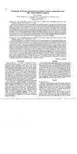
Arabidopsis thaliana (Bartel and Fink, 1995), and SuIfolobus soIfataricus (Colombo, 1995). The nucleotide sequence of hipO C. jejuni TGH9011 has been published (Hani and Chan, 1995) and is shown in figure 2. TAA is used for its termination codon and a AGGAGA Shine-Dalgarno sequence is located at nt-14 to -9 upstrearn of ATG the
methionine initiation codon.
The hipO region is purine rich and the molecular
percentage G+C content is 33.1%. The codon usage is similar to other C.jejuni genes except for the rare usage of UUC (phe), CTC (leu), CTG (leu), GTC (val), TCC (ser), CCG (pro), ACC (th), CGA (arg), and CGG (arg). Codon usage and molecuiar percentage G+C of the hipO open reading fiame within the insert of recombinant plasmid pHIP-O is shown in Table 2. Open reading frarne 2 (OTCFU2) and 3(ORFU3) are upstream of 4(ORFU4), the
hipO.
These three ORFs are continuous on the genome suggesting they may be
cotranscribed as an operon. A single genomic copy of the hipO gene is present in C. jejuni TGH9011. Southern blot hybridization of the C.jejuni genomic DNA probed with a radiolabeled
O II
C - NHCH,
COOH
Hippurate Hydrolase
+ H,O
N-benzoylglycine (Hippuric acid)
+
+ -
Benzoic acid
+
NH,CH,COOH
Glycine
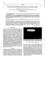
Figure 1. Biochernical reaction of hipourate hvdrolase.
Hippuric acid is formed by condensation of benzoic acid and glycine in human and accumulates in urine. Hippuric acid is hydrolysed ta benzoic acid and glycine in various species of microorganisms, including C. jejuni.
hippurate hydrolase. TTT (phe) TTC ( p h e ) TTA ( l e u ) TTG ( l e u ) CTT ( l e u ) CTC ( l e u ) CTA ( l e u )
20
CTG (leu) ATT ( i l e ) ATC ( i l e ) ATA ( i l e ) ATG (met) GTT ( v a l ) GTC ( v a l ) GTA ( v a l ) GTG ( v a l ) TCT ( s e r ) TCC ( s e r ) TCA ( s e r ) TCG ( s e r ) CCT ( p r o ) CCC ( p r o ) CCA ( p r o ) CCG ( p r o ) ACT ( t h r ) ACC ( t h r ) ACA (thr) ACG ( t h r ) GCT ( a l a ) GCC ( a l a ) GCA ( a l a ) GCG ( a l a ) TAT ( t y r ) TAC ( t y r ) TAA ( e n d ) TAG ( e n d ) CAT (his) CAC (his) (gin) W G (gin) AAT ( a s n ) AAC ( a s n ) ( l y s) AAG ( l y s GAT ( a s p l GAC ( a s p ) GAA ( glu
GAG ( g l u TGT ( w s ) TGC ( w s ) TGA (end)
TGG (t-1 CGT (arg)
CGC (arg) CGA ( a r g ) CGG ( a r g ) AGT (ser)
AGC (ser) AGA ( a r g ) AGG (arg) GGT ( g l y ) GGC ( g l y GGA (@Y) G a(qly) T o t a l codons T o t a l base p a i r s M o l % G+C
6 3
I 11 4 10
2 383 1149 33.1
Figure 2. Nucleotide sequence of C. jejrr~tiTGH9Oll hippurate hvdrolase.
The deduced arnino acid sequence of hippurate hydrolase is given in single Ietter code
above the sequence for those on the "upper" strand. Termination codons are indicated with an asterisk (Hani and Chan, 1995).
V V S W S V D K T H S F T L G F V Y I F GGTAGTATCTTGGAGTGTTGATAAAACCCATAGTTTTACTTTGGGTTTTGTTTATATTTT
v
-81
A L I F I S A I L A Q F V L P R R E N F TGCTTTGATTTTTATTTCAGCGATCTTAGCACAATTTGTTTTACCTAGMGAGAAAAT -21 I
Q
G
E
ORFU4
r
K
M
*
N
L
I
P
E
I
L
D
L
Q
G
E 40
TATACAAGGAGVTAGAATGAATTTmTTCCAGAAATACTAGACTTACMZGCGAAT
F E K I R H Q I H E N P E L G F D E L C TTGAAAAAATTCGTCATCAAATTCATGAAAATCCTGAGCTTGGTTTTGATGAATATGTA
100 hor2
tiprl
T A K L V A Q K L K E F G Y E V Y E E I CTGCAAAATTAGTGGCGCAAAAATTAAAAGAATTAAAAGAATTTGGTTATGAGGTTTATGAGGAAATAG 160 G K T G V V G V L K K G N S D K K I G L GAAAAACAGGCGTTGTGGGGGTTTTAAAAAAGGGGAATAGCGATAAAAAAATAGGACTTC
220
R A D M D A L P L Q E C T N L P Y K S K GTGCAGATATGGATGCTTTGCCTTTGCAAGAATGCACRAATTTGCCTTATAAAAGCAAAA
280
K E N V M H A C G H D G H T T S L L L A AAGAAAATGTAATGCATGCTTGCGGTCATGATGGACATACTACTTCTTTATTGCTTGCTG
340
A K Y L A S Q N F N G T L N L Y F Q P A CAAAGTATTTAGC~GTCAGAATTTTAATGGCACTTTMTCTTTATTTTCMCCTGCTG 4 0 0 E E G L G G A K A M I E D G L F E K F D AAGAGGGTTTGGGTGGTGCTAAGGCAATGATAGAAGATGGATTGTTTGAAAAATTTGATA
460
S D Y V F G W H N M P F G S D K K F Y L GTGATTATGTTTTTGGATGGCACAATATGCCTTTTGGTAGCGATMGMTTTTATCTTA
520
K K G A M M A S S D S Y S I E V I G R G AAAAAGGTGCGATGATGGCTTCTTCGGATAGTTATAGCÀTTGÀAGTTATTGGAAGAGGTG
580
G H G S A P E K A K D P I Y A A S L L V GTCATGGAAGTGCTCCAGWGGCWGATCCTATTTATGCTGCTTCTTTACTTGTTG
640
V
A
L
Q
S
I
V
S
R
N
V
D
P
Q
N
S
A
V
V
S
TGGCTTTACMGCATAGTATCTCGCAATGTTGTTGATCCCCRAIlATTCAGCAGTTGTAAGCA
700
I G A F N A G H A F N I I P D I V T I K TAGGAGCTTTTAATGCAGGACATGCTTTTAATATCATTCCAGATATTGTMCGATT-
760
M
S
V
R
A
L
D
N
E
T
R
K
L
T
E
E
K
I
Y
K
TGAGTGTTAGAGCATTAGATAATGAAACTAGAAAGCTIMCTGMGAnAAAATTTATAAAA
820
I C K G L A Q A N D I E I K I N K N V V TTTGTMGGTCTTGCACAGGCTAATGATATAGAGATTWTCMTWTGTTGTTG
880
A
P
V
T
M
N
N
D
E
A
V
D
F
A
S
E
V
A
K
E
C A C C A G T G A C T A T G A A T A A C G A T G A A G C T G T G G A T T T T T
940
L F G E K N C E F N H R P L M A S E D F T A T T T G G C G ~ T T G T G A A T T T A A T C A T C G T C C T T T A G T G A G G T T T T G1000 G
F
F
C
E
M
K
K
C
A
Y
A
F
L
E
N
E
N
D
I
G h T T T T T T T G C G ~ T G ~ T G T G C C T A C T T ? T T A G T G C A C A T T 1060 T
Y
A
K
L
A
L
K
Y
L
K
4
*
ATGCGAAGCTAGCTTT~TACTTAAAATAAAAACTAATCTAGAATTTCRAGCACAATT
E
S
K
L
G
K
I
Q
I
E
K
L
*
D
L
I
E
L
V
I
K
L
N
S
P
L
N
A
1180
~ G C T C T T T A T T G T T T T T T A T T T G C T T T T T T T G C A C T T A T G G A G A C T A A A A T T ~1300 ~~~TA
L
L
E
K
N
N
K
I
Q
N
K
A
S
I
S
L
L
I
G
R
O
23s rRNA probe IGS rRNA probe
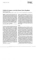
Figure 3. Phvsical map of C. jejuni TGH9011.
C.jejuni TGH90 1 1 produced a 2.1-kb hybridizing band. DNA hybridization detected no hipO gene sequences in C. coli, C. lari, C . upsaliensis, C.fetus, or C . sputorum or the former member of the Campylobacter genus- Helicobacter pylori (Hani and Chan, 1995). This is correlated with the negative hippurate hydrolase activity observed in
Helicobacter, Arcobacter, and other Campylobacter species. Substrate specificity of the C. jejuni HipO protein is highest using N benzoylglycine (N -B-gly), followed by N -benzoylalanine (N -Bz- ala), N benzoylrnethionine (N -B-met), and N -benzoylleucine (N -B-leu) respectively. No activity is detected using N -benzoylhistidine, N -benzoylarginine, or N benzoylthreonine as a substrate. The hipppurate hydrolase gene was mapped by Southern hybridization to the Sac II-A, Sa1 LA, and Sma 1-A fragments which overlap a 450 kb region of the 1800 kb genome (Kim et al., 1993). The hipO gene was localized to the central region of the Smu 1-A fiagrnent by mapping a hipO ::kanamycin mutant of C.jejuni (Figure 3). The argH and ileS genes also have been mapped to these restriction enzyme fragments (Kim et al., 1993, Hong et al., 1995). However the proximity of these genetic markers to each other, is currently unknown.
HomoIogous proteins belong to M40 hvdrolase family
Using a BLAST search, the deduced arnino acid sequence of C. jejuni hippurate hydrolase shows a homology score with the SuIfolobus solfataricus carboxypeptidase (CPSA) (Colombo et al., 1995), the Arabidopsis thdania indole-3acetic acid -amino acid hydrolase (ILR) (Bartel and Fink, 1995; Li et al., 1996), the
Bacillus sterothermophilus N -acyl-L-arnino acid amidohydrolase (AMA)(Sakanyan et
utilizing protein C) (HYUC) (Watabe et al., 1992;
Ishikawa et al.. 1996). the
Synechocystis hypothetical protein (Kaneko et al., 1996) and the Agrobacteriurn tumefaciens cellulose synthase (CELE) (Matthysseet al., 1995) (Table 3).
The Bacillus sterothermophilus N -acyl amino acid aminoacylase enzyme catalyses the hydrolysis of N -acyl arnino acid to yield an arnino acid and acetic acid. The substrate specificity of this enzyme is extremely high with N -choroacetyl-Lphenylalanine and is also high towards N -acetyl-L-ala, N -acetyl-L-tyr, N -acetyl-Lphe, N -acetyl-L-val, N -acetyl-L-gly, N -acetyl-L-leu, IV -acetyl-L-his, N -acetyl-L-met, and N -benzoyl-L-phe. N -benzoylglycine amidohydrolase from Pseudomonas putida C692-3 hydrolyzes N -benzoyl amino acids with different specificity, highest with N -Bz-gly, follow by N -Bz-L-ala and N -Bz-L-aminobutyric acid. It has no enzyme activity towards N-acetyl-glycine, N -acetyl-L- alanine, N -carbobenzoxyglycine, or N carbobenzoxy-L-alanine. Amidohydrolase are able to specifically remove N -1inked amino acids or similar substitutes fiom larger substrates. The
Pseudomonas sp. N -carbamoyl amino acid aminoacylase catalysed
reaction yields an amino acid and carbarnate.
The substrate specificity of the
benzoylamino acid arnidohydrolase of Pseudomonas sp. (strain KT801) is towards Lamino acids and not D-amino acids. N -Bz-Dl-ala is the preferred substrate, followed by N -Bz-DL-met, N -Bz-DL-leu, N -Bz-gly, N -Bz-DL-val, N -Bz-DL- phe, N -Bz-
DL-asp, N -Bz-DL-glu, N 432-DL-trp, and N -Bz-DL-th. No activity is observed on the substrate acetyl-DL-alanine, or phenylacetyl-DL-alanine, indicating a considerable
effect by the acyl group (Kameda et al., 1968).
activity and c m remove amino acid residues fiom benzoyloxycarbonyl derivatives with activity towards benzyloxycarbonyl (Cbz) substrates; Cbz-glyglyphe, Cbz-arg, Cbz-asp, and weakly with Cbz-phe and Cbz-ala. The Arabidopsis thaliana indoxyl-3acetic acid amidohydrolase reaction yields an arnino acid and indole-3-acetic acid. The substrate specificity is toward indole-3-acetic acid arnino acid conjugates: IAAphe, IAA-leu, and weakly towards IAA-ala, IAA-gly, and IAA-ile.
Table 3. Homologous ~roteinsof C.ieiuni hippurate hydrolase.
Sequences with high homolow score
Homolow Score
Campylobacterjejuni hippuricase (HipO) Sulfolobus safataricus carboxypeptidas (CpsA) Arabidopsis thaliana indoxyl-3-acetic acid amidohydrolase (Ill 1) A rabidopsis thaliana (1112) A rabidopsis thaliana (Ilr 1) Synechocystis sp. hypothetical protein Bacillus stearothermophilus N-acyl-L-amino acid amidohydrolase (Amal Bacillus subtilis (YXEP) Agrobacterium tumefaciens (CelE) HaernophiZus infIuenae HipO homolog Psezrdomonas sp. hydantoin utilization protein (hyuC) Escherichia coli hemolysin B (hlyB)
2.1e-248 4.8e-83 7.7e-72 1.8e-70 1.7e-62 4.1 e-47 1.2e-44 3.4e-62 7.8e-2 1 7.6e- 17 0.22 0.52
An extensive study of over 556 strains of thermophilic Cumpylobacter for
species identification was reported by Totten et al., 1987. These members were isolated either from hurnans with diarrhea or from poultry in King County, Washington. They were analyzed by the phenotypic test, after that the classification of the hippurate hydrolase-negative C. jejuni strains was confirmed by a quantitative whole-ce11 DNA hybridization test. The study showed that only 1.6% (9 of 556) of the C. jejuni strains were hippurate hydrolase negative.
Thus, hippurate hydrolase-
negative C.jejuni strains represented a small percentage (1.6%) of C. jejuni strains but a significant portion 20% (9 of 46) of hippurate hydrolase-negative strains in that study. These C jejuni hippurate hydrolase-negative isolates were not only hippurate hydrolase-negative in the rapid tube test but also in more sensitive assays such as gas liquid chromatography (GLC) and thin layer chromatography (TLC) assays. Furthermore, serotyping was also applied to these members, yet hippurate hydrolasenegative C. jejuni isolates show a non-random association with different serotyping systems (Totten et al., 1987). Hippurate hydrolase-negative strains fell into four groups by the Penner serotyping system, four groups by the Lior serotyping system, two auxotypes, and three plasmid patterns, indicating that these strains represented a diverse group of isolates. Hippurate hydrolase-negative isolates of C. jejuni were also examined with respect to the presence of h i p 0 gene sequences in the genome by Southern hybridization analysis (Hani and Chan, 1995). Four hippurate hydrolase-negative isolates D594,D603, D941, and D 19 16 gave a positive 2.1 kb Hind I I I hybridization band when probed with the hipO probe. One hippurate hydrolase-negative isolate,
chromatography assay (Totten et al., 1987) and showed a hybridization band in Southern hybridization blot with the hippurate hydrolase probe (Hani and Chan, 1995).
C. jejuni and C. coli are the most comrnon thennophilic bacteria isolated fiom humans (Ketley, 1995). characteristics.
C. jejuni and C. coli share many cornmon phenotypic
Thus, very few biochernical tests can differentiate between them
(Penner and Hennessy, 1980; Penner et al., 1983; Penner, 1988; Boltan, 1984; Patton et al., 1992). However, they are two distinct species and show only 25 to 58% homology relative to each other by DNA hybridization (Harvey and Greenwood, 1983; Roop et al., 1984; Morris et al., 1985). The hippurate hydrolysis (Hippuricase) assay is the standard bicbzhemical test used to distinguish C. jejuni, C. coli, and other Campylobacter sp. in the clinical laboratory via the detection of the product of hydrolysis by a color reagent system. This asssay was found to correlate closely with species differentiation in taxonomic studies. 80% of cases of Campylobacter mediated enteritis in hurnans are caused by C.jejuni (Ketley, 1997). C. jejuni is the only Campylobacter species that has the hippurate hydrolase gene and is able to hydrolyse hippurate. Recently, the hippurate hydrolase gene from C. jejuni TGH9011 strain has been cloned and characterized (Hani and Chan, 1995). Therefore, it is important to understand the role of the hippurate hydrolase gene in C.jejuni species. Hippurate hydrolase-negative C.jejuni strains have also been identified (Totten et a1.,1987). It is of interest to analyse the mutant hippurate hydrolase gene in these natural isolates. Therefore, the major objective of this study was to determine the molecular nature of hippurate hydrolase-negative C. jejuni clinical isolates from King County, Washington.
We used a radiolabeled single strand conformational
polymorphic assay based on polymerase chain reaction (PCR-SSCP assay) to screen
1
Y
A
A
Y
A 1
-
-
hydrolase-negative C. jejtrni. The S S C P assay based on polymerase chain reaction is a sensitive and simple technique, which is capable of distinguishing single base pair
changes between single DNA strand (Orita et al., 1989; Teleni et al., 1993). It is successfùlly used in various studies in Mycobacteriurn tuberculosis (Orita et al., 1989, Teleni et al., 1993), Mycobacterium leprae (Heyrn et al., 1995), Hepatitis B virus (Yusof et al., 1994), and Parvovirus B19 (Kerr et al., 1995), etc. Oligonucleotide primers were synthesized based on the hippurate hydrolase nucleotide sequence of hippurate hydrolase-positive C .jejuni strain TGH9011 (Hani and Chan, 1995). The primers were used to ampliSr eleven overlapping regions of the hippurate hydrolase gene from each of the five hippurate hydrolase-negative C. jejuni clinical isolates. The
SSCP assay was used to detect point mutations upstream and within the hippurate hydrolase gene sequences. The regions containing the hippurate hydrolase gene of the five diffèrent hippurate hydrolase-negative C . jejuni isolates were cloned and sequenced in order to characterize the exact nature of the sequence heterogeneity within the hippurate hydrolase gene sequence of C.jejuni TGH9011 strain.
Bacterial Strains
The Campylobacter jejuni serotype reference strain for 0:3 [TGH9011 (ATCC 43431) and of Lior serotype 361 was obtained fiom Dr. J. L. Penner, University of Toronto, Canada. C.jejuni TGH9011 was used as a hippurate hydrolase-positive controI strain. Five different hippurate hydrolase-negative clinical strains of C.jejuni (D594, D603, D835, D977, D1713) were received fiom Dr. C. M. Patton, Center for Disease Control and Prevention, Atlanta, Ga, USA.
These hippurate hydrolase-
negative strains were isolated either fiom humans with diarrhea or fiom poultry (Totten et al., 1987). Campylobacter species were grown routinely on Mueller-Hinton agar plates (Merck) or in broth for 48 to 72 hours at 42'C in a carbon dioxide incubator set for 7% CO2. Escherichia coli (JM101) strain [A(lac-proAB ) thi strA supE44 end4 sbcB hsdR4 F' traD36 proA+B+ Zacl A(lacZ) Ml51 was grown routinely on Luria Bertani media aerobically at 37'C and used for making cornpetent cells.
Genomic DNA extraction
Total genomic DNA of C.jejuni TGH9011 was extracted from cells grown on Muller-Hinton a g a plates. A plate of C. jejuni cells was washed three times in 1 ml of saline citrate (SSC:O.1SM NaCl, 0.015M Na citrate [pH7.0]). Suspended cells were pelleted and resuspended in 27% sucrose in 1X SSC at a concentration not higher than 10" cells per ml. The suspended cells were digested with proteinase K to a final
Table 4. Bacterial strains used in this study.
Strains
Genotvpe or Characteristics
References
Campylobacterjejuni ATCC4343 1 serotype reference strain for 0:3, TGH9011
J. L. Penner
hippurate hydrolase negative isolate
C. M. Patton
hippurate hydrolase negative isolate
C. M. Patton
hippurate hydrolase negative isolate
C. M. Patton
hippurate hydrolase negative isolate
C. M. Patton
hippurate hydrolase negative isolate
C. M. Patton
hippurate hydrolase weak isolate
C. M. Patton
A(1ac-pro) thi rspL supE en& sbcB hsdR F' traD36 proAB ZacP ZAM 15
Yanisch-Perron et al.
Esclierickia coli
ATCC:Arnerican Type Culture Collection
concentration of 0.2%.
The lysate was incubated at 50'C for an hour and then
extracted three times with an equal volume of 50 mM Tris-Cl, 10 mM EDTA, pH 8.0 saturated phenol. The aqueous phase of the phenol-extracted lysate was removed and extensively dialyzed in Tris-EDTA (10 mM Tris [pH 8.01-lmM EDTA). DNA was recovered by ethanol precipitation with two volumes of 95% ethanol for 30 minutes, at -70'C.
The precipitated DNA was pelleted by centrifugation, washed with 70%
ethanol and then vacuum dried. The dried pellet was resuspended in 50 pl TE (10mM Tris-Cl pH 8.0, 1 mM EDTA pH8.O) for further use.
Olipronucleotides To scan the hippurate hydrolase gene for the presence of mutations by polymerase chain reaction and single strand conformational polyrnorphism analysis, oligonucleotides were synthesized based on the C. jejuni TGH9011 hippurate hydrolase sequence (Hani and Chan, 1995). These primers were used in polymerase chain reactions and sequencing reactions. The location, direction, size and sequence of primers (PC1, PC3, PC4, H l , H2, H3, H4, H5, H6, H7, H8, H9, H 1 O, H1 1, H12,
H13, H14, H15) designed for the polyrnerase chain reaction, single strand conformational polymorphism analysis and sequencing reactions are s h o w in Table 5 and 6 and Figure 4. The strategy for the amplification of the various regions of the
hippurate hydrolase gene using eleven primer sets is s h o w in Figure 4.
Table 5. Uligonucleotides usecl in mis stuay.
Oligo
Sequence
Purpose
5'-ATCCCGGGTGGTTTCC(A/T)TTTTTCTTTAATCT(T/A)CC-3 ' Pcr 5'-ATGTCGACAAAAAAGAAAATGToATGT(T/A)ATGCATGC/TTGCGGT-3' pcr 5'-ACCCCGGGGC(T/A)GG(T/A)TTTTATGATAAATTCTACAC-3 '
5'-TAGGATCCTCTTGGAGTGTTGATAAAACTCAT-3' 5 '-ATGGATCCTTGATGGCGAATTTTTTCAAATTC-3 ' 5'-GCGGATCCGAATTTGAAAAAATTCGCCATCAA-3' 5'-TTGGATCCAGCAGTACATAATTCATCAAAACC-3' 5'-AAGGATCCAGTTCCATTAAAATTTTGACTAGC-3' 5'-TAGGATCCGCAAGTCAGAATTTTAATGGCACT-3' 5'-AAGGATCCTCCATCTTCAATCATAGCTTTAGC-3' 5'-TTGGATCCTGGCACAATATGCCTTTTGGTAGC-3' 5'-CTGGATCCTCCAATAACTTCAATGCTATAACTATC-3' 5'-ATGGATCCGCTCCTGAAAAAGCTAAAGATCCT-3' 5'-ACGGATCCAGCAGAATTTTGAGGATCAACATT-3' 5 '-CTGGATCCTTTAATGCTGGACATGCTTTTAAT-3 '
5'-TCGGATCCAACAGCTTCATCATTATTCATAGT-3' 5'-GTGGATCCTTAATGGCTAGTGAAGATTTTGGA-3' 5'-AAGGATCCAGCATAAGCACATTTTTTCATTTC-3'
Table 6. Primer pairs used in this study.
Primers
Repions
Sizes
Pcr Pcr Pcr Pcr Pcr Pcr Pcr PCr pcdsequencing Pcr pcr/sequencing pcdsequencing Pcr pcdsequencing Pcr Pcr
Chromosomal DNA of Campylobacter jejuni strains was arnplified by polyrnerase c h a h reaction using Taq DNA polymerase (Boehringer, Mannheim, Germany). The amplification was carried out in a programmable thermal cycler (Gene Arnp PCR 9600 System, Perkin-Elmer, Norwalk, CT, USA) as follows; an initial template denaturation step at 94.C for 5-minutes followed by 25 cycles of 1-minute 30seconds of denaturation at 94C, 1-minute 30-seconds primer annealing at 60C, and 2minutes chain extension at 72'C and then a final extension step at 72'C for 10 minutes. The reaction (50 pl) contained 50 pmol of each primer, 1 pg of genomic DNA, 1.5
mM MgC12, 160 pM each dNTP, 2.5 U Taq DNA polyrnerase and Taq buffer. Sterile distilled water was used to adjust the total reaction volume for the negative control. The reaction mixture was then covered with 70 pl of minera1 oil. One tenth of the arnplified products were electrophoresed on polyacrylarnide gel (PAGE), containing ethidium bromide in TBE buffer (0.45 M Tris-Borate, and 10 mM EDTA). PAGE electrophoresis was performed, as described (Maniatis, 1982).
Briefly, a 10%
acrylarnide gel was cast in a Biorad Mini Protein gel caster. The DNA sarnples were separated in running buffer at 100 V for 2 hours. Aller electrophoresis, the amplified product was exarnined under UV light. Two rnicrograms of DNA from the phage
#X
174 digested with Hae III was used as the size standard. Positive control C. jejuni genomic DNA and negative controls: water instead of DNA, are included in every gel. For the SSCP assay, pairs of PCR primers were chosen to generate fragment sizes of less than 400 bp to enhanced the sensitivity of the SSCP assay.
detection enhancement (non denaturing) gel electrophoresis
For the SSCP assay, double stranded DNA was denatured into single stranded DNA and the products were separated on a mutational detection (MDE) gel under nondenaturation conditions. The amplification reaction was performed in a 50 pl reaction volume containing 10 pl extracted genomic DNA (approximately 1 pg), 200 pM (each) of dTTP, dGTP, and dCTP, 100 pM dATP with 0.5 pl of [alpha-32P] ATP
(5 pCi) instead of total 200 uM dATP was added to the PCR mix, 50 pmole of each oligonucleotide primer, 5 pl 10X Taq buffer and 2.5 U of Taq DNA polymerase (Boehringer, Mannheim, Germany). The SSCP assays were performed as described (Teleni et al., 1993). Briefly, 5 pl of radiolabelled arnplicon was diluted in 100 pl of the SSCP dilution solution (10 mM EDTA, O. 1% SDS). 3 pl of diluted product was mixed with 3 pl of formamide dye (98% formamide, 0.5% bromophenol blue, 0.5% xylene cyanol, and 20 mM EDTA), denatured at 95'C for 5-minutes, cooled on ice, and loaded on a nondenaturing sequencing-format 0.5 X Mutation Detection Enhancement Acrylarnide Gel (MDE gel) (Hydrolink -MDE:AT/Biochern, Malvern, PA, USA). The
MDE gel is composed of 25 ml of MDE gel, 3 ml of 10X TBE buffer, and 22 ml of deionized water polymerized with 300 p1 10% ammonium persulfate and 30 pl
TEMED (Bio Rad, Richmond, Calif., USA). Electrophoresis was done in 90 mM TriBorate (pH 8.3) - 4 mM EDTA in a sequencing gel electrophoresis apparatus (50 by 32 by 0.04 -cm gel) (Bio Rad) at room temperature at a constant power of 8 W for 20 hours. After electrophoresis, the gel was dried for an hour, exposed 3 hours to Kodak film, and the radioactive banding patterns were inspected. Pairs of PCR primers were
Figure 4. Strategv for single strand conformational polvmorphic (SSCP) assay
based on polymerase chah reaction. The positions of eleven sets of primer pairs used are indicated above the hippurate hydrolase gene which are shown as box. Oligonucleotides were synthesized based on the published sequence of hippurate hydrolase-positive C. jejuni strain TGH90 11 (Hani and Chan, 1995). The eleven sets of primers were used to amplify the eleven overlapping regions which are less than 400 bp in size. Each region was overlapping around 100 bp.
assay and at least two SSCP runs were performed for each set of sarnples to assure reproducibility of the assay.
Cloning of the amplified fragments
1 pg PCR product was digested in 20 pl using approximately 2 U of BamHI enzyme per microgram of DNA in BamHI reaction buffer and cloned into a pUC19 vector. Restriction enzymes were obtained fiom Boehringer Mannheim or Pharmacia and used as recomrnended by the manufacturers.
Ligation of DNA fragments The arnplified product, (approximately 1.0 pg), was digested with Barn HI restriction enzyme and purified by ethanol precipitation, as described above. 0.5 pg pUC19 vector DNA was linearized with Barn HI and dephosphorylated using calf intestinal alkaline phosphatase (CIAP). CIAP was used at a concentration of 1U per pg DNA for 15-minutes at 37'C. Alkaline phosphatase activity was terminated by heating at 65'C for 10-minutes with 20 mM EDTA and 0.5% SDS. The volume was adjusted to 240 pl and extracted with phenol in order to remove residual alkaline phosphatase. The top phase was extracted with ether and the DNA precipitated on ice for 30-minutes using 0.3 M sodium acetate (NaOAc) and three volume of ethanol. The pellet was resuspended in TloEi buffer (1 0 mM Tris-HC1, pH 8.0 and 1 mM EDTA). The vector and 100 to 500 ng of insert DNAs were then mixed together in
ligation buffer (50 mM Tris-Cl, pH 7.6, 10 mM MgC12, 1 mM rATP) in a moiar ratio
Gerrnany).
Preparation of competent celis and transformation with recombinant plasmid Competent cells for transformation were prepared. 20 ml of Luria broth was inoculated with 0.5 ml of an ovemight E. coli culture. The cells were then grown at 37C with aeration to early log; OD at 600 nm which was equal to 0.13 - 0.15, taking
approximately 2 hours. The cells were pelleted by centrifugation. The pellet was resuspended in 10 ml of solution A (10 mM 3-(N-Morpholin) propane - sulfonic acid
(MOPS) pH 7.0 and 10 m M rubidium chloride). Cells were again spun down, resuspended in 10 ml of solution B (10 mM MOPS, pH 6.5, 10 mM rubidium chloride, and 50 mM calcium chloride) and incubated on ice for 30-minutes. Afterward, the cells were centrifuged and the ce11 pellet was resuspended again in 1 ml of solution B. For transformation, 0.15 - 0.20 pg of ligated DNA, approximately 10 to 20 pl of ligation mixture was added to 200 pl of the competent cells prepared above. It was mixed and incubated on ice for 30-minutes. The cells were then heat-shocked at 42.C for 90-seconds. 1 ml of Luria broth was added and the mixture was incubated at 37'C for an hour. This allowed for the expression of the antibiotic resistant genes present on the plasmid before transformed cells were plated ont0 selective medium. 100 pl of the transformed cells were added per plate for selection of appropriate transformants.
Recombinant plasmid DNA preparation S,mall scale preparation of plasmid DNA from E. coli was perforrned (Maniatis
pelleted by centrifugation at 1200 rpm for 5-minutes. The bacterial ce11 pellet was resuspended in 100 pl of GTE-lysis buffer: 50 m M glucose, 25 mM Tris-HCl pH 8.0, 10 mM EDTA, 2 mg lysozme per ml and incubated at roorn temperature for 5minutes. The bacterial suspension was treated with 200 pl alkaline SDS buffer (0.2N NaOH, 1% SDS) and mixed by inverting the tubes gently several times and placed on ice for 5-minutes. 150 pl of acetate buffer (3 M potassium acetatelacetic acid) was added mixed by inverting and chilled on ice for 5-minutes.
The samples were
centrifuged for 10-minutes at 4'C , and the supernatant was collected. Plasmid DNA, thus extracted was concentrated by ethanol precipitation with two volumes of 95% ethanol and incubation at -70C for 30-minutes. DNA was pelleted by centrifugation for 20-minutes at 4C, washed with 70% ethanol and vacuum-dried. The dried pellet was resuspended in 50 pl TloEI (1OmM Tris-Cl
-
1 mM EDTA, pH 8.0) for further
use.
Preparation of sequencing grade plasrnid DNA
Bacterial cultures were grown in Luria broth containing ampicillin to ensure the presence of the plasmid within the culture. Afier plasmid DNA was extracted, 18 pl (-1 .O pmol) of DNA was denatured for 5-minutes with 2 pl of 2
N NaOH at room
temperature. Then, the DNA was precipitated by adding 8 pl of 5M ammonium acetate pH 7.4 and 100 pl of absolute ethanol and incubating at -70-C for 10-minutes. The DNA collected by centrifugation was washed twice with 70% ethanol and vacuum dried.
supernatant (after the addition of acetate buffer and centrifugation) from the minipreparation protocol and ethanol precipitation, as mentioned above. The DNA pellet was then resuspended in 50 pl of TE containing RNase (10 pg RNse per ml) and incubated at 37.C for 30-minutes. Then 30 pl of PEG solution (20% PEG-8000,2.5 M NaCl) was added and incubated on ice for an hou. DNA was then centrifuged, washed in 70% ethanol, vacuum dried, and resuspended in H20.
DNA sequencing and polyacwlamide gel electrophoresis The sequence was determined fiom subclones prepared as rnentioned above and by using specific primers. The DNA sequence was detennined by the dideoxy chain termination method (Sanger, 1977). It used the Sequenase sequencing kit (US Biochemical Corp., CleveIand, OH, USA) rnodified version of T7 DNA polmerase with [ ~ c - ~dATP ~ s ] for labeling. For each clone, both strands of insert were cornpletely sequenced. The denatured DNA was annealed to a specific primer and extended products of various lengths were obtained with the use of sequenase. The DNA (3-5
pg of denatured double-stranded DNA or 1-2 pg of the single-stranded DNA per reaction) was mixed with an appropriate primer (0.5 pmol per reaction) in 1X Sequenase Buffer (40 mM Tris-Cl, pH 7.5, 20 mM MgC12, 50 mM NaCl), and annealed by heating the mixture in a capped tube at 65'C for 2-minutes followed by slowly cooling the tube to room temperature for about 30-minutes. After annealing, the DNA was mixed with 6.5 mM DTT. 2 nM of dGTP, dCTP. dTTP, 0.5 pl of ["SI dATP (1000 Ci per mmol), 3 U of Sequenase enzyme and labeled by incubation at
(each mix contains 50 mM NaCI, $0 uM of dNTP/8 p M one of ddATP, ddGTP, ddCTP, or ddTTP) was added to the labeled DNA mixes in each of the four wells, and incubated at 37'C for 5-minutes. After the termination reaction, 4 pl of the stop solution (95% formamide, 20 mM EDTA, 0.05% bromophenol blue, 0.5% xylene cyan01 F F ) was added to each reaction. The sample was heated to 80'C for 5-minutes irnmediately before loading on the 6 % denaturing polyacrylamide sequencing gel. The polyacrylamide mix for sequencing gel was prepared as 35 gm ultrapure urea, 9
ml of a 45% acrylamide stock, 350 pl of 10% ammonium persulfate and 50 pl of T E M E D per 70 ml of polyacrylarnide gel and run at 1700 Volts. After the samples were run on the sequencing gel, the gel was soaked in a solution of 10% acetic acid, 10% methanol, transferred ont0 3MM Whatman filter paper, and dried at 80'C for 45-
minutes. The dried sequencing gel was exposed on X-ray film overnight.
Detection of hippurate hvdrolase sequence heterogeneitv in the hippurate hydrolase-negative C. jejurzi clinical isolates
Genomic DNA of the five hippurate hydrolase-negative C. jejuni clinical isolates was examined by polymerase chain reaction for sequence variations. The hippurate hydrolase-positive C. jejuni TGH9011 strain was used as a control (Hani and Chan, 1995). The PCR strategy used in this study is shown in Figure 4. The specific sequence primers, H l , H2, H3, H4, H5, H6, H7, H8, H9, H l 0 and H l 1, with three degenerate primers, PCl, PC3 and PC4, were used in the polyrnerase chain reaction. The positions of the eleven primer sets used, are indicated above the hippurate hydrolase gene which is depicted as a box in Figure 4. The degenerate primers, PC1,3 and 4 were previously designed with the minimal number of possible degenerate oligonucleotides required to account for al1 variations of a particular arnino acid residue (Hani, 1997). Primers were used to ampli@ the eleven overlapping regions of the hippurate hydrolase gene from five different hippurate hydrolase-negative C. jejuni clinical isolates, D594, D603, D835,D 1713, Dg77 and hippurate hydrolase-positive C. jejuni TGH9O 11. Eleven primer pairs were used in polymerase chain reactions that
covered from upstream nt -259 to nt 1317 downstream of the hippurate hydrolase gene. The adjacent amplified products overlapped each other by about 100 bp. The amplified products were electrophoresed in an 8% polyacrylamide gel (PAGE) with
4
X 174 Hae III fragment as markers. The results of the amplified products from eleven overlapping fragments of the five different C.jejuni hippurate hydrolase-negative
that were identical to that of positive control C. jejzrni TGH9011. These findings indicate that al1 tested hippurate hydrolase-negative C. jejuni isolates do not have major sequence variations such as gross deletions or insertions at their hippurate hydrolase gene. However, a standard gel analysis of the arnplified products would be unable to detect minor sequence variations within the hippurate hydrolase gene of hippurate hydrolase-negative isolates.
Single strand conformational polvmorphic analysis of the natural mutant hippurate hydrolase Eene
The flanking regions and the coding sequence of the hippurate hydrolase loci from hippurate hydrolase-negative C. jejzrni isolates were analyzed by
3 2 labeled ~
single strand conformational polymorphism (SSCP) assay based on polymerase chain reaction. The F7 and F8 overlapping regions spanning from nucleotides 473 to 912 of the hippurate hydrolase gene showed polymorphisms in al1 five different hippurate hydrolase-negative C. jejuni isolates compared with those of control C. jejuni TGH 9011 (Figure 5).
Other regions, F4, F5, F6, F9, F10 and F11, showed no
polymorphism in their SSCP profiles. They were indistinguishable from each other and also similar to those of the control C, jejuni TGH9011. The polymorphic regions,
F7 and F8, may presumably account for the hippurate hydrolase-negative phenotype of the five negative C. jejuni isolates in the hippurate hydrolysis test. The promoter area is covered by the overlapping amplified regions F1, F2 and F3 spanning from nucleotides -259 to 150 bp. The SSCP analysis of promoter regions
Table 7. Surnmarv of PCR analvsis in the hippurate hvdrolase gene from the five hippurate hydrolase-ne~ativeC. jejuni isolates. Vertically presents control C. jejuni TGH9011 strain (top) along with the hippurate hydrolase-negative C .jejuni isolates. Horizontally presents the arnplified products of eleven overlapping regions F1 through F11.
+ sign represents normal
arnplified product to its respective region.
Strains
PCR amplified products
and identical
Figure 5. PCR-SSCP analvsis of F7 and FS regions of the hippurate hydrolase pene of five different hippurate hydrolase-negative C.ieiuni isolates. Figure 5(A). The sarnples anaIyzed for F7 were lane 1, C. jejuni TGH9011; lane 2,
D594; lane 3, D603; lane 4, TGH9011; lane 5, D835; lane 6 , D977; lane 7, Dl 7 13. Figure 5(B). The sarnples analyzed for F8 were lane 1, C.jejuni TGH9011; lane 2. D977;
lane 3, D1713; lane 4, D835; lane 5, TGH9011; lane 6, D603; lane 7,
TGH90 1 1; lane 8, D594; lane 9, TGH90 1 1. Mobility shifts are found in fragments F7 and F8 of the five hippurate hydrolasenegative C.jejuni isolates when compared to the respective regions of control C. jejuni
TGH90 11.
Figure 5(A).
Figure 5 (BI.
Figure 6 (A, B, and C). PCR-SSCP analvsis of FI, F2 and F3 regions of the hippurate hydrolase gene of five different hippurate hvdrolase-negative C. iejuni isolates.
Figure 6(A). The sarnples analyzed for FI were lane 1, C. jejuni TGH9Oll; lane 2, D594; lane 3, D603; lane 4,TGH90I 1; lane 5, D835; lane 6,D1713; lane 7, D977; lane 8, TGH9011. Figure 6(B). The sarnples analyzed for F2 were lane 1, C. jejuni TGH9011; lane 2, D594; lane 3, D603; lane 4, D835;lane 5, D1713; lane 6, TGH9011. Figure 6(C). The sarnples analyzed for F3 were lane 1, C. jejuni TGH9011; lane 2 , D594;
lane 3, D603; lane 4, TGH9011; lane 5, D835; lane 6, D1713; lane 7,
TGH9011; lane 8, D977. The PCR-SSCP analyses of the promoter region from the five hippurate hydrolasenegative C. jejuni isolates which were covered by the FI, F2, and F3 amplified fragments. The assays show unaitered strand mobility compared with that of control C.jejuni TGH90 I 1.
Figures 6 (A).
Figures 6 (BI.
Figures 6 (C).
regions of the hippurate hydrolase-negative C.jejuni isolates. They were also identical to the respective regions of C.jejuni TGH9011 (Figure 6).
These results suggest that the promoter regions of the hippurate hydrolasenegative isolates are unlikely to be the site of sequence variations.
Cloning of SSCP polymorphic regions and DNA sequencing
To establish the precise molecular nature in the five hippurate hydrolasenegative isolates, the DNA coding regions which displayed the abnormal SSCP profiles were cloned, sequenced and compared with the hippurate hydrolase sequence from C. jejuni TGH9011.
The amplified fragments of F7 and F8 regions from the five
hippurate hydrolase-negative C. jejuni isolates were cloned into
pUC 19
vector
(Pharmacia) and sequenced by the dideoxynucleotide chain termination procedure using T7 sequenase (USB) with specific primers. The nucleotide sequences of the insert in pUC19 were detennined in both directions.
The sequence of each polymorphic region was almost completely
homologous with the respective region of the C. jejuni TGH90 1 1 sequence. The respective numbers of nucleotide substitutions from the published hippurate hydrolase C. jejzrni TGH9011 sequence for each hippurate hydrolase-negative sample are listed in Table 8. Most of the mutations detected were silent in nature and they do not affect the amino acid alterations. They were TTA
+
+ TTG at nucleotide (nt) position 633, TTA
TTG at nt position 648, AGC + AGT at nt position 654, CCC
+ CCA
at nt
position 678, and TCA + TCT at nucleotide position 687. Except the nt substitution
(Val) codon to alanine (Ala) at residue 250 (Figure 7). This valine at residue 250 change was common to al1 of the hippwate hydrolase-negative C.jejuni isolates.
Sequenice comparison
The hippurate hydrolase homologous sequences were obtained from a cornputer search of the Protein Data Base GeneBank and the Swiss Protein Database. The Clustal 1.4W program was used to compare the deduced amidohydrolase sequence of C. jejuni TGH9011 (HipO) with those of the Bacillus sterothermophilus arninoacylase
(Ama)
(Sakanyan et al., 1993),
utilizing protein C
the
Pseudamonas sp. amidohydrolase hydmtoin
- HyuC) (Watabe et al.,
1992; Ishikawa et al., 1996) and the
Synechocystis sp. hypothetical arnidohydrolase protein (Kaneko et al., 1995) of the AMA/HIPO/HYUC M40 hydrolase family (Figure 8). The valine at residue 250 of C. jejuni hippurate hydrolase is located as a conserved residue in these homologous
proteins from the M40 hydrolase family.
Predicted hydrophathy profiles A hydrophobicity plot is a current method for predicting the structure of
proteins. It is a graph displaying the distribution of polar and apolar residues along sequences. The sequences are scanned with a moving window of a given length and computed for each position of the window, the mean hydrophobicity of al1 residues in the window.
The predicted hydropathy and flexibility profiles of the
hippurate
hydrolase protein were plotted by the Kyte-Doolittle scale with a window size of 7
Table 8. Nucleotide substitutions between nucieotide 473 and 912 of the hipO genc of C. iejuni hippurate hvdrolase-negative isolates.
HipO Sequence from C. jejrtni TGH90 1 1
Isolates
Nucleotide #
Codon
Codon Substitutions in isolates
(Hani et al., 1995)
D594
TTA TTA AGC GTA
TTG TTG AGT GCA
D603
TTA TTA AGC CCC GTA
TTG TTG AGT CCA GCA
D835
TTA TTA AGC GTA
TTG TTG AGT GCA
TTA TCA AGC GTA
TTG TTG TCT AGT GCA
TTA TTA AGC GTA
TTG TTG AGT GCA
D977
Dl713
Phenotvpe Condition
Hippurate Hydrolysis
TGH 901 1
215
positive
ALQSIVSRNVDPQNSAWSIGAFNAGHAFNI 1PDIVRTIKMSVRALDNET
negative
ALQSIVSRNVDPQNSAWSIGAFNAGHAFNI 1 PDIARTIKMSVRALDNET
11111111111111111111111111111111111 D835
1 1 1 1 1 ~ 1 1 1 1 1 1 1 1
Hipput-ate Hydroiysis TGH 901 1
D835
...................
positive
KLTEEKIYKICKGLAQANDIEIKINKNW. Illllllllllllltllll1 1 IlII1III
neetive
KLTEEKIYKICKGLAQANDIEIKINKNWA.
.................
Figure 7. The deduced amino acid sequence alignment of the mutated region of hippurate hvdrolase-positive C. jejuni TGH9011 and the hippurate hvdrolasenegative strain DS35. A single arnino acid substitution at valine to alanine at residue 250 was found in al1 the
hippurate hydrolase-negative proteins.
Figure 8.
AIignment of C.jejuni hi~puratehvdrolase and related proteins.
The complete polypeptide deduced from gene hipO for N-benzoyl-arnino acid amidohydrolase (hippurate hydrolase) was aligned using the Clustal W (1.4) program with the complete polpeptides of related proteins. The top line gives the C. jejuni Nbenzoyl-arnino acid amidohydrolase (HipO) protein sequence, second line shows the Bacillus stearothermophilus N-acyl amino acid arnidohydrolase (arninoacylase) (Ama)
protein sequence and the third line is the N-carbamyl-L-amino acid amidohydrolase (DL-hydantoinase) fiom Pseudomonas sp. (hydantoin utilizing protein C) (hyzlC ) and lastly Synechocystic hypothetical protein. Residues involved in conserved areas are s h o w in bold face. Identical residues are marked with stars, sirnilar residues are indicated by dots and hyphens indicate gaps introduced to maximize alignment. Val-250 is located as an identical residue in the sequence of the
AMNHIPOMYUC M40 hydrolase family.
CLUSTAL W (1.4) Multiple Sequence Aliqnment
H ~ P O------- MNLIPEILDLQGEFEKIRHQIHENPELGFDELCTAKLVAQKLKEFGYEVYEEI
Am=
----MTKEEIKRLVDEVKTDVIAWRRHLHAHPELSFQEEKTAQFYETLQSFGHLELSRP
HyuC MKTVTISKERLRIHIEQLGEIGKTKDKGVQRLALSKEDREATLLVSEWMREAGLTVTHDH S ~ P------- MELKNLAQTLLPRLVEIRRHLHAHPELSGQEYQTAAWAGVLSSCGLHVEEAI
. .
HipO Ama HyuC shp HipO Ama
HyuC shp
HipO HyuC shp HipO Ama
HyuC shp
. .. *
*
GKTGVVGVLKKG--NSD-KKIGLRADMDALP-------- LQECTNLPYK--SKKENVMHA TKTSVMARLIGQ--QPG-RVVAIRADMDALP-------- IQEENTFEFA--SKNPGVMMA FGN-LIGRKEGE--TPSLPSVMIGSHIDSVRNGGKFDGVIGVLAGIEIVHAISEAMNHE GKTGVVGQLSGKGDDP--RLLAIRTDMDALP-------- IEEMVSLPFA--SRHPGVMHA
.
. . . .*..
.*
.*.*
--------c---G--- HDGHTTSLLLAAKYLASQ--NFNGTLNLYFQPAEEGLGG-AKAM --------c---G--- HDGHTAMLLGTAKIFSQLRDDIRGEIRFLFQHAEELFPGGAEEM HSIEVVAFCEEEGSRFNDGLFGSRGMVGKVKPEDLQKVDDNNVTRYEALKTFGFGIDPDF --------c---G--- HDIHTTLGLGTAMVLSQMGHRLPGDVRFLFQPAEEIAQG-ASWM
* Ama
*
*
..
.*
IEDGLFEKFDSDYVFGWHN--MPFGSDK--KFYLKKGAMMSSDSYSIEVIGRGGH-GSA VQAGVMDGVD--VVIGTHL--WSPLERG--KIGIVYGPMMAAPDRFFIRIIGKGGH-GAM THQSIREIGDIKHYFEMHIEQGPYLEKNNYPIGIVSG--IAGPSWFKVRLVGEAGHAGTV IQDGAMKGVS--HILGVHV--FPSIPAQ--QVGIRYGALTMDDLEIFIQGESGH-GAR
*
. *
*
. . * ** * .
PEKA-KDPIYAASLLVVALQSIVSRNVDPQNSAVISIGAF~IAGH-A~I 1 PDIVTIKMSV PHQT-IDAIAIGAQVVTNLQHIVSRYVDPLEPLVLSVTQFVAGT-AHNVLPGEVEIQGTV PMSLRKDPLVGAAEVIKEVETLCMN--DPNAPWGTVGRIAAFPGGSNIIPESVEFTLDI PHEA-IDAIWIAAQVITALQQAISRTQNPLRPMVLSLGQISGGR-APNVIADQVR~GTV
*
* .
*
. . . ..
* ..
*. .
*
RALDNETRKLTEEKIYKICKGLAQANDIEIKINKNVVAPVTMNNDEAV-EFASEVAKELF RTFDETLRRTVPQWMERIVKGITEAHGASYEFRFDYGYRPVINYDEGDPRHGGNGVRAVR H ~ u C RDIELERRNKIIEKIEEKIKLVSNTRGLEYQIEKNElAAVPVKCSENLI-NSLKQSCKELshp RSLHPETHAQLPQWIEGIVANVCQTYGAKYEVNYRRGVPSVQNDAQLN-KLLENAVREAW HipO
Ama
*
. .
.. .
HpC ohp
GEKNCEFNHRPLMASEDFGFFCEMKKCAYAFLEN----- ENDIYLHNSSYVFNDKLLARA RRGSGPLETEH--GRRRFLRLFAKSARQLFLRRRGQCRKRHRLPAPPPALYD--------EIDAPIIVSG--AGHDAMFLAEITEIGMVFVRC------ RNGISHSPKEWAEI DDILTG GESALQIIPEPSLGAEDFALYLEHAPGAMFRLGTGFGDRQMNHPLHHPRFEADEAAILTG
HipO
ASYYAKLALKYLK----
HipO Ama
Ama
-----------------
HyuC shp
TKVLYESIIKHI----VVTLSYAAWQYWQNIAI
surface-exposed regions (Hopp and Woods, 198 1). The predicted hydrophathy profile of C. jejuni hippurate hydrolase (HIPO) is generally
simiiar to the
predicted
profiles
of the amidohydrolase
of B.
stevothermophiIus (AMA), Pseudornonas sp.(HYUC), and Synechocystis (Figure 9). Hydrophilic regions are located at the outside of the protein structure, and hydrophobic regions are buried inside the protein or within other hydrophobic environment such as membrane. Val-250 is located in the hydrophobic region of the predicted hippurate hydrolase protein profile of C. jejuni (Figure 10). The flexibility blot is used to identify the specific sites that are protruding or highly mobile regions on the surface, and Val-
250 is also situated in the rigid region of the protein profile (Figure 10). When alanine is substituted in place of valine, a change in the predicted secondary structural from beta pleated sheet (p-slieet) to alpha helix (a-helix) and also a reduction in hydrophobicity of the protein were observed (Figure 11).
the
CL*
LOZ
1 OC
LO*
I l
I
1
L 0 1 1
L
100. s-
OLE
00 ' S00
'
fr-
5.00
1. î S 1 .20
HydrophilicityWindowSize= 7
,
Scale = Kyte-Doolittle 1
.....................*...;................................................. ................ - ....................... ........................ ,..................*.....;.......................*+; ........................ ...................*.....:......................*..;................................................... ........................ ................ ;*
;
;
;
i
;
;
...........
.......... ........
........................i........................i.............
..........*.;................ ...... .........
........................................................... ....................................................i ....................... ........................ ............. 0 .85 - ................................................6.........................i.........................i..................................................6..............*.....**.. & .............*.. ......................... &.......................*&................ 0 .80 - ........................9........................6.........................i................. .........*............... O . 75
1
I
1
1
l
1
1
50
1O0
150
200
250
300
350
Figure 10. Predicted hvdropathv (hvdro~hobiciwand flexibiliw) profiles of C. jeirrni hippurate hydrolase. The hydropathy and flexibility profiles of the HIPO protein with the KyteDoolittle scale and a window size of 7. Numbers on the bottom indicate arnino acid positions. This profiles graph the local hydrophilicity of a protein along with its amino acid sequence. Hydrophobic regions deflect downward (negative value). In flexibility profile of HIPO, the region deflect downward (negative value) which represents the rigid region. Val-250 is predicted to locate at rigid hydrophobic region of hippurate hydrolase.
Kyet & Uoolittle Hydrophobicity Protïles
Figure 11. Cornparison of the predicted secondary structure and hvdrophobicity
profiles of C. iejuni hippurate hvdrolase (Val-250) and its modified sequence (substitution of Ala-250).
Hippurate hydrolase is a key enzyme used to identifi C. jejuni and to differentiate C. jejuni from other species of Campylobacter. The role of hippurate hydrolase in C. jejuni is unclear at this moment.
However, kanarnycin cassette
insertion mutants of C. jejuni hippurate hydrolase have been constructed and they appear to grow normally in Mueller-Hinton medium (Hani, 1997). Thus, the hippurate hydrolase gene is not essential for growth under such conditions. The prevalence of hippurate hydrolase-negative C. jejuni in poultry and humans is very rare.
The
hippurate hydrolase-negative isolates which hydrolyzed low levels of hippurate could be detected by a sensitive method, such as gas liquid chromatography (Totten et al., 1987). Characterization of the defective hippurate hydrolase gene and the encoded hippurate hydrolase enzyme could provide valuable information on its structure and fünction. In this study, we characterized five different hippurate hydrolase-negative C. jejuni clinical isolates (D594, D603, D835, D1713, and D941) isolated fiom chicken and humans. These strains fell into different Lior and Penner serotypes and have different plasmid patterns. Therefore, they represent a diverse group of isolates (Totten et al., 1987). A radiolabeled single strand conformational polymorphic (SSCP) assay based on polymerase chain reaction was used to detect the putative mutated regions of the hippurate hydrolase gene from five hippurate hydrolase-negative C. jejurzi isolates. The polymorphic regions detected in the SSCP assay from each isolate were cloned and sequenced to determine the exact nature of the mutations. We observed that valine at
hydrolase-negative clinical C .jejuni isolates. Although valine and alanine are both nonpolar hydrophobic arnino acids and have alkanes as side chains, stereochemical representation shows that valine has a larger hydrocarbon side chain than alanine. The bond between the alpha-carbon and beta-carbon of a valine residue in a polypeptide emphasizes the steric interactions between the peptide backbone and the carbons in the two garna-positions of this arnino acid and, by extension, the steric interactions for each of the others. One or two substitutes might be attached to the beta-carbon, and this places one or two substituents at either or both of the positions of the two methyl groups of valine. This does not happen in alanine. Therefore, the change of valine to alanine may have some impact on both the structure and fùnction of the C.jejuni hippurate hydrolase. Sequence cornparisons of proteins between species are frequently used to establish conserved elements which may have importance for protein function. Multiple sequence alignment of the C. jejuni hippurate hydrolase enzyme with homologues from
M40 hydrolase family (AMA/HIPOMYUC), identified conserved sequences within the hydrolase proteins of different species:Bacillus sterotherrnophilus, Psedornonas species, and Synechocystis. Homologous enzymes share the cornmon feature of the splitting action, which is similar to the enzyme that cleaves or hydrolyzes hippurate. Significantly, Val-250 is identified as a conserved amino acid based on sequence alignment of the AMA/HIPO/HYUC deduced amino acids.
The presence of a
particular arnino acid at a conserved location in homologous proteins across different species suggests a functional role associated with that site. This reinforces the common identity of the protein in that farnily. Hippurate hydrolase protein from C. jejuni is
characteristic and important positions in the structure of the C. jejuni hippurate hydrolase protein may also be characteristic and important in the other hydrolase members. The conservation of Val-250 in al1 these homologous proteins suggests that it may potentially be critical for hippurate hydrolase function in C.jejuni. Particular positions in the amino acid sequence that are buried in hydrophobic clusters are the most invariant. Moreover, the hydrophobic effect contributes favorably to protein folding. There are usually a number of locations in the structure of a protein where difficulties resulting fiom the packing of the backbone of the polypeptide arise. The preference for particular amino acids rnay reflect the constraints of an intricate, interlocking stereochemistry in the interior of protein structure.
The hydropathy
profile of the C.jejuni hippurate hydrolase protein revealed that Val-250 is located in a hydrophobic region of the C. jejuni hippurate hydrolase protein. Furthemore, the nearest-neighbor effects are taken into account by assigning different flexibility values to a given kind of residue depending on whether it is surrounded by "rigid" residues or not. The flexibility profile identified that Val-250 is located in a rigid region of the hippurate hydrolase. The difference in standard fiee energy of folding between valine and alanine is +5 kJ mol-' (Salahuddin and Tanford, 1970). The replacement of valine by alanine rnay result in a change of standard free energy and this will also change the hydrophobic effect. Therefore the change could result in destabilizing the protein. Most likely the reason is that an empty space wiil be created in the protein, unless the native structure is rearranged to fil1 it. Therefore, the particular residue Val-250 may be required and essential to maintain C. jejuni hippurate hydrolase structure and enzymatic function.
iI
1
Y
1
ir
- -
--
alpha helix (a-helix), in place of beta pleated sheet (P-sheet) if alanine is substituted in the place of vaiine. Although both valine and alanine have closely related aliphatic side chains of the building blocks, alanine with a methyl group in its side chain favors the formation of an a-helix by a higher relative frequency of 1.29. The a-helix is tightly coiled and it tends to reside on the periphery of the protein structure. Unlike alanine which has one carbon side chain, valine has an additional methyl g o u p in its side chain
and this enhances the P-pleated sheet in a relative fiequency of 1.49 (Lim, 1974; Garnier et al., 1978). In general, a polypeptide chain is almost fully extended in the
B-
pleated sheet and tends to reside in the center of crystallographic molecular models of proteins. This indicates that the tendency of a polypeptide to adopt a regular secondary structure depends largely on its arnino acid composition and may have some intrinsic preferences among the amino acids for certain secondary structures. Therefore, the conserved arnino acids have a critical role in folding or general stability. The amino acid substitutions may be tolerated and have no deleterious effect on the protein structure. Yet mutation of a critical structural residue might modi@ the protein and consequently cause the catalytic ability, and the apparent conformation and stability of mutated protein to become less efficient. Three continuous overlapping upstream regions of the hippurate hydrolase gene from the five hippurate hydrolase-negative C. jejuni clinical isolates revealed no polymorphism in the SSCP assay. This indicates that there is possibly no sequence variations in these upstream regions. A conclusive picture wouId require nucleotide sequencing of the upstream regions of the hippurate hydrolase genes from hippurate
multiple copies of transcripts produced by these five hippurate hydrolase-negative C. jejuni isolates (Hani, 1997). The above study did not show a defect in transcription of
the hippurate hydrolase gene in the five hippurate hydrolase-negative C. jejuni isolates. At this stage, the fimction of hippurate hydrolase in C. jejuni is not clear. There is no specific information available on differences in disease severity in hurnans relative to either hippurate hydrolase-positive or -negative C. jejuni isolates.
The only
distinguishing feature of C.jejuni from a closely related farnily member, C. d i , is the presence of a hippurate hydrolase gene and its ability to hydrolyse hippurate.
In
addition, C.jejuni is fiequently isolated from cases o f human diarrhea and fiom poultry. The active sites of hippurate hydrolase in C .jejuni need to be defined. It is a subject for future studies. Residue substitution consequently occurring o n the catalytic component Ieads to an enzyme with dramatically reduced activity. Valine and alanine have closely related aliphatic amino acids with the only difference in side chain residues being carbon chain length. Mowever, this change may affect or disturb the folding of the enzyme structure or change the amino acid size, resulting in a nonfunctional or reduced stability of hippurate hydrolase protein. The active sites of hippurate hydrolase need to be determined to see whether this specific residue is essential for hippurate hydrolase function.
Analysis of the naturally mutated hipO genes from the five different C. jejuni isolates suggests that valine at residue 250 may potentially be a key residue of hippurate hydrolase in C. jejuni. Therefore, we can examine if this valine at residue 250 is at the catalytic site of hippurate hydrolysis by site-directed mutagenesis and enzyme assay.
In addition, other conserved arnino acids have been identified based on a multiple alignment of the four amino acid sequences of the M40 hydrolase farnily. We can also examine the putative roles of other conserved residues by site-directed mutagenesis. The enzyrnatic properties can be exarnined using a chromogenic substrate or the hydrolysis ability by using a color reagent system. Our lab has constmcted an isogenic hipO mutant of C. jejuni by gene replacement with a kanamycin-insertion hippurate hydrolase mutated gene (Hani,
1997). Therefore, the correlation between the hipO gene and C.jejuni pathogenicity can be investigated using kmamycin-inserted isogenic mutants in animal models or the
study of invasiveness with human intestinal cell lines.
In addition, there is no report on the disease severity related to particular Cumpylobacter species and it will be interesting to reevaluate the association of hippurate hydrolase-negative C.jejuni strains with infection.
Alderton, M.R., V. Korolik, P.J. Coloe, F.E. Dewhirst, and B.J. Paster. 1995. Campylobacter hyoilei sp. nov., Associated with Porcine Proliferative Enteritis. Int. J. Syst. Bacteriol. 45(1):61-66. Allos, B.M., and M.J. Blaser. 1995. Campylobacter jejuni and the expanding spectnim of related infections. Clin. Infect. Dis. 20: 1092-1 101. Alm, R.A., P. Guerry, M.E. Power, H. Lior, and T.J. Trust. 1991. Analysis of the role of flagella in the heat-labile Lior serotyping scheme of thermophilic Campylobacters by mutant allele serotyping scheme of thermophilic Campylobacters by mutant allele exchange. J. Clin. Microbiol. 8-2445. Bartel, B., and G.R. Fink. 1995. ILR1, an amidohydrolase that releases active indol3-acetic acid from conjugates. Scienes. 268(52 18): 1745- 1748. Black, R.F., M.H. Merson, 1. Huq, A.R.M.A. Alim, M.D. Yunus. 1981. Incidence and severity of rotavirus and Escherichia coli diarrhea in rural Bangladash. Implication for vaccine development. Lancet. i: 141-43. Blaser, M.J., I.D. Berkowitz, F.M. LaForce, J. Cravens, L.B. Reller, and W-L,L, Want. 1979. Campylobacter enteritis:clinical and epidemiologic features. Ann Intem. Med. 91 :179-85. Blaser, M.J., R.I. Glass, M.I. Huq, B. Stoll, G.M. Kibriya, and A.R.M.A. Alim. 1980. Isolation of Campylobacter fetus subsp. jejuni fiom Bangladeshi children. J. Clin. Microbiol. 12:744-47. Blaser, M.J., P.F. Smith, J.A. Hopkins, J. Bryner, 1. Heinzer, and W-L.L. Wang. 1987. Pathogenesis of Campylobacter fetus infections:serum resistance associated with high-moiecular-weight surface proteins. J. Infect. Dis. 155:696-706 Bolton, C.F. 1995. The changing concepts of Guillian-Barre syndrome. N. Engl. J. Med. 333:1415-1417 Bolton, F.J., A.V. Holt, and D.N. Hutchinson. 1984. Campylobacter biotyping scheme of epidemiological value. J. Clin. Pathol. 37:677-68 1. Brunton, W.A.T., and D. Heggie. Edinburgh. Br. Med. J. ii:956. Butzler, J.P., and M.B. Skirrow. Gasteroenterol. 8:737-65.
1977. Campylobacter associated diarrhea in
1979.
Campylobacter enteritis.
Clin
xnvasiveness in ce11 cultures cointected with other bacteria. InIect. Immun. 55:28 16282 1. Burnens, A., U. Stucki, J. Nicolet, and J. Frey. 1995. Identification and Characterization of an irnrnunogenic outer membrane protein of Campylobacterjejuni. J . Clin. Microbiol. 33(11):2826-2832. Caldwell, M.B., P. Guerry, E.C. Lee, J.P. Burans, and R.I. Walker. 1985. Reversible expression of flagella in Campylobacter jejuni. Infect. Immun. 50:941943. Chan, V.L., and H. Bingham. 1991. Complete sequence of the Campylobacter jejuni glyA gene encoding serine hydroxymethyltransferase. Gene. 10 1(1 ):5 1-5 8. Chan ,V.L.,H. Bingham, A. Kibue, P.R. Nayudu, and J.L. Penner. 1988. Cloning and expression of the Campylobacter jejuni glyA gene in Escherichia coli. Gene. 73(1): 185-191 Chan, V.L., and H.L. Bingham. 1992. Lysyl-tRNA synthetase gene of Campylobacter jejuni. J. Bacteriol. 174:695-70 1. Chan, V.L., H. Louie, D. Ng, and S. Al Rashid. 1997. Structure and Expression of a Third Flagellin Gene of Campylobacter jejtïni TGH9011 (Sumrnitted). Chan, V.L., H. Louie, and H. Bingham. 1995. Cloning and transcriptional regulation of the ferric uptake regulatory gene of Campylobacterjejuni TGH 901 1. Gene. 164(1): 25-3 1 Chan, V.L., N.W. Kim, B. Brourke, E. Hani, D. Ng, R. Lombardi, H. Bingham, Y. Hong, T. Wong, and H. Louie. 1997. Physical map of Campylobacter jejuni TGH9011 in "Bacterial Genomes:Physical structure and Analysis". In F.J. de Bruijin, J.R. Lupski, and G. Weinstock (eds) Chapman and Hall N.Y. Chang, N., and D.E. Taylor. 1990. Use of pulse field agarose gel electrophoresis to size genomes of Campylobacfer species and to construct a San map of Campylobacter jejuni UA58O. J. Bacteriol. 172:5211-5217. Colombo, S., G . Toietta, L. Zecca, M. Vanoni, and P. Tortora. 1995. Molecular cIoning, nucleotide sequence, and expression of a carboxypeptidase-encoding gene from the archaebacteriurn Sulfolobus solfataricus. J. Bacteriol. 177(19):5561-5566. de Melo, M. A., and J-C. Pechere. 1990. Identification of Campylobac/er jejtrni surface proteins that bind to eucaryotic cells in vitro. Infect. Immun. 58: 1749-1756. de Mol, and P. Bosmans E. Lancet. i :6O4.
1978. Campylobacter enteritis
in Central Africa.
and characterization of the enterotoxin produced by Campylobacter jejuni.
Infect.
Immun. 58:2414-24 19. Drake, A.A., M.J.R. Gilchrist, J.A. Washington, K.A. Huizenga, and R.E. Van Scoy. 1981. Diarrhoea due to Campylobacter fetus subspecies jejuni. A clinical review of 63 cases. Mayo Clin. Proc. 56:414-23. Fernandez, H., U.F. Neto, F. Fernandes, M. de Almeida Pedra, and L.R. Trabulsi. 1983. Culture supernatants of Campylobacter jejuni induce a secretory response in jejunal segments of adult rats. Infect. Immun. 40:429-43 1. Fisher, S.H., and 1. Nachrnkin. 1991. Cornmon and variable domains of the flagellin gene,flaA in Campylobacterjejuni. Mol. Microbiol. 5 :1151-1 158. Garvis, S.G., S.L. Tipton, and M.E. Konkel. 1997. Identification of a functional homolog of the E. coli and Salmonella typimuriurn CysM gene encoding Oacetylserine sulfhydrylase B in Campylobacterjejuni. Gene. 185,63-67. Garnier, J., D.J. and Osguthorpe, B. Robson. 1978. Analysis of the accuracy and implications of simple methods for predicting the secondary structure of globular proteins. J. Mol. Biol. 120:97-120. Grant, C.C.R., M.E. Konkel, W. Cieplak Jr., and L.S. Tompkins. 1993. Role of flagella in adherence, intemalization, and translocation of Campylobacter jejuni in nonpolarized and polarized epithelial ce11 cultures. Infect. Immun. 6 1:1764-177 1. Grant, KA., and S. F. Park. 1995. Molecular characterization of katA from Campylobacter jejuni and generation of a catalase-deficient mutant of Campylobacter coli by interspecific allelic exchange. Microbiology. 141:1369-1 376. Griffiths, P.L., G . Dougan, and I.F. Connerton. 2996. Transcription of the Campylobacter jejuni ce11 division geneftsA. FEMS Microbiol. Lett. 14333-87. Green, E.S., N.E. farker, A.R. Gellert, and E.R. Beck. 1984. Carnpylobacter infection mimicking Corhn's disease in an immunodeficient patient. Br. Med. J. 289: 159-160. Griffiths, P.L., R.W.A. Park, and 1.F. Connerton. 1995. The gene for carnpylobacter trigger factor: evidence for multiple transcription start sites and protein products. Microbiology. 141:1359-1367. Guerrant, R.L., C.A. Wanke, R.A. Ennie, L.J. Barrett, A.A.M. Lima, and A.D.O. Brien. 1987. Production of a unique cytotoxin by Campylobacfer jejuni. Infect. Immun. 55:2526-2530.
111
-
1
-
-
-
-
-
---
- --
-
7
- - ------
7
--- - - ----
ru,
-a--
A *.Y.
-VU1
6bV.J.
1994. Development and characterization of recA mutants of Campylobacter jejuni for inclusion in attenuated vaccines. Infect. Immun. 62(2):426-432. Gurwith, M.J., W. Wenman, D. Hinde, S. Felthan, and H. Greenberg. 1981. A prospective study of rotavirus infection in infants and young children. J. Infect. Dis. 144:218-24.
Hani, E. K., and V.L. Chan. 1994. Cloning, charcaterization, and nucIeotide sequence analysis of the argH gene from Campylobacter jejuni TGH9011 encoding argininosuccinate lyase. J. Bacteriol. 176(7): 1865- 1871.
Hani, E.K., and V.L. Chan. 1995. Expression and characterization of CampyIobacter jejuni benzoylglycine amidohydrolase (hippuricase) gene in Escherichia coli. J. Bacteriol. 177(9):2396-2402. Hani, E.K. 1997. Hippurate hydrolase gene of Campylobacferjejuni. Ph.D. Thesis. University of Toronto. Harris, L.A., S.M. Logan, P. Guerry, and T.J. Trust. 1987. Antigenic variation of Carnpylobacter flagella. J. Bacteriol. 169:5066-5071. Hawey, S.M. 1980. Hippurate hydrolysis by Campylobacter ferus. Microbiol. 11:43 5-437.
J. Clin.
Hawey, S. M., and J. R. Greenwood. 1983. Relationships arnong catalase-positive carnpylobacters determined by deoxyribonucleic acid-deoxyribonucleic acid hybridization. Int. J. Syst. Bacteriol. 33(2):275-284. Heym, B., P.M. Alzari, N. Honore, and S. T. Cole. 1995. Missense mutations in the catalase-peroxidase gene, KatG, are associated with isoniazid resistance in Mycobacterium tuberculosis. Mol. Microbiol. 15(2), 235-245. Ho, T.W., B. Mishu, C.Y. Li, C.Y. Gao, D.R. Cornblath, J.W. Griffin, A.K. Asbury, M.J. Blaser, and G.M. McKhann. 1995. Guillian-Barre syndrome in North China. Relationship to Campylobac~erjejuni infection and antiglycolipid antibodies. Brain. 118:597-605. Hoffman, P.S., and T.G. Goodman. 1982. Respiratory physiology and energy conservation efficiency of Campylobacter jejuni. J. Bacteriol. 150(1):319-326. Hong, Y., T. Wong, B. Bourke, and V.L. Chan. 1995. Isolation and sequence analysis of isoleucyl-tKVA synthetase gene from Canlpylobacter jejuni. Microbiol. 141:2561-2567. Hopp, T.P., and K.R. Wood. 1981. Prediction of protein antigenic determinants from arnino acid sequences. Proc. Natl. Acad. Sci. U.S.A. 78:3 824-3828.
Mutatrons in Mycobacferium tuberculosis and Mycobacferium Leprae. Ishikawa, T., K. Watabase, Y. Mukohara, and H. Nakamura. 1996. N-CarbamylL-amino acid arnidohydrolase of Pseudornonas sp. Strain NS671:Purification and some properties of the enzyme expressed in Escherichia coli. Biosci. Biotech. Biochem. 6O(4)6 12-615. Johnson, K., M. Ostensen, A.C.S. Melby, and K. Melby. 1983. HLA-B27-negative arthritis related to Campykobacter jejuni enteritis in three children and two adults. Acta. Med. Scand. 2 14:165- 168. Johnson, W.M., and H. Lior. 1986. Cytotoxic and cytotonic factors produced by Campylobacter jejuni, Campylobacter coli, and Campykobacter laridis. J. Clin. Microbiol. 24:275-28 1. Johnson, W.M., and H. Lior. 1988. A new heat-lablile cytolethal distending toxin (CLDT) produced by Campylobacter spp. Microb. Patho. 4: 1 15- 126. Kaidor, J., and B.R. Speed. 1984. Guillain-Barre syndrome and Campylobacfer jejuni: a serological study. British Medical Journal. 288: 1867-1870. Kameda, Y., T. Kuramoto, K. Matsui, and T. Ebara. 1968. Studies on acylase activity and microorganisms. XXV. Purification and properties of benzoylamino-acid amidohydrolase in KT 801 (Psedornonas sp.). Chem. Pharm. Bull. 16(6):1023- 1029. Kapikian A.Z., H.W. Kim, and R.G. Wyatt. 1976. Human retroviruslike agent as the major pathogen associated with "winter" gastroenteritist in hospitalized infants and young children. N. Engl. J. Med. 294:965-72. Kerr, J.R., M.D. Curran, J.E. Moore, D.D. Erdman, P.V. Coyle, T. Nunoue, D. Middleton, and W. P. Ferguson. 1995. Genetic diversity in the non-structural gene of parvovirus B 19 detected by single-stranded conformational polymorphism assay (SSCP) and partial nucleotide sequencing. J. Virol. Methods. 53:2 13-222. Ketley, J.M. 1995. Virulence of Carnpylobacter species: a molecular genetic approach. J. Med. Microbiol. 42:3 12-327. Ketley, J.M. 1997. Pathogenesis of enteric infection by Carnpylobacter. Microbiol. 143:5-21. Khawaja, R., K. Neote, H.L., Bigham, J.L. Penner, and V. L. Chan. 1992. Cloning and sequence analysis of the flagellin gene of Campylobacter jejuni TGH9011. CUIT.Microbiol. 24:2 13-22 1. Kim, N.W., and V.L. Chan. 1989. Isolation and characterization of the ribosomal RNA genes of Campylobacter jejuni. Curr. Microbiol. 19:247-252.
A
4
-
-
-.
jejuni by field inversion gel electrophoresis. Curr. Microbiol. 22: 123-127. Kim, N.W., R. Gutell, and V.L. Chan. 1995. Complete nucleotide sequences and the organization of rrnA nbosornal RNA operon in Campylobacter jejuni TGH9011 (ATCC 4343 1). Gene. 164:lOl-l06.
Kim, N.W., W. Bingham, R. Khawaja, H. Louie, E. Hani, K. Neote, and V.L. Chan. 1992. Physical map of Campylobacterjejuni TGH9011 and localization of 10 genetic markers by use of pulse-field gel electrophoresis. J. Bacteriol. 174:34943498.
Kim, N.W., R. Lombardi, H. Bingham, E. Hani, H. Louie, ID. Ng, and V. L. Chan. 1993. Fine mapping of the three ribosomal RNA operons on the updated genomic map of Campylobacterjejuni TGH9011 (ATCC 4343 1). J. Bacteriol. 175:7468-7470. Klipsteoin, F.A., and R.F. Engert. 1984. Properties of crude Campylobacter jejuni heat-labile enterotoxin. Infect. Immun. 45 :3 14-3 19. Klipsteoin, F.A., R.F. Engert, and H.B. Short. 1986. Enzyme-linked immunosorbentassays for virulence properties of Campylobacter jejuni clinical isolates. J . Clin. Microbiol. 23 :1O39- 1043. Konkel, M.E., D.J. Mead, and W. Cieplak Jr. 1996. Cloning , Sequencing, and Expression of a Gene fiom Campylobacter jejzrni Encoding a Protein (Omp18) with Similarity to Peptidoglycan Associated Lipoprotein. Infect. Irnmu. 64(5): 1850- 1853. Konkel, M.E., R.T. Marconi, D. J. Mead, and W. Cieplak Jr. 1994. Cloning and expression of the hup gene encoding a histone like protein of Campylobacferjejuni. Gene. 146:83-86. Kyte, J., and R.F. DoolittIe. 1983. A simple method for displaying the hydropathic character of a protein. J. Mol. Biol. 157: 105-132. Labigne-Roussel, A., P. Courcoux, and L. Tompkins. 1988. Gene disniption and replacement as a feasible approach for mutagenesis of Campylobacter jejuni. J. Bacteriol. 170:1704-1708. Labigne-Roussel, A., P. Courcoux and L. Tompkins. 1992. Cloning of Campylobacterjejuni genes required for leucine biosynthesis, and construction of leunegative mutant of C. jejuni by shuttle transposon mutagenesis. Res. Microbiol. 143:15-26. Lee, E.C., R.I. Walker, and P. Guerry. 1985. Expression of carnpylobacter genes for proline biosynthesis in Escherichia coli. Can. J . Microbiol. 3 1:1064-1067.
massinosteroids in Light-Dependent Development of Arabidopsis. 272:398-401.
Scinence.
Lim, V.I. 1974. Algorithrns for prediction of alpha-helical and beta-structural regions in globular proteins. J. Mol. Biol. 88:873-894 Lim, V.I. 1974. Structural principles of the globular organization of protein chains. A stereochernical theory of globular protein secondary structure. J. Mol. Biol. 88:85 7872 Lin, J.Y., K.C.S. Chen, J. Hale, P.A. Totten, and K.K. Holmes. 1986. Twodimentional thin-layer chromatography for the specific detection of hippurate hydrolysis by microorganisms. J. Clin. Microbiol. 20:636-640. Louie, H., and V. L. Chan. 1993. Cloning and characterization of the garnrnaglutarnyl phosphate reductase gene of CampyIobacter jejuni. Mol. Gen. Genet. 240:29-35. Mahajan, S., and F.G. Rodgers. 1990. Isolation, characterization, and host-cellbinding properties of a cytotoxin from Campyhbacter jejuni. J. Clin. Microbiol. 28: 1314-1320. Matthysse, A.G., S. White, and R. Lightfoot. 1995. Gene required for cellulose synthesis in Agrobacterium tumefaciens. J. Bacteriol. 177(4):1069-1075. Mc Cardell, B.A., J.M. Madden, and E.C. Lee. 1984. Production of chloera like toxin by Campylobacterjejuni lcoli. Lancet i:448-449.
McFadean, J., and S. Stockman. 1913. G. Brit. Board of Agric. and Fish., Rept. Dept. Comm. Epiz.Abortion, London, p. 22 and Appendix part III. McSweegan, E., and R.I. Walker. 1986. Identification and characterization of two Campylobacter jejuni adhesins for cellular and mucous substrates. Infect. Immun. 53:141-148. Miller, S., E.C. Pesci, and C.L. Pickett. 1994. Genetic oragnization of the region upstream from the Campylobacter jejuni flagellar geneflhA. Gene. l46:3 1-38.
Miyagana, E., J. Yano, T. Hamakado, Y. Kido, K. Nishirnoto, and Y. Moto Ki. 1985. Crystallization and properties of N-Benzoylglycine Arnidohydrolase from Pseudomonas purida. Agric. Biol. Chem. 49 (1O):288 1-2886. Molnar, G.K., J. Mertsola, and M. Erkko. 1983. Guillain-Barre syndrome associated with campylobacter infection. Br. Med. J. 288:652. Morris, G.K., M.R.E.I. Sherbeeny, C.M. Patton, H. Kodaka, G.L. Lombard, P. Edmonds, D.G. Hollis, and D.J. Brenner. 1985. Comparison of four hippuricase
Nuijten, P.J.M., F.J.A.M. van Asten, W. Gastra, and B.A.M. van der Zeijst. 1990. Structural and functional analysis of two Campylobacter jejuni flagellin genes. J. Biol. Chem. 265: 17798-17804. Nuijten, P.J.M., C. Bartels, N.M.C. Bleumink-Pluym, W. Gaastra, and B.A.M. van der Zeijst. 1990. Size and physical map of the Campylobacter jejuni chromosome. Nuc. Acids Res. 183621 1-6214.
On, S.L.W. 1996. Identification Methods for Carnpylobacter, Helicobacters, and Related Organisms. Clin. Micro. Rev. 9(3):405-422. Orita, M., Y. Suzuki, T. Sekiya, and K. Hayasbi. 1989. Rapid and sensitive detection of point mutations and DNA polymorphisms using the polmerase chain reaction. Genomics. 523744379. Owen, R.J. 1983. Nucleic acids in the classification of Carnpylobacters. Eur. J. Clin. Microbiol. 2:367-377.
Park, S.F., and P.T. Richardson. 1995. Molecular chatracterization of a Campylobacter jejuni lipoprotein with homologyto periplasmic siderophore-binding proteins. J. Bacteriol. 177(9):2259-2264. Paster, B.J., and F.E. Dewhirst. 1988. Phylogeny of Campylobacters, Wolinellas, Bacteriods gacilis and Bacteroides urelyticus by 16s ribosomal ribonucleic acid sequencing. Int. J. Syst. Bacteriol. 38:56-62. Patton, C.M., and I.K. Wachsmuth. 1992. Typing schemes: are current methods usefûl?. pl 10-128. In 1. Nacharnkin, M.J. Blaser and L.S. Tompkins (eds), Carnpylobacter jejuni current status and future trends. American Society for Microbiology, Washington DC. Pai, G.H., S. Sorger, K. Lackman, R.E. Sinai enteritis in children. 1979. J. Pediatr. 94:589-9 1.
, M.I.
Marks.
Campylobacter
Penner, J.L. 1988. The genus Campylobacter: a decade of progress. Clin. Microbiol. Rev. 1(2):157-172. Penner, J.L. 1991. Carnpylobacter, Helicobacter, and related Spiral Bacteria. In A. Balows (ed). Mannual of Clinicai Microbiology 5th ed. Ameriacn Society for Microbiology, Washington, D.C. 402-409. Penner, J.L., and J.N. Hennessy. 1980. Passive hemagglutination technique for serotyping Campylobacter fetus subsp. jejuni on the basis of soluble heat-stable antigens. J. Clin. Microbiol. 12:732-737.
jejuni and Campylobacter coli on the basis of thermostable antigen. Eur. J. Clin. Microbiol. 2:378-383. Perez-Perez, G.I., D.L. Coln, R.L. Guerrant, C.M. Patton, L.B. Reller, and M.J. Blaser. 1989. Clinical and irnrnunologic significance of Cholera-like toxin and cytotoxin production by Campylobacter species in patients with acute inflamrnatory diarrhoea in the USA. J. Infect. Dis. 1601460-467. Pesci, E.C., D.L. Cottle, and C.L. Pickett. 1994. Genetic, enzyrnatic, and pathogenic studies of the iron superoxide dismutase of Campylobacter jejuni. Infect. Immun. 62:2687-2694. Pickering, L.K., D.J. Evans, and 0. Munoz. 1978. Prospective study of enteropathogens with children with diarrhoea in Houston and Mexico. J. Pediatr. 93:383-88. Pickett, C.L., E.C. Pesci, D.L. CottIe, G. Russell, A.N. Erdem, and H. Zeytin. 1996. Prevalence of cytolethal distension toxin production in Campylobacter jejuni and relatedness of campylobacter sp. cdtB gene. Infect. Immun. 64:2070-2078. Rashtchian, A., and M. Shaffer. 1986. The nucleoide sequence of two tRNA genes from Campylobacterjejuni. Nuc. Acids Res. 14:231. Rees, J.H., N.A. Gregson, and R.A. Hughes. 1995. Anti-ganglioside antibodies in patients with Guillain-Barre syndrome and Campylobacterjejuni infection. J. Infect. Dis. 172;605-606. Roop, R.M., II, R.M. Smibert, J.L. Johnson, and N.R. Krieg. 1984. Differential characteristic of catalase-posiive Campylobacters correlated with DNA homology groups. Can. J. Microbiol. 3O:% 8-95 1. Ruiz-Palacios, G.M., J. Torres, N.I. Escamilla, B. uiz-Palacios, and J. Tamayo. 1983. Cholera-like enterotoxin produced by Campylobacterjejuni. Lancet. ii:250-251. SaIahuddin, A., and C. Tanford. 1970. Thermodynamics of the Denaturation of Ribonuclease by Guanidine HydrochIoride. Biochem. 9: 1342-1347. Sakanyan, V., L. Desmarez, C. Legrain, D. Charlier, 1. Mett, A. Kochikyan, A. Savchenko, A. Boyen, P. Falmagne, A. Pierard, and N. Glansdorff. 1993. Gene cloning, sequence analysis, purification, and characterization of a thermostable arninoacylase from Bacillus sterotherrnophilus. Appl. Environ. Microbiol. 59(11):3878-3888. Sanger, F., S. Nicklen, and A.R. Coulson. 1977. DNA sequencing with chahterminating inhibitors. Proc. Natl. Acad. Sci. USA. 745463-5467.
vibrions. Am. Inst. Pasteur. 1O5:8%'-9 10. Simbert, R.M. 1984. Genus Campylobacter (Sebald and Veron 1963). pl 1 1-1 18. In N.R. Krieg and H.G. Holt (ed.), Bergeys' manual of systemic bacteriology. vol. 1. The Williams and Wilkins Co., Baltimore. Skirrow, M.B. 1977. Campylobacter enteritka "new" disease. Br. Med.J. ii:9-11. Skirrow, M.B., and J. Benjamin. 1980. ' 100' Campylobacters:cultural characteristics of intestinal campylobacter from man and animals. J. Hyg. Camb. 85:427-442. Skirrow, MB., and J. Benjamin. 1981. The classification of "themophilic" campylobacters and their distribution in man and domestic animals, p. 40-44. In D.G. newell (ed.,), carnpylobacter. MTD Press Unlimited, Boston. Smith, CL., S.R. Klco, and C.R. Cantor. 1988. Pulse field gel electrophoresis and the technology of large DNA molecules, p. 41-72. In K. Davies (ed), Genome analysis: A Practical Approach. Oxford, Washington D.C., IRL Press. Sninsky, C.A., R. Ramphal, D.L. Gaskins, D.A. Goldberg, and J.R. Mathias. 1985. Alterations of myoelectric activity associated with Campylobacter jejuni and its cell-fi-eefiltrate in the small intestine of rabbits. Gastroenterology. 89:337-344. Stucki, V., J. Frey, J. Nicolet, and A.P. Burnens. 1995. Identification of Campylobacter jejuni on the Basis of a Species-Specific Gene that Encodes a membrane Protein. J. Clin. Micro. 33(4):855-859. Svedhem, A., and B. Kaijser. 1980. Campylobacter fetus subsp. jejuni. A common cause of diarrhea in Sweden. J. Infect. Dis. 142:353-59. Tauxe, R.V.
1992. Epidemiology of Campylobacter jejuni infection in the United States and other industrialized nations. p.9-19. In: 1. Nachamkin, M.J. Blaseer, and L.S. Tompkins (ed.), Campylobacter jejuni:current status and future trends. American Society for Microbiology, Washington D.C.
Taylor, D.E. 1986. Plasmid-mediated tetracyline resistance in Campylobacrer jejuni: Expression in Escherichia coli and identification of homology with Streptococcal Class M deterrninant. J. Bacteriol. 165:1037-1039. Taylor, D.E., M. Eaton, W. Yan, and N. Chang. 1992. Genome maps of Campylobacter jejuni and Campylobacter coli. J. Bacteriol. 174:2332-23 3 7. Taylor, D.E., M. Simons, and N. Chang. 1991. Construction of Genomic maps of Campylobacter jejuni and Campylobacter coli. Microbial Ecology in Health and Disease. 4:S35.
kanamycin resistance gene, aphA-7, fiom Campylobacter jejuni and cornparison with other kanamycin phosphotransferase genes. Plasmid. 2252-58. Totten, P.A., C.M. Patton, F.C. Tenover, T.J. Barrett, W.E. Stamm, A.G. Steigerwalt, J.Y. Lin, K.K. Wolmes, and D.J. Brenner. 1987. Prevalence and Characterization of Hippurate-Negative Campylobacter jejuni in King Co-, Washington. J. Clin Microbiol. X(9) 1747-1752. Teleni, A., P. Imboden, F. Marchesi, T. Schmidhini, and T. Bodmer. 1993. Direct, Automated Detection of Rifarnpin-resistant Mycobacterium tuberculosis by Polyrnerase Chain Reaction and Single-strand Conformation Polymorphism Analysis. Antimicrob. Agents Chemother. 37(10) 2054-2058. Thompson, L.M., R.M. Smibert, J.L. Johnson, and N.R. Krieg. 1988. Phylogenetic study of the genus Carnpylobacter. Int. J. Syst. Bacteriol. 38: 190-200. Trust, T.J., S.M. Logan, C.E. Gustafson, P.J. Romaniuk, N.W. Kim, V.L. Chan, M.A. Ragan, P. Guerry, and R.R. Gutell. 1994. Phylogenetic and molecular characterization of a 23s rRNA gene positions the genus Carnpylobacter in the episolon subdivision of the Proteobacteria and shows that the prescen of transcribed spacers is common in Campylobacter spp. J. Bacteriol. 176(150):4597-4609. Ursing, J.B., H. Lior, and R.J. Owen. 1994. Proposa1 of minimal standard for describing new species of the farnily Carnpylobacteraceae. Int. J. Syst. Bacteriol. 44(4):842-845. Vandamme, P., E. Falsen, R. Rossau, B. Hoste, P. Segers, R. Tytgat, and J.de Ley. 1991. Revision of Carnpylobacter, helicobacter, and Wolinella taxonomy:Emendation of generic descriptions and proposa1 of arcobacter gen. nov.. Int. J. Syst. Bacteriol. 41 :88-103. Vandamme, P., M.I. Danashvar, F.E. Dewhirst, B.J. Paster, K. Kersters, W. Goossens, and C.W. Moss. 1995. Chemotaxonomic analyses of Bacteroides gracilis and Bacteroides ureolyricus and reclassi fication of B. gracilis as Campylobacter graciiis comb. nov. Int. J. Syst. Bacteriol. 45: 145-152. Veron, M., and R. Chatelain. 1973. Taxonomie study of the genus Carnpylobacter Sebald and Veron and designation of the neotype strain for the type species, Campylobacter fetus (Smith and Taylor) Sebald and Veron. Int. J. Syst. Bact. 23(2): 122-134. Walker, R.I., M.B. Caldwell, E.C. Lee, P. Guerry, T.J. Trust, and G.M. ruizPalacios. 1986. Pathophysiology of campylobacter enteritis. Microbiol. Rev. 5O(l):8 1-94. Wassenaar, T.M., N.M.C. Bleumink-Pluym, and B.A.M. van der Zeijst. 1991. Inactivation of Campylobacter jejuni flagellin genes by homologous recombination demonstrates that flaA but not flaB is required for invasion. EMBO. J. 1O:2OS5-2061.
Watabe, K., T. Ishikawa, Y. Mukohara, and H. Nakamura. 1992. Identification and Sequencing of a Gene encoding a Hydantoin Racemase from the Native Plasmid of Pseudomonas sp. Strain NS67l. J. Bacteriol. l74(l 1):3461-3466. Wooldridge, K. G., P. H. Willikams, and J. M. Ketley. 1994. Iron-responsive genetic regulation in Campylobacter jejuni:cloning and characterization of a fur homolog. J. Bacteriol. l76(18):5852-5856. Wosten, M.M.S.M., H.J. Dubbink, and B.A.M. van der Zeijst. 1996. The aroA gene of Campylobacter jejuni. Gene. 181:109-1 12. Yao, R., D.H. Burr, P. Doig, T.J. Trust, H. Niu, and P. Guerry. 1994. Isolation of rnotile and non-motile insertional mutants of Campylobacterjejuni:the role of motility in adherence and invasion of eukaryotic cells. Mol. Microbiol. 14:883-893. Yao, R., and P. Guerry. 1996. Molecular cloning and site specific mutagenesis of a gene involved in arylsulfatase production in Campylobacter jejuni. J . Bacteriol. 178(11):3335-3338 Yanish-Perron, C., J. Vieira, and J. Messing. 1985. Improved Ml3 phage cloning vectors and host strains:nucleotide sequence of the M 13mp18 and pUC 19 vectors. Gene. 33 :103- 119. Young, J.R., P. Callahan, W.L. Drew, and W.K. Hadley. 1980. Diarrhoea disease in adults associated with isolation of Campylobacterfetus subsp. jejuni. p339-40. In: Current chemotheraphyand infectious disease:proceedings of the 19th Interscience Conferrence on Antimicrobial Agents and Chemotheraphy, Washington DC.Arnerican Society for Microbiology. Yusof, J.H.M., A.J.E. Flower, and C.G. Teo. 1994. Transmission of hepatitis B. Virus Analysed by Conformation-dependent Polymorphisms of single-Stranded Viral DNA. J. Infect. Dis. 169:62-7.
APPLIED
- I M G E .lnc
-.------
1653 East Main Street ,Rochester, NY 14609 USA Phone: i l 61482-0300 Fax: 7161288-5989
Q
1993,Applied Image, Inc., All Righls R e s e ~ e d