Case Reports
Toxic epidermal necrolysis in a patient receiving concurrent phenytoin and whole brain and thoracic radiotherapy Imtiaz Ahmed, MBBS, MD, Ahitagni Biswas, MD, DNB, Sapna Krishnamurthy, MBBS, MD, Pramod K. Julka, MBBS, MD.
ABSTRACT إن انحالل البشرة السمي هو متالزمة فرط احلساسية – النوع الرابع الناجم عن األدوية والذي ينتج عن األدوية املضادة للصرع خصوص ًا يتلقى أكثر املرضى.مضادات الصرع االروماتية مثل الفينيتوين املصابني بنقائل الدماغ العالج اإلشعاعي للدماغ بأكمله مع تدابير .مضادات الوذمة ومضادات الصرع للوقاية أو السيطرة على األعراض ، في مجموعة من املرضى.ًويعد الفينيتوين من أكثر األدوية استخداما العالج االشعاعي للجمجمة يعد مبثابة عامل مساعد مع مضادات نستعرض في هذا التقرير حالة.الصرع لتطور انحالل البشرة السمي عام مصاب بسرطان الرئة النقيلي غير54 مريض يبلغ من العمر صغير اخلاليا وعولج بالعالج التلطيفي للدماغ كله والعالج االشعاعي للنصف مع الفينيتوين املصاحب والذي بدأ من داخل بوابات اإلشعاع ، على الرغم من أن املضاعفات نادرة ولكنها خطيرة.ًوانتشر الحقا والتي اشتملت على جتنب استخدام الفينيتوين بالتزامن مع العالج ، لتحل محل الفينيتوين مع أحدث مضادات الصرع،اإلشعاعي والتحكم باألعراض والوعي بهذه املضاعفات احملتملة،والتعرف املبكر .استعرضت جميعها في هذه التقرير Toxic epidermal necrolysis (TEN) is a severe drug induced type IV hypersensitivity syndrome that can be caused by anticonvulsant drugs, especially the aromatic anticonvulsants such as phenytoin. Most patients with brain metastasis receive whole brain radiotherapy along with anti-edema measures and anticonvulsants either as prophylactic or for symptom control; phenytoin being the most commonly used drug. In a subset of patients, cranial irradiation may act as a precipitating factor along with anticonvulsants for the development of TEN. We report a 54-yearold patient with metastatic non-small cell lung cancer treated with palliative whole brain and mediastinal radiotherapy with concurrent phenytoin-developing TEN, which started within the radiation portals with subsequent generalization. Though a rare, but serious complication, avoidance of the use of phenytoin concurrent with radiotherapy, replacing phenytoin with newer anticonvulsants, early recognition, aggressive management and awareness of this possible complication has been implied upon in this report.
OPEN ACCESS
Saudi Med J 2014; Vol. 35 (11): 1393-1395 From the Department of Radiation Oncology, All India Institute of Medical Sciences, New Delhi, India. Received 22nd June 2014. Accepted 15th September 2014. Address correspondence and reprint request to: Dr. Imtiaz Ahmed, Department of Radiation Oncology, All India Institute of Medical Sciences, New Delhi 110029, India. Tel. +91 (112) 9575241. E-mail:
[email protected]
S
teven Johnson Syndrome (SJS), Steven Johnson Syndrome-toxic epidermal necrolysis (SJS-TEN), and toxic epidermal necrolysis (TEN) are a spectrum of type IV hypersensitivity drug-induced disorders, which are characterized by blisters and epidermal detachment resulting from epidermal necrosis in the absence of substantial dermal inflammation often associated with anticonvulsant medications.1,2 Patients with brain metastasis, irrespective of the primary tumor location are often symptomatic. They require active treatment in the form of palliative radiotherapy to the whole brain and symptomatic management, which includes anti-edema measures with systemic steroids and use of anticonvulsants either for seizure control or prophylactic use. Phenytoin is the most common drug used for seizure control. A subgroup of patients receiving this combination of whole brain radiotherapy and phenytoin develop severe cutaneous hypersensitive drug reaction that may manifest as SJS-TEN spectrum of systemic syndrome.3,4 In this report, we describe one case of TEN developing in a patient with metastatic lung cancer receiving palliative whole brain and thoracic radiotherapy (RT) with concurrent phenytoin, emphasizing the need for anticipation, early recognition, and aggressive management of this potentially fatal medical emergency.
www.smj.org.sa
Saudi Med J 2014; Vol. 35 (11)
1393
Phenytoin and RT included TEN ... Ahmed et al
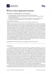
Case Report. A 54-year-old male patient presented with a history of shortness of breath and multiple episodes of seizure of one week duration. On evaluation, he was diagnosed with right lung non-small cell lung cancer (squamous cell carcinoma) with metastasis to brain and superior vena caval syndrome (stage IV). He was started on tab phenytoin 300 mg at bedtime and anti-edema measures with steroids (tab dexamethasone 8 mg 3 times daily during RT and tapered over 2 weeks post RT). He received palliative RT to the mediastinum and whole brain with a total dose of 20 Gray in 5 fractions over one week with Co-60 gamma rays. Two weeks post radiotherapy, his general condition improved and he received one cycle of palliative chemotherapy with paclitaxel-carboplatin combination. Nearly 3 weeks after completing the RT, he presented with painful, erythematous lesions in the scalp, which subsequently generalized. During examination, he was afebrile with pulse rate 98/min, respiratory rate 28/min, blood pressure 109/83 mm Hg, and features of mild dehydration. Examination of the skin revealed erythematous, tender macules over the scalp, face, trunk, and limbs with areas of confluent epidermal detachment and blistering involving almost 30% of his body surface area (Figure 1). Conjunctivitis, hemorrhagic crusting on lips, erosions over buccal and nasal mucosa and over the glans penis were noted, strongly suggesting TEN caused by phenytoin. Hemogram and biochemical parameters were normal except for hyponatremia. Phenytoin was immediately discontinued, and he was managed with intravenous fluid replacement, electrolyte correction, systemic antibiotics, steroids (dexamethasone 16 mg/ day tapered at 2 mg/day over one week), and local skin care with antibiotic and antifungal dressings. After primary supportive care, his condition worsened and he died due to septicemia on the seventh day of hospital admission. Discussion. Patients with TEN, initially present with acute onset, painful skin lesions, fever >39°C (102.2°F), sore throat, oral and ocular mucosal complications, with rapidly spreading confluent and extensive epidermal detachment, dehydration, dyselectrolytemia, which rapidly evolves into systemic disease with 25-35% mortality.2 The risk of death of patients with TEN can be accurately predicted by TEN specific severity-of-illness score, which considers 7 independent risk factors such as age >40 years, malignancy, heart rate >120/ min, initial percentage of epidermal detatchment over 10%, serum urea
1394
Saudi Med J 2014; Vol. 35 (11)
www.smj.org.sa
Figure 1 - An image showing areas of: A) dusky eruptions over the entire scalp; B) extensive epidermal detachment over the scalp, nape of the neck, and upper back.
>10 mmol/litre, serum glucose >14 mmol/litre, and bicarbonate