Editorial
Transesophageal echocardiography: Rapid expansion in perioperative medicine in emerging economies – Are we ready for a safe and effective practice? Sheila Rajashekara, Balachundhar Subramaniam1 Clinical Fellow in Anesthesiology, 1Assistant Professor of Anesthesiology, Director of Cardiac Anesthesia Research, Harvard Medical School, Beth Israel Deaconess Medical Center, Boston, MA, USA
Access this article online
Website: www.annals.in PMID: *** DOI: 10.4103/0971-9784.83982 Quick Response Code:
Transesophageal echocardiography (TEE) is a relatively recent modality in anesthesia and intensive care. The initial development of TEE occurred in the late 1970s, when Oka et al., investigated the utility of attaching an m-mode probe to an esophageal stethoscope to evaluate left ventricular (LV) function.[1] By the mid-1990s, TEE was being used routinely in cardiac surgery, and had just begun to expand into high-risk non-cardiac surgery and the intensive care unit (ICU). Clinical application of TEE, organized TEE education and outpouring of publications regarding its specific uses has led to this explosion. Furthermore, many countries have quickly adapted TEE in the last decade. However, while it has been shown that TEE can be used to diagnose and manage both anticipated and unanticipated complications during a high-risk surgery, it has been hard to quantify the impact of TEE on patient outcome. How is it possible that TEE has exploded as a significant armamentarium of a practicing anesthesiologist without such hard data on its impact on perioperative outcome? To answer this question, we need not look further than the utility of the pulse oximeter in our routine practice. While every practicing senior anesthesiologist will proudly mention that they grew up with a hand on the pulse and eye on the skin color, none will choose to practice without a pulse oximeter today.
Dr. Mayfield, in his article “The Impact of Intra-operative Monitoring on Patient Safety”, notes that prior to the standardization of such monitors, the mortality rate due to anesthetics could be as high as 11.7 per 10,000. [2] In 20,000 patients undergoing surgery, pulse oximetry had little measurable change on the incidence of general cardiovascular or respiratory complications.[3-4] Yet no one today would question the necessity of using pulse oximetry for early recognition of respiratory events. Thus, while it is not always possible to show outcome improvement with a monitor, TEE can be extremely useful either when it diagnoses a cause for instability or when it excludes an important differential diagnosis. Practicing echo in context certainly is a faster, efficient way of practicing perioperative highrisk medicine. With advances in technology, and industry competition it is only a matter of time before TEE becomes a cheaper and cost-effective diagnostic and monitoring modality. We need to make sure we train future anesthesiologists for a safe and effective use of this modality. Although TEE is a semi-invasive monitor, esophageal perforation is a rare (0.1%) but potentially fatal complication.[5] Despite this margin of safety, we routinely ask patients preoperatively of any swallowing difficulty. We also pass an orogastric tube to empty gastric contents that may or may not enhance
Address for correspondence: Dr. Balachundhar Subramaniam, Assistant Professor of Anesthesiology, Harvard Medical School, Director of Cardiac Anesthesia Research, Beth Israel Deaconess Medical Center, Boston, MA, 02215. USA. E-mail:
[email protected]
Annals of Cardiac Anaesthesia Vol. 14:3 Sep-Dec-2011
171
Rajashekara and Subramaniam: TEE and perioperative medicine
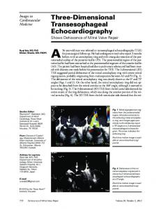
imaging[6] but ensures a patent pathway before we pass the TEE. Any such simple questionnaire and/ or a clinical strategy should be employed suited to individual practices to reduce unforeseen TEE-related complications. In cardiac surgery, TEE in addition to being a routine monitoring and diagnostic modality, has immensely helped surgeons to perform minimally invasive valvular or coronary procedures. Without the ability to visualize the heart directly as in open-heart surgery, physicians must increasingly depend on image acquisition through TEE to guide their numerous cannulae to appropriate cardiac locations, and evaluate the success of surgical or percutaneous valve repair. With advances such as 3D TEE,[7] accurate localization of perivalvular leaks[8] is a norm. Furthermore, cardiac anesthesiologists have made significant early steps to predict appropriate valves for a given patient,[9] and possibly predict functional recovery of wall motions. We must simply take such opportunities with both hands and become leaders in these exciting areas of research. Intraoperative TEE has long been a domain of the cardiac anesthesiologist. Yet in terms of acquiring and interpreting the images, does it seem likely that anesthesia’s rather recent experience with obtaining and reading echocardiography has similar conclusions as that of other physicians? Mathew et al., opted to study this question by comparing the results obtained by anesthesiologists, cardiologists and radiographers.[10] He came to the conclusion that anesthesiologists who had more than five years of training with TEE had similar diagnostic capabilities as that of the cardiologist and/or the radiologist, and therefore, was as capable of using TEE to make intraoperative decisions. The Society of Cardiovascular Anesthesiologists (SCA) has combined with the American Society of Echocardiography (ASE) and has established standards of training for perioperative practice of echocardiography. With rapid expansion of TEE in emerging economies, most notably in India, the Indian Association of Cardiothoracic Anesthesiologists, spearheaded by notable echocardiographers who have been practicing TEE for more than a decade in India, has started the fellowship examination in perioperative TEE in India (FIACTA). Furthermore, the need for basic echo training to enable practice of TEE in a non-cardiac setting is exemplified by the basic TEE examination started by the American Association of Anesthesiologists last year. There is a growing need for TEE education that can be accomplished in multiple ways including simulator technology.[11] 172
As anesthesia expanded into different areas of medicine such as pain management, so too did TEE become a more accessible tool for rapid clinical decision-making. In no other place is this more evident than in the ICU. TEE can differentiate between several causes of persistent hypotension, ranging from pericardial tamponade to hypovolemia, aiding in the rapid diagnosis and treatment of the patient.[12] TEE and its use in high-risk non-cardiac surgery such as vascular surgery, liver transplants, orthopedic surgery amongst other areas has been described previously.[13] All of the previous examples serve to illustrate the point that TEE has a variety of uses that expands from the cardiac operating rooms into all areas of medicine. Yet this is still in keeping with the Category I indications of perioperative TEE as set forth by the American Society of Anesthesiologists (ASA) / SCA taskforce.[14] In no particular order, the indications include: assessment of ejection fraction, evaluation of valvular stenosis, restrictive, hypertrophic and dilated cardiomyopathies, aortic dissection/aneurysm, cardiac arrhythmias, and neurologic disease with specific focus on cardioembolic disease. It would seem, therefore, that TEE has many advantages, perhaps on a grand scale, increasing the value of life but also giving the impetus to develop new technologies on a more affordable scale. We only need to review the short history of TEE to remember that its initial beginnings were marked by fits and starts, most notably, in creating a machine that would simultaneously transmit images without interrupting the surgery and prolonging the time under anesthesia. It has now evolved into a tool that can be used in many different fields of medicine, to aid in clinical decision-making and provide the best care for our patients. From this point, it can only progress to becoming a standard of care in all areas of the world. REFERENCES 1. 2. 3.
4.
5.
Oka Y. The evolution of intraoperative transesophageal echocardiography. Mt Sinai J Med 2002;69:18-20. Mayfield JB. The impact of intraoperative monitoring on patient safety. Anesthesiol Clin 2006;24:407-17. Moller JT, Pedersen T, Rasmussen LS, Jensen PF, Pedersen BD, Ravlo O, et al. Randomized evaluation of pulse oximetry in 20,802 patients: I. Design, demography, pulse oximetry failure rate, and overall complication rate. Anesthesiology 1993;78:436-44. Moller JT, Johannessen NW, Espersen K, Ravlo O, Pedersen BD, Jensen PF, et al. Randomized evaluation of pulse oximetry in 20,802 patients: II. Perioperative events and postoperative complications. Anesthesiology 1993;78:445-53. Hilberath JN, Oakes DA, Shernan SK, Bulwer BE, D'Ambra MN, Eltzschig HK. Safety of transesophageal echocardiography. J Am Soc Echocardiogr Annals of Cardiac Anaesthesia Vol. 14:3 Sep-Dec-2011
Rajashekara and Subramaniam: TEE and perioperative medicine 2010;23:1115-27. Roscher C, Reidy C, Augoustides JG. Progress in perioperative echocardiography: Focus on safety, clinical outcomes, 3-dimensional imaging, and education. J Cardiothorac Vasc Anesth 2011;25:559-64. 7. Vegas A, Meineri M. Core review: Three-dimensional transesophageal echocardiography is a major advance for intraoperative clinical management of patients undergoing cardiac surgery: A core review. Anesth Analg 2010;110:1548-73. 8. Karthik S, Sundar S, Lerner A, Panzica P, Subramaniam B, Mahmood F. Intraoperative assessment of perivalvular mitral regurgitation: Utility of three-dimensional echocardiography. J Cardiothorac Vasc Anesth 2008;22:431-4. 9. Mahmood F, Subramaniam B, Gorman JH 3rd, Levine RM, Gorman RC, Maslow A, et al. Three-dimensional echocardiographic assessment of changes in mitral valve geometry after valve repair. Ann Thorac Surg 2009;88:1838-44. 10. Mathew JP, Fontes ML, Garwood S, Davis E, White WD, McCloskey G, et al. Transesophageal echocardiography interpretation: A comparative analysis between cardiac anesthesiologists and primary echocardiographers. Anesth Analg 2002;94:302-9. 6.
11. Bose R, Matyal R, Panzica P, Karthik S, Subramaniam B, Pawlowski J, et al. Transesophageal echocardiography simulator: a new learning tool. J Cardiothorac Vasc Anesth 2009;23:544-8. 12. Subramaniam B, Talmor D. Echocardiography for management of hypotension in the intensive care unit. Crit Care Med 2007;35:S401-7. 13. Subramaniam B, Park KW. Impact of TEE in noncardiac surgery. Int Anesthesiol Clin 2008;46:121-36. 14. Cheitlin MD, Alpert JS, Armstrong WF, Aurigemma GP, Beller GA, Bierman FZ, et al. ACC/AHA Guidelines for the Clinical Application of Echocardiography. A report of the American College of Cardiology/ American Heart Association Task Force on Practice Guidelines (Committee on Clinical Application of Echocardiography). Developed in collaboration with the American Society of Echocardiography. Circulation 1997;95:1686-744. Cite this article as: Rajashekara S, Subramaniam B. Transesophageal echocardiography: Rapid expansion in perioperative medicine in emerging economies - Are we ready for a safe and effective practice?. Ann Card Anaesth 2011;14:171-3. Source of Support: Nil, Conflict of Interest: None declared.
Announcement
“QUICK RESPONSE CODE” LINK FOR FULL TEXT ARTICLES The journal issue has a unique new feature for reaching to the journal’s website without typing a single letter. Each article on its first page has a “Quick Response Code”. Using any mobile or other hand-held device with camera and GPRS/other internet source, one can reach to the full text of that particular article on the journal’s website. Start a QR-code reading software (see list of free applications from http://tinyurl.com/yzlh2tc) and point the camera to the QR-code printed in the journal. It will automatically take you to the HTML full text of that article. One can also use a desktop or laptop with web camera for similar functionality. See http://tinyurl.com/2bw7fn3 or http://tinyurl.com/3ysr3me for the free applications. Annals of Cardiac Anaesthesia Vol. 14:3 Sep-Dec-2011
173