L. A. (1991) in Methods in Molecular Biology (Murray, E. J., and Walker,. 8. Bradford, M. M. (1976) Anal. Bwchem. 72,248-254. 9. Chirgwin, J. M., Przybyla, A. E., ...
Communication
THE JOURNAL OF BIOLOGICAL CHEMISTRY Vol. 266, No. 24, Issue of August 25, pp. 15559-15562, 1991 0 1991 by The American Society for Biochemistry and Molecular Biology, Inc. Printed in U.S.A.
Transgenic Mice Demonstrate a Testis-specific Promoter for Angiotensin-converting Enzyme*
-
A somaticACE 11
12
15
1 1 1 1 NCOI
Ne01
1
Testis ACE
(Received for publication, April 18, 1991) Kimberly G. Langford, Shaw-Yung Shai, Tom E. Howard$, MichaelJ. Kovacp, Paul A. OverbeekO, and KennethE. Bernsteinl
Ncd
2
NE01
IOLSP
B
ACEPromoter-Lac2Construct ItACE-DLacII
From the Departmentof Pathology and Laboratory Medicine, Emory University, Atlanta,Georgia 30322 and of Cell the §Howard Hughes Medical Institute, Department Biology and Institute of Moleculnr Genetics, BaylorCollege of Medicine, Houston, Texas 77030
Ncol
Ncol
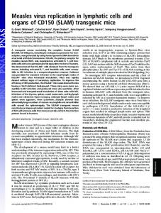
There are two isozymes of angiotensin-converting enzyme (ACE), one producedby somatic tissues and a smaller protein synthesized by developing spermatozoa (testis ACE). To investigate the molecular control of testis ACE, we generated mice transgenic for a construct containing a putative testis-specific ACE promoterlinked to the Escherichiacoli reportergene encoding @-galactosidase. The transgenic mice express FIG. 1. A , somatic ACE and testis ACE are encoded by a single &galactosidase protein and RNA only within the testis. gene (4). Both lines in A represent the same stretch of mouse genomic DNA. The top schematic shows the positions of the l l t h , 12th, and Histochemical analysis of the transgenic mice shows co-localization of @-galactosidase protein and endoge-13th exons of somatic ACE (solid boxes).The second schematic shows the identical region of genomic DNA with the 1stexon of testis ACE nous ACE within elongating spermatozoa. These stud- indicated by the stippled box. This testis-specific exon is encoded by ies demonstrate that transcription testis of ACE is con- DNA treated as an intron by somatic tissues. The second exon of trolled by a strong intragenic testis-specific promoter testis ACE is identical to the13th exon of somatic ACE. The remainthat is contained within a 698-base pair fragment im- der of testis ACE corresponds to the carboxyl portion of somatic mediately upstream from the transcription start site of ACE. A shows the NcoI restrictionsites used to clone a 698-bp testis ACE. Characterizationofthe testis ACE pro- restriction fragment into the vector pLacI to create tACE-pLacI. B , 5’ of the moter may provideinsights into the molecular mecha- tACE-pLacI contains 698bp of mousegenomicDNA nisms controllingcell stage-specific gene expression in initiator ATG of testis ACE. In tACE-pLacI, this is followed by the coding region of the E. coli lacZ gene, plus an intron and a poly(A) the male germline.
Angiotensin-converting enzyme (ACE)’ is a cell membrane peptidase that influences blood pressure through at least two mechanisms, conversion of angiotensin I to thevasoconstrictor angiotensin I1 and the degradation of bradykinin, a vasodilator (1). Pharmacologic inhibitors of ACE reduce blood pressure and are widely used to treathypertension and heart failure (2). There are two isozymes of ACE, one produced by
addition site from the mouse metallothionein I1 gene (5). An XbaIPstI fragment was used for microinjection. C , the sequence of the mouse genomic DNA cloned into tACE-pLacI. The 12th exon of somatic ACE is indicated by capital letters. The 3’ NcoI restriction site, CCATGG, encodes the initiating methionine (ATG) of testis ACE. Previous work (4) has demonstrated that the start of transcription of testis ACE begins 16 or 17 bases upstream of the initiator methionine (double underline). A region resembling a TATA box is underlined.
endothelium and several other somatic tissues (somatic ACE) and a second smaller protein only produced by developing spermatozoa (testis ACE) (3). In the mouse, the NH2-terminal *This workwas supported by Public Health Service Grant DK39777 from the National Institutes of Health and by the Howard 66 amino acids of testis ACE are unique to this isozyme. Hughes Medical Institute (grants toM. J. K. and P. A. 0.).The costs Previous studies have shown that these 66 amino acids are of publication of this article were defrayed in part by the payment of completely encoded by the first exon of testis ACE which is page charges. This article must therefore be hereby marked “adver- located within the 12th intron of the somatic ACE gene (see tisement’’ in accordance with 18U.S.C. Section 1734 solelyto indicate Fig. L 4 ) (4). Thus, the transcription of testis ACEbegins this fact. approximately 7000 base pairs downstream of the somatic The nucleotide sequence(.)reported in this paper has been submitted ACE promoter, within a region that is treated as an intron by totheGenBankTM/EMBLDataBankwith accession numberfs) somatic tissues. To address the origin of thetestis ACE M61094. $ Supported by the HarrisFamily Scholarship and theHelen Miller isozyme, we hypothesized that developing germ cells utilize a Scholarship Fund. distinct promoterlocated immediately upstream of the testis(I Established Investigator of the American Heart Association. To specific first exon. To test thishypothesis, a genomic fragment whom correspondence and reprintrequests should be addressed Rm. containing 682 bp immediately upstream from the transcrip711 WMB, Dept. of Pathology, Emory University, Atlanta, GA 30322. tion start site was isolated and linked to the Escherichia coli Tel.: 404-727-3134;Fax: 404-727-3133. The abbreviations used are: ACE, angiotensin-converting enzyme; lac2 reporter gene. We report that this genomic fragment drives testis-specific &galactosidase expression in transgenic bp, base pair(s); X-gal, 5-bromo-4-chloro-3-indolyl-~-~-galactopyranoside; GAPDH, glyceraldehyde-3-phosphate dehydrogenase. mice. We conclude that the synthesis of testis ACE is con-
15559
Test&-specificPromoter for ACE
15560
F ~ G2.. The testis on the right of the figure was removed froma mouse transgenic for tACE-pLacI, brieflyfixed, and then soakedovernightinan 1 mg/ml solution of X-gal. The blue color results from 8-galactosidase-catalyzed hydrolysis of X-gal into an insoluble blue product. The testis on the kft of the figure wasfrom anon-transgenic littermate control treated in an identical tpanner. The epididymis ( E ) contains endogenous8-galactosidase and thus stgins blue. The control testis remains unstained and thus contains little @-galactosidase activity.
F
"
-
p - Galactosidase Activity
lo
1
IACE-pbrl
Wtml
RNA Transcription
FIG. 3. Analysis of 8-galactosidase expression. Upper panel, tissues were removed from a mouse transgenic for tACE-pLacI and homogenized (7). After centrifugation, $galactosidase activityin the supernatant wasmeasuredcolorimetricallyusing the substrate ONPG. An identical studywas performed using tissues from a nontransgenic, control mouse. Only the testis of the mouse transgenic for tACE-pLacI expressed elevated levelsof @-galactosidase. Lower panel, RNA expressionof the E. coli kacZ gene encoding @-galactosidase was detectedusing an RNase protection protocol (11).A 418-base antisense RNA probe was synthesized from the E. coli lac2 gene. This was hybridizedto total RNA prepared from organs of animals transgenicfortACE-pLacI.RNAencoding E. coli @-galactosidasewill protect a 318-base band. Only RNA from the testis of micetransgenic for tACE-pLacI protected such a fragment. The integrity of the RNA was verified by RNaseprotection analysis for the ubiquitous enzyme GAPDH (12). The probe for GAPDH was derived from the rat gene; RNA from mouse protects several regions including the doublet of 110 and 105 bases shown in the lower panel.
trolled by an intragenic promoter that is utilized exclusively by developing spermatogeniccells. MATERIALS AND METHODS
Transgenic Mice-The plasmid tACE-pLacI (see Fig. 1)was created by cloninga 698-bp NcoIrestriction fragment of mouse genomic
15561
RESULTS
Testis-specificPromoter for ACE
15562
later stages of spermiogenesis (Fig. 4A).These are the same cell stages that contain testis ACE as detected by reaction with an anti-ACE antisera (Fig. 4B).Thus, thetestis-specific ACE promoter region appears to maintain its in uiuopattern of cell type-specific transcription even when linked to the /3galactosidase reporter gene. Experiments are in progress to assess at what stage of spermatogenesis promoter activity first becomes detectable. DISCUSSION
Angiotensin-converting enzyme is produced by several different tissue types. Analysis of cDNA encoding the somatic form of the enzyme has demonstrated that somatic ACE is a single polypeptide chain composed of two homologous domains, each about half the size of the parent molecule and each containing a potential catalytic site (14, 15). We and others (4,16-18) have postulated that the gene encoding the somatic form of ACE resulted from a tandem duplication of a primordial ACE gene. The function of ACE within developing spermatozoa is controversial. It is postulated to participate in capacitation, sperm motility or in aspects of sperm development (19). TestisACE is roughly half the size of the somatic isozyme. While the amino-terminal 66 amino acids of testis ACE are unique to this isozyme, the remaining portion of the molecule (600 amino acids) exactly duplicates the carboxyl domain of somatic ACE. Werecently determined that testis ACE mRNA extends only 16 or 17 bases upstream of thetestis ACE translation start site (4).This finding suggestedtwo hypotheses: either that testisACE transcription begins at thesomatic ACE promoter and RNA splicing results in themature testis ACE mRNA or, alternatively, that developing spermatocytes recognize a unique promoter located immediately upstream of the first exon of testis ACE. The experiments reported here support the second hypothesis and conclusively demonstrate that there is an intragenic testisspecific promoter for angiotensin-converting enzyme. The testis-specific promoter is contained within a 682-bp fragment immediately upstream of the transcriptional start site of testis ACE; this region of the ACE gene is located almost exactly between the duplicated domains of somatic ACE. The promoter described here is active in elongating spermatids of transgenic mice, in concordance with the pattern of expression of the endogenous testis-specific protein. The 698-bp fragment shown to contain the testis ACE promoter containsvery few ofthe structuralfeatures typically associated with an RNA polymerase I1 promoter. Studies are in progress to localize more precisely the regulatory regions that provide the testis-specific activity. Other promoters that drive testis-specific expression in
transgenic mice have been described. These include the promoters for the protamine-1 andprotamine-2 genes, which are spermatid-specific promoters, and thecontrol region for phosphoglycerate kinase-2 (PGK-2), which is active in both spermatocytes and spermatids (20-22). The testis ACE promoter described here appears to be particularly strong. This is demonstrated bothby the uniformly dark blue staining of the transgenic testes upon treatment with X-gal and by the large amounts of endogenous testis ACE mRNA synthesized in vivo (4).The testis ACE promoter may provide a useful tool for future research on factors that control male germ cell differentiation andfactors that areintegral to theprocess of fertilization. Acknowledgments-We wish to thankGerri Hanten for the mouse embryo microinjections and Dr. Grant MacGregor for assistance with the X-gal staining procedures. Dr. Cynthia Cohen, Patricia B. DeRose, and Bonnie Whitaker greatly assisted in the preparation of immunocytochemical preparations. MarkMainiero prepared the photographs used in Fig. 4. REFERENCES 1. Patchett, A. A,, and Cordes, E. H. (1985) Adu. Enzymol. Relat. Areas Mol. Biol. 67,1-84 2. Hollenberg, N. K. (1985) Am. J . Med. 7 9 , Suppl. 3c, 1-2 3. Cushman, D. W., and Cheung, H. S. (1971) Bwchim. Biophys. Acta 2 6 0 , ')Gl"xc, I "
. . " "
4. Howard, T. E., Shai, S-Y., Langford, K. G., Martin, B. M., and Bernstein, H. E. (1990) Mol. Cell. Biol. 10,4294-4302 5. Searle, P. F., Davison, B. L., Stuart, G. W., Wildie, T. M., Norstedt, G., and Palmiter. R. D. (1984) Mol. Cell. Biol. 4.1221-1230 6. Taketo, M., Schroeder; A. C . , Mabraaten, L. E., Gunning, K. B., Hanten, G., Fox, R. R., Roderick, T. H. Stewart, C. L. Lilly F., Hansen, C. T., and Overheek, P. A. (1991) Pro:. Natl. Acad. Sii. U. 8.A. 88,2065-2069 7. MacGregor, G. R., Nolan, G. P., Fiering, S., Roederer, M., and Herzenherg, L. A. (1991) in Methods in Molecular Biology (Murray, E.J., and Walker, J. M., eds) Vol. 7, pp. 217-235, Humana Press, Inc., Clifton, NJ 8. Bradford, M. M. (1976) Anal. Bwchem. 72,248-254 9. Chirgwin, J. M., Przybyla, A. E., MacDonald, R. J., and Rutter, W. J. (1979) Biochemistry 18.5294-5299 10. Glisin, V., Crkvenjakov, R., and Byus, C. (1974) Biochemistry 1 3 , 26332631 11. Ausubel, F. M., Brent, R., Kingston, R. E., Moore, D. D., Seidman, J. G., Smith, J. A., and Struhl,K. (eds) (1989) in CurrentProtocols in Molecular Biology Vol. 1, pp. 4.7.1-4.7.8, John Wiley and Sons, New York 12. Fort, P. H., Marty, L., Piechaczyk, M., El Sahrouty,S., Dani, Ch., Jeanteur, P., and Blanchard,J. M. (1985) Nucleic Acids Res. 13,1431-1442 13. Unger, E. R., and Brigati, D.J. (1989) Curr. Top. Microbiol. Immunol. 1 4 3 , 21-31 14. Souhrier, F., Alhenc-Gelas, F., Hubert, C., Allegrini, J., John, M., Tregear, G., and Corvol, P. (1988) Proc. Natl. Acad. Sci. U. S. A. 86,9386-9390 15. Bernstein, H. E.,Martin, B. M., Edwards, A. S., and Bernstein, E.A. (1989) J. Biol. Chem. 264,11945-11951 16. Kumar, R. S., Kusari, J., Roy, S. N., Soffer, R. L., and Sen, G. C. (1989) J. Biol. Chem. 264,16754-16758 17. Lattion, A-L., Soubrier, F., Allegrini, J., Hubert,C., Corvol, P., and AlhencGelas, F. (1989) FEES Lett. 262,99-104 18. Ehlers, M. R. W., Fox, E. A., Strydom, D. J., and Riordan, J. F. (1989) Proc. Natl. Acad. Sci. U. S. A. 86, 7141-7745 19. Ehlers, M. R. W., and Riordan, J. F. (1989) Baochemistry 28,5311-5318 20. Peschon, J. J., Behringer, R. R., Brinster, R. L., and Palmiter,R. D. (1987) Proc. Natl. Acad. Sci. U. S. A. 84,5316-5319 21. Stewart, T. A,, Hecht, N. B., Hollin shead, P. G , Johnson, P. A., Leong, J. C., and Pitts, S. L. (1988) Mol. b U . Biol. 8, i748-1755 22. Robinson, M. O., McCarrey, J. R., and Simon, M. I. (1989) Proc. Natl. Acad. Sci. U. S. A. 86,8437-8441