Reviews and Overviews
Treatment Advances for Cocaine-Induced Ischemic Stroke: Focus on Dihydropyridine-Class Calcium Channel Antagonists Bankole A. Johnson, M.D., Ph.D. Michael D. Devous, Sr., Ph.D. Pedro Ruiz, M.D. Nassima Ait-Daoud, M.D.
Objective: The authors reviewed the pathogenesis of cocaine-related cerebral ischemia, appraised current knowledge of its sequelae, and assessed the role of putative therapeutic agents, particularly dihydropyridine-class calcium channel antagonists. Method: The authors performed an OVIDbased literature review of all indexed journals between 1966 and 2000. Results: Cocaine abuse significantly increases the risk of ischemic stroke. The principal mechanism of cocaine-induced cerebral ischemia is vasospasm of large cranial arteries or within the cortical microvasculature. Increased levels of extracellular monoamines, particularly dopamine, mediate vasospasm. Neuroanatomical and labeling studies also have shown that dopamine-innervated neurons may regulate cerebral blood flow. Indeed, dopamine-rich brain regions appear to be relatively specific targets for cocaine-induced cerebral ischemia. Neuroimaging studies show that cocaine-induced hypoperfusion can persist even after 6 months of abstinence. Hypoperfusion can result in
deficits on complex and simple psychomotor tasks but perhaps not on memory or attention. Severe cerebral ischemia can directly precipitate neuronal death and degradation, a condition exacerbated by liberation of the excitatory amino acid glutamate. Dihydropyridine-class calcium channel antagonists inhibit cocaine-mediated dopamine release on neurons involved in vasospasm and the control of cortical circulation. Other causes of cerebral ischemia include thrombogenesis and vasculitis. Although antithrombotic agents have potential in alleviating cocaine’s neurotoxic effects, their use may be limited by the risk of spontaneous hemorrhage. Conclusions: Cocaine abuse can result in stroke, neuroischemia, and cognitive deficits that can persist even after prolonged abstinence. Dihydropyridine-class calcium channel antagonists, such as isradipine, show promise as therapeutic agents for preventing cocaine-induced cerebral ischemia. (Am J Psychiatry 2001; 158:1191–1198)
C
ocaine abuse can cause strokes (1, 2). The relative likelihood of developing a stroke in cocaine users may be as much as 14 times greater than that in age-matched non-cocaine-using comparison subjects recruited from the same patient pool (3). Between 25% and 60% of cocaine-induced strokes can be attributed to cerebral ischemia (4–6). About 80% of the infarcts occur in the regional distribution of the middle cerebral artery (7), typically in young adults without preexisting vascular malformations (6). Nevertheless, increased polysubstance abuse among cocaine addicts has complicated attribution and accurate characterization between amount and duration of cocaine use and its relationship with global and regional hypoperfusion abnormalities (8). Perfusion deficits also can occur in the absence of clinically detectable symptoms in the central nervous system (9, 10) and may persist in abstinent cocaine addicts for 6 months (11) or more (12). Mechanistic processes that underlie cocaine’s site-specific effects on the cerebral vasculature are, thereAm J Psychiatry 158:8, August 2001
fore, imperfectly understood. Advanced neuroimaging techniques offer a vehicle for elucidating these mechanisms and ascertaining the effectiveness of putative therapeutic agents for treating cerebral ischemia. Preclinical research suggests that the etiology of cocaine-induced brain ischemia is multifactorial and involves the stimulation of vasospasm (13), platelet aggregation (14), and pathological changes in the cerebral vasculature that can impair cellular oxygenation (14). Within all of these processes, calcium-channel-mediated interactions, particularly with dopamine, play an important role. Dihydropyridine-class calcium channel antagonists, by counteracting cocaine-induced vasospasm, have potential as putative therapeutic agents (15). This review article is subdivided into three major parts. First, we critically appraise the neurobiology of cocaineinduced cerebral ischemia. Second, we summarize current knowledge on the sequelae of cocaine’s neuroischemic effects. Third, we assess the role of various puta-
1191
COCAINE-INDUCED STROKE
tive therapeutic agents for treating cocaine-induced cerebral ischemia, with a particular focus on dihydropyridine-class calcium channel antagonists.
Neurobiology of Cerebral Ischemia Cerebral ischemia is associated with a variety of cocaine’s neurovascular effects. We will review the relative importance of various mechanistic processes attributable to cerebral ischemia.
Vasospasm Cocaine, the alkaloid benzoylmethylecgonine, by being highly lipid soluble, traverses the blood-brain barrierrapidly. As in the periphery, cocaine blocks monoamine reuptake, particularly those related to dopamine (7). Cocaine-induced changes in dopamine level are most prominent in mesocorticolimbic neurons (16, 17). Augmentation of mesocorticolimbic dopamine function may be associated with cerebral ischemia by means of two mechanisms. First, dopamine may control local blood flow by inducing vasospasm of smooth muscles lining the cerebral vessels (18–21), particularly those of the middle cerebral artery (19). Second, cocaine-induced reductions in cerebral metabolism may lead to feedback down-regulation of blood flow (22). Rapid reperfusion of these previously ischemic areas may, conversely, result in hemorrhage (23). More recently, the role of dopamine in the regulation of cortical blood flow has been better elucidated. First, my colleagues and I (24) established that cocaine-induced cerebral vasospasm was relatively specific to dopamine-rich brain areas and hypothesized that dopamine pathways play a central role in controlling cerebral blood flow (CBF). Later, Krimer and colleagues (25) provided a mechanistic basis for these findings by showing that dopamine-containing neurons were located adjacent to blood vessels in the frontal lobe and prefrontal cortex. These vessels constricted in response to the perivascular application of dopamine with the maximal calculated reduction of up to 58% of pretreatment flow. Hypothetically, in the living brain, pericytes containing contractile elements located proximal to dopamine nerve endings, which are also responsive to vasoactive substances in the endothelium, may be mechanistically responsible for this process (26, 27). Essential to this process appears to be an initial transmembrane loss of magnesium followed by a rapid rise in intracellular calcium concentration. Severity of the perfusion deficit is, therefore, typically correlated with the relative strength of these neurochemical changes (28). Nevertheless, cocaine’s vasospastic effects can persist beyond its half-life because its major metabolites (benzoylecgonine and norcocaine) also are potent vasoconstrictors (29), and bradykinin-mediated endothelium-dependent relaxation is impaired in chronic cocaine abusers (30, 31).
1192
Although intracortical vessels are preferentially innervated by dopamine, those located extraparenchymally receive their neuronal supply from norepinephrine-containing cells in the superior cervical ganglia (25). Thus, multifactorial mechanisms may be associated with the development and extension of cerebral vasoconstriction. Deep cortical brain regions may be more susceptible to dopamine’s vasospastic effects. In contrast, reductions in CBF subsequent to constriction of large cranial arteries may be more dependent on norepinephrine facilitation (32, 33). The vasospastic effects of serotonin (5-HT), levels of which are also increased by cocaine, also may be greater at large and medium-sized cranial arteries. Cocaine’s vasospastic effects can, therefore, be associated with generalized global reductions in CBF (34).
Thrombosis Cocaine-induced cerebral ischemia also can be caused by thrombus formation in the vasculature (29). Chronic cocaine use increases platelet levels, enhances adenosine diphosphate platelet activation, and augments sporadic release of platelet-bound α granules. Platelet activation and aggregation, precursors of thrombus formation, may be mediated by cocaine-induced increases in monoamine levels, particularly of 5-HT. Release of α granules containing growth factors and platelet-specific agglutinating proteins, β thromboglobulin and platelet factor 4, together with enhanced elastase-mediated atherosclerotic damage to the cerebral vasculature, all contribute to cocaine’s thrombogenic potential (35, 36). Thrombus formation also may be facilitated by cocaine-induced cardiomyopathy (14). In vitro studies also have demonstrated cocaine’s ability to facilitate the platelets’ response to arachidonic acid, thereby stimulating thromboxane production and, consequently, their aggregation (37–39). Protein C antithrombin III depletion and enhanced prostaglandin release are additional contributors to cocaine’s procoagulant effects (40–42). In sum, a gallimaufry of cocaine’s platelet aggregating and cardiomyopathic effects contribute to thrombus formation.
Vasculitis Apart from the atherosclerotic changes attributable to chronic cocaine use that contribute to thrombus formation, there also is evidence of other pathological structural alterations in cerebral vasculature. For example, Martinez and co-workers (14) found that long-term cocaine use, due to fluctuations between vasospasm and reperfusion, may result in vessel damage. This vasculitis, not attributable to arteriosclerosis or infection, has also been described by others (2, 42). Chronic cocaine-induced vasculitis exacerbates nonlaminar blood flow and sludging in the vessels. This decreases cellular oxygenation, thereby increasing platelet aggregation and consequent thrombus formation. Am J Psychiatry 158:8, August 2001
JOHNSON, DEVOUS, RUIZ, ET AL.
Consequences of Cerebral Ischemia Cerebral ischemia can result in a variety of quantifiable structural and functional deficits. These deficits contribute to the pathogenesis of cocaine dependence and can impair an addict’s recovery. Although the focus of this review relates to the quantification of dynamic changes in CBF, it is noteworthy that structural defects also may be a long-term consequence of this abnormality. Ischemic cerebral damage can result in cellular death identifiable by magnetic resonance imaging (MRI). Cocaine-induced cerebral damage appears to have similar hallmarks to other diseases that result in global damage. For instance, as brain cells die, there are generalized atrophic changes with concomitant ventricular enlargement and sulcal widening (42, 43). On a cellular level, ischemia-induced neuronal degeneration is associated with the extracellular leakage of glutamate. This is due to inactivation of the adenosine diphosphate-dependent glutamate reuptake pump, which results in the accumulation of intracellular sodium and consequent neuronal engorgement (44). Glutamate’s neurotoxic effects are potentiated by the long-term influx of intracellular calcium. Excessive intracellular calcium concentration produces direct necrotic cellular changes and promotes the activity of calcium-sensitive proteolytic enzymes. Consequently, apoptotic processes may be triggered prematurely (45). In the aggregate, cerebral ischemia can provoke or potentiate a sequence of events resulting in structural damage due to direct or programmed neuronal cell death. Functional deficits represented as decreases in cerebral perfusion can exist in the absence of structural damage. These perfusion deficits have mainly been studied using two types of neuroimaging: single photon emission computed tomography (SPECT) and positron emission tomography (PET); however, future studies may make use of developing techniques in functional MRI. The relationship between chronic cocaine abuse and reported cognitive deficits, hypothetically due to perfusion abnormalities in the striatum (46), remains controversial. Methodological problems, such as small group size, use of published cognitive test norms instead of appropriately matched comparison subjects, differences in population characteristics (e.g., social class, treatment-seeking versus incarcerated status, presence or absence of concomitant drug abuse or dependence, varying lengths of abstinence, and level of care in its verification), have resulted in conflicting findings (47, 48). Generally, controlled studies of cocaine abusers with several months of abstinence support an impairment of response in complex and simple psychomotor tasks (47, 49) and to auditory tones or discrimination tasks (50–52) but less consistently demonstrate decrements in memory and attention (47, 48). Future studies of the relationship between cocaine abuse and cognitive dysfunction need to address two current limitations in our knowledge. First, we know of no longterm longitudinal studies charting the relationship beAm J Psychiatry 158:8, August 2001
tween the progression of disease, as identified by neuroimaging studies, and a comprehensive set of standardized cognitive tests. Second, it is important for us in more fully characterizing the relationship between cognitive function and functional brain abnormality to determine how these parameters are affected by whether or not the individual is actively taking cocaine, has recently withdrawn from it, or has entered into a period of prolonged abstinence. Development of accurate methods for characterizing functional changes in CBF, therefore, remains at the cornerstone of quantifying cerebral ischemia and its consequent neuropsychological sequelae.
Neuroimaging Findings Neuroimaging studies have provided a vehicle for understanding cocaine’s ischemic effects on the brain. Early studies vary in the reported distribution of regional cerebral blood flow (rCBF) abnormalities found in cocaine users (10, 53, 54); however, these rCBF deficits can be severe (46, 55, 56). Although most abused substances (including cocaine) reduce both global and regional cerebral glucose metabolism during acute use, rCBF can be either increased or decreased, perhaps related to the vasoactive characteristics of particular compounds (57). The distribution of both rCBF and abnormalities in regional cerebral glucose metabolism also depends on whether the individual was currently using the drug, was exposed to cues, had recently withdrawn from cocaine, or was in long-term detoxification (53, 58, 59). A typical report is that of Holman et al. (60), who studied cocaine abusers with 99m Tc-labeled exametazime ([99m Tc]hexamethylpropyleneamine oxime) and highresolution SPECT. Sixteen of 18 cocaine-dependent users had abnormal perfusion characterized primarily as small focal defects involving the infraparietal, temporal and anterofrontal cortex, and basal ganglia (the results of psychometric testing were abnormal in all 18). There was no relationship between the severity of SPECT abnormalities and the mode of cocaine administration or the frequency or length of cocaine use. It is important, however, that these results of global and regional hypoperfusion were consistent with the findings of others (61, 62). Later, Holman and colleagues (58) reported that such findings can be confounded by similar vascular abnormalities observed in HIV-positive and AIDS patients because of the frequent comorbidity of HIV infection and drug abuse. Pearlson and co-workers (63) advanced these findings in a well-controlled study of acute cocaine dosing. They suggested that cocaine’s ischemic effects were most prominent in dopamine-rich brain regions such as the caudate, inferior cingulate, and frontal areas; however, these sitespecific effects could not be established by this experiment because a quantitative approach to SPECT was not employed and the cocaine dose was not manipulated. Pearlson and co-workers (63) also suggested that cocaine-
1193
COCAINE-INDUCED STROKE FIGURE 1. Effect of Cocaine on Brain Regional Cerebral Blood Flow (rCBF)a Decreased rCBF
Low-Dose Cocaine
Increased rCBF
Inferior frontal
Anterior temporal/perisylvian High-Dose Cocaine
Cerebellum Inferior/orbital frontal
Hippocampus
Cingulate
Thalamus Cerebellum a
Parametric images of significant rCBF changes (cocaine versus placebo) from nine subjects studied after administration of placebo, low-dose cocaine (0.33 mg/kg), and high-dose cocaine (0.65 mg/kg) as shown by single photon emission computed tomography (SPECT). Areas of significant change are shown as solid objects overlaid on a model SPECT brain image. Images are oriented with the subject’s left on the viewer’s right. Low-dose cocaine produced rCBF reductions (relative to placebo) in bilateral anterior temporal (perisylvian) sites, in left mesial/anterior temporal sites, and in a right inferior orbital frontal site (Brodmann’s area 11). Additional decreases were observed in superior antero- and posterolateral frontal regions (not shown). Increased rCBF was observed in the brainstem and cerebellum. High-dose cocaine produced more extensive decreases to bilateral anterior temporal (perisylvian) sites and to the right inferior orbital frontal site. New areas of significant rCBF decline (relative to low-dose cocaine) were observed in the inferior cingulate, right mesial temporal lobe (hippocampus), and left inferior/mid temporal lobe (not hippocampus). Furthermore, high-dose cocaine led to increases in left thalamic and left hippocampal sites not seen with low-dose cocaine and to the cerebellar changes seen with low-dose cocaine.
induced reductions in CBF were associated with increased positive subjective mood and abuse liability. These findings differ from those of London and co-workers (54), who, using PET and [18F]fluorodeoxyglucose, did not find an association between regional cerebral glucose metabolism and subjective mood. An advantage of the study of Pearlson and co-workers (63) was that imaging and the peak of cocaine’s behavioral effects were synchronized carefully, thereby increasing the predictive power of the findings. While a detailed description of the relationship between cocaine-induced subjective mood and other (i.e., non-flow-related) aspects of brain function is outside the scope of this review, further studies are clearly needed. Notably, however, this field of inquiry has been advanced by the demonstration that dopamine transporter function may be closely correlated with the expression of cocaine’s subjective effects (64). More recently, we studied the effects of cocaine on global and rCBF using a novel approach to quantified SPECT (24). In that study, nine cocaine-dependent men and women (six men) with no evidence of structural brain abnormality (verified by an MRI at screening) were intravenously administered low and high doses of cocaine (0.33 mg/kg and 0.65 mg/kg, respectively) or placebo in a double-blind crossover fashion. We established that cocaine administration, compared with placebo, was associated with significant reductions in global and rCBF, which was relatively specific for dopamine-rich brain areas. Also,
1194
these CBF reductions were mostly dose dependent (i.e., the high dose led to greater involvement of cortical sites than the low dose), and the areas with greatest deficits were the prefrontal, frontal, temporal, and subcortical gray matter (Figure 1). This study elucidated the site-specific effects of cocaine related to CBF changes and established the utility of this quantified approach to SPECT. In sum, cocaine abuse, either acute or long-term, is associated with hypoperfusion-related CBF abnormalities. Despite some variance in the literature concerning the anatomical regions most affected, the current evidence suggests that dopamine-rich brain areas are particularly vulnerable. The correlation between regional blood flow reductions in dopamine-rich areas and cocaine abuse was intriguing and, as was later demonstrated, provided strong evidence for a role of cortical dopamine neurons in the regulation of the arteriolar microcirculation. Thus, medications that antagonize these dopamine-mediated effects on CBF offer promise as potential treatments for cocaine-related ischemic stroke.
Pharmacological Approaches Until recently, despite the growing epidemic of cocainerelated ischemic stroke, the development of medications specifically targeted at this indication remained in its infancy. The next section describes the potential role of some promising medications aimed at reducing cocaine’s vasospastic, thrombogenic, or neurotoxic effects. Am J Psychiatry 158:8, August 2001
JOHNSON, DEVOUS, RUIZ, ET AL.
Antivasospastics Animal studies support the use of the dihydropyridineclass calcium channel antagonist isradipine for the reversal of cerebral ischemia and the consequent neuronal cell damage (65–67). In a landmark study, Sauter and colleagues (68) examined the relative effectiveness of various dihydropyridine-class calcium channel antagonists (i.e., darodipine, nimodipine, nitrendipine, and isradipine) in reducing the ischemic damage produced by occlusion of the middle cerebral artery in the rat. CBF was estimated by using the [14C]iodoantipyrine method for blood flow quantification. In that study, isradipine (2.5 mg/kg given subcutaneously) had the greatest anti-ischemic effect. The doserelated vasodilator response to isradipine was wider than for the other dihydropyridine-class calcium channel antagonists and was correlated with a reduction in infarct size. These antivasospastic effects of isradipine were attributed to the blockade of dopamine release from cortical neurons involved in the control of the cerebral vasculature. In another example, 60 minutes of cerebral ischemia was induced in spontaneously hypertensive male rats by carotid artery occlusion. Hydrogen clearance was used to measure CBF, and in vivo microdialysis was used to quantify extracellular increases in dopamine and glutamate levels. Although the vehicle-treated group experienced 180and 24-fold increases in extracellular dopamine and glutamate levels, respectively, isradipine significantly reduced the dopamine increase but had no effect on glutamate levels (69). Ooboshi and colleagues (70) confirmed these results. They showed that pretreatment with isradipine (200 µg/ml) did not alter ischemia-induced dopamine release in the striatum but produced a 37% decrease in dopamine levels in the forebrain. Additionally, isradipine significantly reduced forebrain ischemia (i.e., improved blood flow) but had no effect on striatal blood flow. Together, these results suggest that isradipine is a potent cerebral vasodilator with selectivity for dopamine-innervated brain regions. Most recently, we extended these results to humans using a quantified SPECT approach by demonstrating that isradipine (10 mg given orally) administered 60 min before intravenous cocaine (0.33 mg/kg) prevented both global and regional ischemia in dopamine-rich brain areas (15). This was the first clear demonstration that any medication, including isradipine, could prevent cocaine’s vasospastic effects on CBF. One caveat to this study was that there was no condition using isradipine alone. Thus, the relative strength of isradipine’s effectiveness at antagonizing cocaine-induced cerebral ischemia is unknown. In sum, isradipine appears to be a promising therapeutic medication for the prevention of cocaine-related cerebral ischemia. It is important, however, that we await the results of larger-scale confirmatory neuroimaging studies and the results of clinical testing with the longitudinal evaluation of cerebral perfusion. Am J Psychiatry 158:8, August 2001
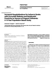
FIGURE 2. Thrombosis-Promoting Effects of Cocaine a Cocaine
(+)
Arachidonic acid Prostaglandin
2 Prostacyclin (+)
Release
(–)
Thromboxane; adenosine diphosphate (+)
Gp IIb–IIIa complex
3 Phospholipid complex
Platelet synthesis Thrombus formation
1
von Willebrand factor
Endothelium
Vascular injury
Extrinsic Pathway
Intrinsic Pathway Ca2+
Common Pathway Stimulate phases 2 & 3
Thrombin Ca2+
Fibrinogen
Fibrin
Consolidate platelet aggregation a
Platelets play an important role in the formation of a thrombus. They undergo three phases: 1) adhesion, 2) secretion, and 3) aggregation. Thromboxane and adenosine diphosphate set up an autocatalytic reaction leading to the buildup of an enlarging platelet aggregate (the primary hemostatic plug). Cocaine enhances thromboxane release, the synthesis of platelets, and thrombus formation.
Antithrombogenic Agents Recently, there has been interest in the use of aspirin to reduce cocaine’s thrombogenic effects. This is predicated on the finding that prostaglandin synthesis, which is enhanced by cocaine (Figure 2), can be blocked by aspirin. Aspirin blocks this process by irreversibly inhibiting the action of cyclo-oxygenase in platelets released from bone marrow. These platelets, derived from the bone marrow, constitute about 15% of the body’s total daily circulation (71, 72). Preliminary results indicate that aspirin (325 mg/day for 4 weeks) may improve CBF in chronic cocaine users; however, the results of a more definitive analysis are awaited (73). Aspirin treatment may, however, increase the risk of spontaneous or uncontrolled bleeding from wounds. Thus, implementation of long-
1195
COCAINE-INDUCED STROKE
term aspirin treatment for cocaine addicts is impractical, because of their propensity for exposure to high-risk situations. Also, acute aspirin administration is unlikely to result in reversal of cerebral ischemia or substantial CBF improvement in individuals with an evolving ischemic episode of recent onset. There may be potential for using the potassium-sparing diuretic amiloride for the prevention of ischemia caused by blockade of platelet α granule release in cocaine addicts (73, 74). Again, the results of more definitive studies are awaited. Although the 5-HT2 receptor antagonist ritanserin has demonstrated antiplatelet aggregation properties, its use for this indication in cocaine addicts remains unexplored (75, 76).
Antineurotoxic Agents One approach that has been suggested to reduce cocaine’s neurotoxic effects is the use of glutamate antagonists, such as phencyclidine and MK-801. Although these medications should, potentially, reduce the effects of excitatory amino acids released during neuronal degeneration at N-methyl-D -aspartate receptors, their other psychopharmacological effects limit their extensive use in humans. For instance, not only are these medications hallucinogenic, but they can also reduce the seizure threshold. Medications with antiglutamate properties and a more benign side effect profile, such as lamotrigine, or those that facilitate γ-aminobutyric acid neurotransmission, such as gabapentin, may have greater ability to prevent or treat cocaine-induced cerebral ischemia. Evidence for the use of kappa opiate agonists for preventing ischemia because they modulate glutamate expression is conflicting (77; cf. reference 78). Although decreases in cocaine-induced perfusion abnormalities have been reported with the kappa opiate antagonist buprenorphine, that study’s results were limited by the lack of a placebo control group (59). Furthermore, research is needed to elucidate the role of kappa opiate antagonists in treating cocaine-induced cerebral ischemia.
ated with the enhancement of cellular processes that augment platelet aggregation and the enhancement of nonlaminar vascular flow after the development of cardiomyopathy. Cocaine-induced cerebral ischemia can result in marked hypoperfusion abnormalities. These abnormalities may themselves be associated with deficits in the manipulation of complex and simple psychomotor tasks, but the evidence supporting memory and attentional deficits is less established. Little is known about the pathological development of these hypoperfusion abnormalities, but the prevailing evidence suggests that they persist even after a prolonged period of cocaine abstinence. With repeated cerebral ischemic episodes and subsequent reperfusion, vascular walls are weakened, and the likelihood of hemorrhagic episodes increases. Severe cerebral ischemia can result in direct neuronal death and degradation with the liberation of excitatory amino acids, particularly glutamate, which itself potentiates this process. Antithrombotic agents or medications that can ameliorate cocaine’s neurotoxic effects may have use in preventing or treating cocaine-induced cerebral ischemia. Promising recent data provide strong support for the use of dihydropyridineclass calcium channel antagonists in preventing cocaineinduced vasospasm, the principal cause of cerebral ischemia. Thus, the use of dihydropyridine-class calcium channel antagonists may herald a new vista in the prevention of cocaine-induced cerebral ischemia. Received Dec. 22, 1999; revision received Nov. 2, 2000; accepted Nov. 10, 2000. From the Departments of Psychiatry and Pharmacology and the Southwest Texas Addiction Research and Technology (START) Center, University of Texas Health Science Center at San Antonio; the Nuclear Medicine Center, University of Texas Southwestern Medical Center, Dallas; and the Department of Psychiatry and Behavioral Sciences, University of Texas Medical School at Houston. Address reprint requests to Dr. Johnson, START Center, 3939 Medical Dr., San Antonio, TX 78229;
[email protected] (e-mail). Funded by grants DA-13002-01 and DA-12191 from the National Institute on Drug Abuse.
References
Conclusions Cocaine abuse significantly increases the risk of ischemic stroke. Current evidence shows that the genesis of cocaine-related ischemia is multifactorial. Principally, cocaine’s vasospastic effects at large cranial arteries or within the cortical microvasculature are mediated by increased levels of extracellular monoamines, particularly dopamine. Indeed, dopamine-rich brain areas appear to be relatively specific targets for cocaine-induced cerebral ischemia. The development of various neuroimaging tools, particularly PET and SPECT, has greatly advanced our knowledge about cocaine’s global and site-specific effects on CBF. Other mechanistic processes involved with the development of cerebral ischemia include thrombogenesis and vasculitis. Cocaine’s thrombogenic potential is associ-
1196
1. Klonoff DC, Andrews BT, Obana WG: Stroke associated with cocaine use. Arch Neurol 1989; 46:989–993 2. Krendel DA, Ditter SM, Frankel MR, Ross WK: Biopsy-proven cerebral vasculitis associated with cocaine abuse. Neurology 1990; 40:1092–1094 3. Petitti DB, Sidney S, Quesenberry C, Bernstein A: Stroke and cocaine or amphetamine use. Epidemiology 1998; 9:596–600 4. Daras M, Tuchman AJ, Koppel BS, Samkoff LM, Weitzner I, Marc J: Neurovascular complications of cocaine. Acta Neurol Scand 1994; 90:124–129 5. Levine SR, Brust JC, Futrell N, Ho KL, Blake D, Millikan CH, Brass LM, Fayad P, Schultz LR, Selwa JF, Welch KMA: Cerebrovascular complications of the use of the “crack” form of alkaloidal cocaine. N Engl J Med 1990; 323:699–704 6. Bartzokis G, Beckson M, Hance DB, Lu PH, Foster JA, Mintz J, Ling W, Bridge P: Magnetic resonance imaging evidence of “silent” cerebrovascular toxicity in cocaine dependence. Biol Psychiatry 1999; 45:1203–1211
Am J Psychiatry 158:8, August 2001
JOHNSON, DEVOUS, RUIZ, ET AL. 7. Fessler RD, Esshaki CM, Stankewitz RC, Johnson RR, Diaz FG: The neurovascular complications of cocaine. Surg Neurol 1997; 47:339–345 8. Withers NW, Pulvirenti L, Koob GF, Gillin JC: Cocaine abuse and dependence. J Clin Psychopharmacol 1995; 15:63–78 9. Volkow N, Mullani N, Gould KL, Adler S, Krajewski K: Cerebral blood flow in chronic cocaine users: a study with positron emission tomography. Br J Psychiatry 1988; 152:641–648 10. Tumeh SS, Nagel JS, English RJ, Moore M, Holman BL: Cerebral abnormalities in cocaine abusers: demonstration by SPECT perfusion brain scintigraphy: work in progress. Radiology 1990; 176:821–824 11. Strickland TL, Mena I, Villanueva-Meyer J, Miller BL, Cummings J, Mehringer CM, Satz P, Myers H: Cerebral perfusion and neuropsychological consequences of chronic cocaine use. J Neuropsychiatry Clin Neurosci 1993; 5:419–427 12. Bell KM, Milne N, Lyons KP: Regional cerebral blood flow and cocaine abuse. West J Med 1994; 161:412–413 13. Kaufman MJ, Levin JM, Maas LC, Rose SL, Lukas SE, Mendelson JH, Cohen BM, Renshaw PF: Cocaine decreases relative cerebral blood volume in humans: a dynamic susceptibility contrast magnetic resonance imaging study. Psychopharmacology (Berl) 1998; 138:76–81 14. Martinez N, Diez-Tejedor E, Frank A: Vasospasm/thrombus in cerebral ischemia related to cocaine abuse (letter). Stroke 1996; 27:147–148 15. Johnson BA, Baron B, Fang B, Lamki L, Wagner L, Wells L, Kenny P, Overton D, Dhother S, Abramson D, Chen R, Kramer L: Isradipine prevents global and regional cocaine-induced changes in brain blood flow: a preliminary study. Psychopharmacology (Berl) 1998; 136:335–341 16. Koob GF, Bloom FE: Cellular and molecular mechanisms of drug dependence. Science 1988; 242:715–723 17. Kuhar MJ, Ritz MC, Boja JW: The dopamine hypothesis of the reinforcing properties of cocaine. Trends Neurosci 1991; 14: 299–302 18. Anday EK, Lein R, Goplerud JM, Kurth CD, Shaw LM: Pharmacokinetics and effect of cocaine on cerebral blood flow in the newborn. Dev Pharmacol Ther 1993; 20:35–44 19. He GQ, Zhang A, Altura BT, Altura BM: Cocaine-induced cerebrovasospasm and its possible mechanism of action. J Pharmacol Exp Ther 1994; 268:1532–1539 20. Kurth CD, Monitto C, Albuquerque M, Feuer P, Anday E, Shaw L: Cocaine and its metabolites constrict cerebral arterioles in newborn pigs. J Pharmacol Exp Ther 1993; 265:587–591 21. Madden JA, Konkol RJ, Keller PA, Alvarez TA: Cocaine and benzoylecgonine constrict cerebral arteries by different mechanisms. Life Sci 1995; 56:679–686 22. Rogers KJ, Nahorski KJ: Depression of cerebral metabolism by stimulant doses of cocaine. Brain Res 1973; 57:255–258 23. Caplan L: Intracerebral hemorrhage revisited. Neurology 1988; 38:624–627 24. Johnson B, Lamki L, Fang B, Barron B, Wagner L, Wells L, Kenny P, Overton D, Dhother S, Abramson D, Chen R, Kramer L: Demonstration of dose-dependent global and regional cocaine-induced reductions in brain blood flow using a novel approach to quantitative single photon emission computerized tomography. Neuropsychopharmacology 1998; 18:377–384 25. Krimer LE, Muly ECI, Williams GV, Goldman-Rakic PS: Dopaminergic regulation of cerebral cortical microcirculation. Nat Neurosci 1998; 1:286–289 26. Shepro D, Morel NML: Pericyte physiology. FASEB J 1993; 7: 1031–1038 27. Haefliger IO, Zschauer A, Anderson DR: Relaxation of retinal pericyte contractile tone through the nitric oxide-cyclic guanosine monophosphate pathway. Invest Ophthalmol Vis Sci 1994; 35:991–997
Am J Psychiatry 158:8, August 2001
28. Zhang A, Cheng TP-O, Altura BT, Altura BM: Acute cocaine results in rapid rises in intracellular free calcium concentration in canine cerebral vascular smooth muscle cells: possible relation to etiology of stroke. Neurosci Lett 1996; 215:57–59 29. Konzen JP, Levine SR, Garcia JH: Vasospasm and thrombus formation as possible mechanisms of stroke related to alkaloidal cocaine. Stroke 1995; 26:1114–1118 30. Copeland JR, Willoughby KA, Police RJ, Ellis EF: Repeated cocaine administration reduces bradykinin-induced dilation of pial arterioles. Am J Physiol 1996; 271(4, part 2):H1576–H1583 31. Havranek EP, Nademanee K, Grayburn PA, Eichhorn EJ: Endothelium-dependent vasorelaxation is impaired in cocaine arteriopathy. J Am Coll Cardiol 1996; 28:1168–1174 32. Langner RO, Bement CL, Perry LE: Arteriosclerotic toxicity of cocaine. NIDA Res Monogr 1988; 88:325–336 33. Bement CL, Cohen L, Nielson SW, Langner RO: Arterial injury in rabbits following alternate day injections of cocaine (abstract). FASEB J 1989; 3:A297 34. Kaufman MJ, Levin JM, Ross MH, Lange N, Rose SL, Kukes TJ, Mendelson JH, Lukas SE, Cohen BM, Renshaw PF: Cocaine-induced cerebral vasoconstriction detected in humans with magnetic resonance angiography. JAMA 1998; 279:376–380 35. Heesch CM, Wilhelm CR, Ristich J, Adnane J, Bontempo FA, Wagner WR: Cocaine activates platelets and increases the formation of circulating platelet containing microaggregates in humans. Heart 2000; 83:688–695 36. Ross R, Vogel A: The platelet-derived growth factor. Cell 1978; 14:203–210 37. Togna G, Tempesta E, Togna AR, Dolci N, Cebo B, Caprino L: Platelet responsiveness and biosynthesis of thromboxane and prostacyclin in response to in vitro cocaine treatment. Haemostasis 1985; 15:100–107 38. Monga M, Chmielowiec S, Andres RL, Troyer LR, Parisi VM: Cocaine alters placental production of thromboxane and prostacyclin. Am J Obstet Gynecol 1994; 171:965–969 39. Zurbano MJ, Heras M, Rigol M, Roig E, Epelde F, Miranda F, Sanz G, Escolar G, Ordinas A: Cocaine administration enhances platelet reactivity to subendothelial components: studies in a pig model. Eur J Clin Invest 1997; 27:116–120 40. Cook JL, Randall CL: Cocaine does not affect prostacyclin, thromboxane or prostaglandin E production in human umbilical veins. Drug Alcohol Depend 1996; 41:113–118 41. Isner JM, Chokshi SK: Cocaine and vasospasm (editorial). N Engl J Med 1989; 321:1604–1606 42. Ohki S, Ogino N, Yamamato S, Hayaishi O: Prostaglandin hydroperoxidase, an integral part of prostaglandin endoperoxide synthetase. J Biol Chem 1979; 254:829–836 43. Amass L, Nardin R, Mendelson JH, Teoh SK, Woods BT: Quantitative magnetic resonance imaging in heroin- and cocaine-dependent men: a preliminary study. Psychiatry Res 1992; 45: 15–23 44. Strock T, Schutle S, Hofmann K, Stoffel W: Structure expression and functional analysis of a Na+-dependent glutamate/aspartate transporter from rat brain. Proc Natl Acad Sci USA 1992; 89:10955–10959 45. Berridge MJ, Bootman MD, Lipp P: Calcium—a life and death signal. Nature 1998; 395:645–648 46. Woods SW, O’Malley SS, Martini BL, McDougle CJ, Price LH, Krystal JH, Hoffer PB, Kosten TR: SPECT regional cerebral blood flow and neuropsychological testing in non-demented HIV-positive drug abusers: preliminary results. Prog Neuropsychopharmacol Biol Psychiatry 1991; 15:649–662 47. Robinson J, Heaton R, O’Malley S: Neuropsychological functioning in cocaine abusers with or without alcohol dependence. J Int Neuropsychol Soc 1999; 5:10–19 48. Horner M: Attentional functioning in abstinent cocaine abusers. Drug Alcohol Depend 1999; 54:19–33
1197
COCAINE-INDUCED STROKE 49. O’Malley SS, Gawin FH: Abstinence symptomatology and neuropsychological impairment in chronic cocaine abusers. NIDA Res Monogr 1990; 101:179–190 50. Roberts LA, Bauer LO: Reaction time during cocaine versus alcohol withdrawal: longitudinal measures of visual and auditory suppression. Psychiatry Res 1993; 46:229–237 51. Bauer LO: Vigilance in recovering cocaine-dependent and alcohol-dependent patients: a prospective study. Addict Behav 1994; 19:599–607 52. Bauer LO: Motoric signs of CNS dysfunction associated with alcohol and cocaine withdrawal. Psychiatry Res 1993; 47:69–77 53. London ED, Cascella NG, Wong DF, Phillips RL, Dannals RF, Links JM, Herning R, Grayson R, Jaffe JH, Wagner HN Jr: Cocaineinduced reduction of glucose utilization in human brain: a study using positron emission tomography and [fluorine 18]fluorodeoxyglucose. Arch Gen Psychiatry 1990; 47:567–574 54. London ED, Broussolle EP, Links JM, Wong DF, Cascella NG, Dannals RF, Sano M, Herning R, Snyder FR, Rippetoe LR, Toung TJK, Jaffe JH, Wagner HN Jr: Morphine-induced metabolic changes in human brain: studies with positron emission tomography and [fluorine 18]fluorodeoxyglucose. Arch Gen Psychiatry 1990; 47:73–81 55. Jenssen R, Olsen TS, Winther BB: Severe non-occlusive ischemic stroke in young heroin addicts. Acta Neurol Scand 1990; 81: 354–357 56. Woods SW: Regional cerebral blood flow imaging with SPECT in psychiatric disease: focus on schizophrenia, anxiety disorders, and substance abuse. J Clin Psychiatry 1992; 53(suppl):20–25 57. Fowler JS, Volkow ND: PET imaging studies in drug abuse. J Toxicol Clin Toxicol 1998; 36:163–174 58. Holman BL, Garada B, Johnson KA, Mendelson J, Hallgring E, Teoh SK, Worth J, Navia B: A comparison of brain perfusion SPECT in cocaine abuse and AIDS dementia complex. J Nucl Med 1992; 33:1312–1315 59. Holman BL, Mendelson J, Garada B, Teoh SK, Hallgring E, Johnson KA, Mello NK: Regional cerebral blood flow improves with treatment in chronic cocaine polydrug users. J Nucl Med 1993; 34:723–727 60. Holman BL, Carvalho PA, Mendelson J, Teoh SK, Nardin R, Hallgring E, Hebben N, Johnson KA: Brain perfusion is abnormal in cocaine-dependent polydrug users: a study using technetium99m-HMPAO and ASPECT. J Nucl Med 1991; 32:1206–1210 61. Mena I, Giombetti RJ, Miller BL, Garrett K, Villanueva-Meyer J, Mody C, Goldberg MA: Cerebral blood flow changes with acute cocaine intoxication: clinical correlations with SPECT, CT, and MRI. NIDA Res Monogr 1994; 138:161–173 62. Miller BL, Mena I, Giombetti R, Villanueva-Meyer J, Djenderedjian AH: Neuropsychiatric effects of cocaine: SPECT measurements. J Addict Dis 1992; 11:47–58 63. Pearlson GD, Jeffrey PJ, Harris GJ, Ross CA, Fischman MW, Camargo EE: Correlation of acute cocaine-induced changes in local cerebral blood flow with subjective effects. Am J Psychiatry 1993; 150:495–497 64. Volkow ND, Wang GJ, Fowler JS, Logan J, Gatley SJ, Hitzemann R, Chen AD, Dewey SL, Pappas N: Decreased striatal dopaminer-
1198
gic responsiveness in detoxified cocaine-dependent subjects. Nature 1997; 386:830–833 65. Sauter A, Rudin M, Wiederhold KH: Reduction of neural damage in irreversible cerebral ischemia by calcium antagonists. Neurochem Pathol 1988; 9:211–236 66. Sauter A, Rudin M: Calcium antagonists reduce the extent of infarction in rat middle cerebral artery occlusion model as determined by quantitative magnetic resonance imaging. Stroke 1986; 17:1228–1234 67. Lepakhin VK, Ivleva A, Romashova MF, Udotova SA, Zubovskii LG: [The effect of isradipine on the central and cerebral hemodynamics of patients with circulatory encephalopathy and hypertension.] Eksp Klin Farmakol 1992; 55:24–26 (Russian) 68. Sauter A, Rudin M, Wiederhold KH, Hof RP: Cerebrovascular, biochemical, and cytoprotective effects of isradipine in laboratory animals. Am J Med 1989; 86:134–146 69. Nakane H, Ooboshi H, Ibayashi S, Yao H, Sadoshima S, Fujishima M: Isradipine, a calcium channel blocker, attenuates the ischemia-induced release of dopamine but not glutamate in rats. Neurosci Lett 1995; 188:151–154 70. Ooboshi H, Sadoshima S, Ibayashi S, Yao H, Uchimura H, Fujishima M: Isradipine attenuates the ischemia-induced release of dopamine in the striatum of the rat. Eur J Pharmacol 1993; 233:165–168 71. Patrignani P, Filabozzi P, Patrono C: Selective cumulative inhibition of platelet thromboxane production by low-dose aspirin in healthy subjects. J Clin Invest 1982; 69:1366–1376 72. Patrignani P, Canete-Soler R: Biosynthesis, characterization and inhibition of leukotriene B4 in human whole blood. Prostaglandins 1987; 33:539–551 73. Kosten TR: Pharmacotherapy of cerebral ischemia in cocaine dependence. Drug Alcohol Depend 1998; 49:133–144 74. Thibonnier M, Bayer AL, Simonson MS, Douglas JG: Effects of amiloride analogues on AVP binding and activation of V1-receptor-expressing cells. Am J Physiol 1992; 262(1, part 1):E76– E86 75. McBride PA, Mann JJ, Nimchinsky E, Cohen ML: Inhibition of serotonin-amplified human platelet aggregation by ketanserin, ritanserin, and the ergoline 5HT2 receptor antagonistsLY53857, sergolexole, and LY237733. Life Sci 1990; 47:2089– 2095 76. Wagner B, Schneider B, Blochl-Daum B, Speiser W, Brenner B, Eichler HG, Lechner K, Kyrle PA: Effect of ritanserin, a 5hydroxytryptamine2-receptor antagonist, on platelet function and thrombin generation at the site of plug formation in vivo. Clin Pharmacol Ther 1990; 48:419–423 77. McGinty JF: Introduction to the role of excitatory amino acids in the actions of abused drugs: a symposium presented at the 1993 annual meeting of the College on Problems of Drug Dependence. Drug Alcohol Depend 1995; 37:91–94 78. Faden AI: Dynorphin increases extracellular levels of excitatory amino acids in the brain through a non-opioid mechanism. J Neurosci 1992; 12:425–429
Am J Psychiatry 158:8, August 2001