Childs Nerv Syst (2010) 26:129–132 DOI 10.1007/s00381-009-0958-8
CASE REPORT
Trilateral retinoblastoma: an institutional experience and review of the literature Elżbieta Jurkiewicz & Iwona Pakuła-Kościesza & Olga Rutynowska & Katarzyna Nowak
Received: 3 July 2009 / Published online: 31 July 2009 # Springer-Verlag 2009
Abstract Background Retinoblastoma is the most common pediatric intraocular neoplasm. The association of uni- or bilateral retinoblastoma with synchronic or metachronic ectopic midline intracranial tumor [trilateral retinoblastoma (TRB)] is uncommon. Materials and methods MR examinations of 202 children with retinoblastoma treated at our institute were retrospectively reviewed. MR images and clinical data of children with TRB were evaluated for the patient’s age at diagnosis of the intracranial tumor and intraocular lesions, tumor size, signal characteristics, and further course in follow-up MR examinations. Results There were three patients with TRB in our group of patients. All three children had had a negative family history. Two of them had a primary midline intracranial tumor and intraocular lesions at the time of the first diagnosis. In the third case, the first diagnosis was intracranial midline primitive neuroectodermal tumor. Diagnosis of lesions in both eyes was confirmed in ophthalmologic examination 1 month later. In one case, the intracranial tumor was in the pineal region and, in the other two cases, in the sellar and suprasellar regions. There was no evidence of leptomeningeal spread of the tumors in any patient. Conclusions Patients with uni- and bilateral intraocular tumors should receive brain screening by MR imaging. We E. Jurkiewicz (*) : I. Pakuła-Kościesza : K. Nowak Department of Radiology, MR Unit, The Children’s Memorial Health Institute, 04-730 Warsaw, Poland e-mail:
[email protected] O. Rutynowska Department of Oncology, The Children’s Memorial Health Institute, Warsaw, Poland
also recommend that patients under the age of 4 years with midline tumors should be carefully diagnosed for ocular neoplasms. Keywords Intraocular tumors . Trilateral retinoblastoma . Intracranial midline tumors . Infants
Introduction Retinoblastoma is the most common pediatric intraocular neoplasm, accounting for about 3% of pediatric malignant tumors and occurring in approximately one in 20,000 live births. According to the literature, about 35% of these tumors are bilateral [1]. Retinoblastoma is a highly malignant tumor of the primitive neural retina. Its cause is inactivation of both copies of a tumor suppressor gene (Rb1) that participates in the control of cell cycling [2]. This tumor occurs in germline (40%) and sporadic (60%) forms. Germline disease includes patients with a positive family history (hereditary disease) and those who have inherited a germline mutation from an unaffected parent. The hereditary predisposition to retinoblastoma is caused by a mutant allele occurring in the q14 band of chromosome 13 (13q14.2) [2, 3]. Occasionally, patients with ocular hereditary retinoblastoma have an associated independent primary midline intracranial neuroblastic tumor. This syndrome is called trilateral retinoblastoma (TRB). The combination of bilateral intraocular retinoblastoma and a midline primary intracranial neoplasm was described by Jakobiec in 1977 [4]. Later, the term of TRB was used to indicate bilateral intraocular tumors associated with an intracranial tumor of similar histopathologic features [5]. Midline intracranial tumors are typically found in the pineal region, but sellar and suprasellar regions are also affected.
130
Localization in the third or fourth ventricle has been described, too [6–9]. The survival rate for intraocular retinoblastoma is generally good because of early diagnosis. Survival rates of 92–93% at 5 years have been reported. The prognosis for TRB is markedly worse, with a high rate of subarachnoid tumor spread. The mean survival in this group of patients after treatment is about 9 months [10]. The purpose of this study is to review MR brain examinations and tumor characteristics in patients with TRB treated at our institute.
Materials and methods We retrospectively reviewed 202 patients with uni- and bilateral retinoblastoma treated at our institute between 1996 and 2008. The median age at the time of diagnosis was 12 months for bilateral and 22 months for unilateral tumor. In this group of patients there were only three cases of TRB. We present the MR examinations of one boy and two girls with this syndrome, aged 16, 5, and 10 months at the time of diagnosis, respectively. All MR examinations were performed with a 1.5-T scanner using a phased-array eight-channel head coil. T1W and T2W sequences before and T1W sequence after contrast administration in three planes were obtained. MR images of the brain were reviewed for the detection of a primary neoplasm (pineal and suprasellar region) or metastatic lesions (evidence of leptomenigeal spread as curvilinear or nodular areas of contrast enhancement).
Case reports In cases 1 (a 16-month-old boy) and 2 (a 5-month-old girl), ocular symptoms led to ophthalmologic examination and diagnosis of bilateral retinoblastoma (Fig. 1a). Subsequently, MRI examinations were performed in both patients as a standard diagnostic procedure to evaluate brain structures. Intracranial midline lesions were found in both cases, ultimately leading to the diagnosis of TRB. Case 1 The intracranial mass was detected in the pineal region. No neurological signs were observed. The size of the pineal tumor was relatively small in the first MR examination. The homogenous, slightly enhanced lesion measured 11× 14 mm and disappeared in the next MR follow-up after chemotherapy (Fig. 1b). Due to disease progression 3 months from treatment completion, the patient underwent right eye globe enucleation and conformal pineal tumor
Childs Nerv Syst (2010) 26:129–132
irradiation. Cryosurgical therapy and brachytherapy were applied to the left eye because of progression of bulbar lesions. The boy is in a good clinical condition 18 months after diagnosis with no signs of intracranial tumor progression. Case 2 In this patient, the intracranial mass was found in the sellar and suprasellar regions. No neurological or endocrine signs were observed. The tumor was unhomogenous and densely enhanced after contrast administration. It measured 19× 13 mm. After two courses of chemotherapy, the brain lesion diminished to 7×6 mm and remains stable to date. Lesions in both eyes completely regressed after five courses of chemotherapy. The patient underwent the full chemotherapy protocol for primary CNS malignant tumors for children under 3 years of age. The girl is in complete remission 19 months after diagnosis and 3 months after treatment completion. Case 3 The third patient, a 10-month-old girl, presented severe neurological symptoms—failure to thrive, vomiting, and cachexy—and a sellar and suprasellar tumor at diagnosis. Biopsy revealed neuroblastic tumor [primitive neuroectodermal tumor (PNET)]. The treatment started with the CNS tumor chemotherapy protocol. The intrabulbar tumor of the right eye was evaluated in control CT examination 1 month after diagnosis of the intracranial mass. Strabismus developed in the further course. The patient underwent ophthalmologic examination, in which diagnosis of intrabulbar lesions in both eyes was confirmed. In this case, we observed a spectacular decrease of the tumor mass, which was about 100 mm in the first examination and diminished to 25 mm after 17 courses of chemotherapy (Fig. 1c). The brain lesion remains stable to date. The patient underwent right eye bulb enucleation due to severe retinal detachment with ophthalmologic signs of still active retinoblastoma focus. She is being prepared for radiation therapy to the region of the brain tumor. The intracranial mass is not resectable because of invasion of the hypothalamus. The child is in good clinical condition 18 months after diagnosis. To date, there is no evidence of leptomeningeal spread in any of our cases. None of the patients have a positive family history.
Discussion The median age of our patients at diagnosis of TRB was 10 months, earlier than the median age for TRB patients
Childs Nerv Syst (2010) 26:129–132
131
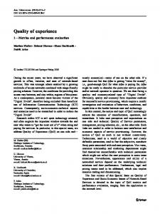
Fig. 1 T1-weighted image, after contrast enhancement. a Bilateral intrabulbar lesions. b Slightly enhanced lesion in a pineal region. c Densely enhanced tumor in the sellar/suprasellar localizations
reported in the literature (26 months) because there was no lag period between detection of the retinoblastoma and appearance of the brain tumor [1, 11, 12]. In other reports, bilateral RB and intracranial tumor were found at the same time in 13 of 93 children [12] and in two of eight children [1]. The median age of those patients was also 10 months. However, according to several studies, a lag period between the diagnosis of the ocular disease and the intracranial lesion is usually observed. Ocular lesions are generally detected earlier than the intracranial lesion. The mean time from diagnosis of retinoblastoma to detection of the associated primary intracranial tumor is about 23–24 months [1]. To date, in our retinoblastoma patient group, we have not observed metachronic intracranial tumor after RB treatment. In our two patients with TRB (cases 1 and 2), intrabulbar and intracranial lesions were found simultaneously in the first MR examinations. In one patient (case 3), a huge sellar and suprasellar tumor (PNET) was detected earlier than intrabulbar lesions, and TRB was diagnosed after detection of the midline tumor. A similar case was reported by Amoaku et al. [13]. The authors described a 4-month-old infant also with PNET in the suprasellar region treated as a primary intracranial tumor. In this case, calcified RB was found in both eyes 13 months after diagnosis of the intracranial lesion. In our case, as in the one mentioned above, the intracranial tumor was situated in the suprasellar region. This also supports previous reports of other authors that the diagnosis of primary midline intracranial tumor may predate detection of intraocular retinoblastoma and that the brain tumor is usually located in the suprasellar region in these cases [13]. No explanation is currently available for this phenomenon. We strongly recommend that a very careful ophthalmologic examination be performed in all children below 4 years of age with intracranial midline primary tumor.
All of our patients had bilateral RB associated with an intracranial mass. In previously published papers, 86–89% of children with TRB had bilateral RB and 11–12% children with TRB had unilateral RB [11, 12, 14]. Intracranial tumors associated with intraocular retinoblastoma usually occur in the pineal and sellar–suprasellar regions. The pineal region is the more common localization [1, 3]. Other localizations like primitive neuroectodermal tumor in the fourth ventricle are very rare but also possible [9]. In our cases, we found the brain lesion in the pineal region in one case and in the sellar and suprasellar regions in two cases. In all our cases, the size of the intracranial masses markedly diminished after chemotherapy, and in all patients, tumor size correlates well with clinical outcome to date. In case 1, the size of the pineal tumor was relatively small in the first examination. The mass measured 11×14 mm and disappeared in the next follow-up after chemotherapy. In case 2, the sellar and suprasellar tumor diminished from 19 to 7 mm after chemotherapy. In case 3, we observed a spectacular decrease of the huge sellar and suprasellar mass from 100×90 mm to 15×16 mm after chemotherapy. All lesions have remained stable to date, and there is no evidence of metastases. This correlates well with the good clinical status of the patients. In one report, the size of the intracranial masses also diminished to variable degrees after chemotherapy or radiation therapy, but correlated poorly with clinical outcome. In three of these patients, despite the tumor size decreasing markedly, leptomeningeal dissemination with a fatal outcome occurred [1]. In the other reports, the tumor size at the time of diagnosis was similar. The largest was from 30 mm [11] to 90 mm [12], but no information about changes in tumor size over time was gives. In our patients, we observed different patterns of the tumor signal. In case 1, the midline lesion was relatively
132
isointense compared with gray matter on T2-weighted images and homogenously enhanced after contrast administration. In cases 2 and 3, the brain tumors showed heterogeneous signal intensity on T2-weighted images and variable contrast enhancement. We found similar reports of tumor images in the literature [3, 10]. MR imaging has become a very useful diagnostic tool in evaluation of patients with retinoblastoma. Although ophthalmoscopy and US are of great value in the diagnosis of intraocular lesions, MRI is the method of choice in the evaluation of primary, intracranial tumors that can associate with retinoblastoma and for detection of leptomeningeal spread of the tumor. According to other studies, the mean survival of patients with TRB is about 9–13 months from diagnosis [1, 3, 12]. The short survival is caused mainly by cerebrospinal metastases, especially in patients with pineal tumors [10]. All of our patients are alive from 18 months (case 3) to 19 months (cases 2 and 3) from diagnosis, and to date, no leptomeningeal metastases have been found. This is longer than the mean survival rate in literature, even in case 3 with the PNET in the suprasellar region, for which the median survival rate is 6 months [11, 15]. It is well known that children with a positive family history are particularly predisposed to TRB syndrome and should be examined very carefully. The authors of many studies recommend neuroimaging screening in patients with familial bilateral RB until the age of 4 years to detect early asymptomatic pineal and suprasellar tumors because the prognosis for such patients is much better than that for symptomatic patients [11, 13, 16]. However, in our patients no evidence of familial retinoblastoma was found, so as in several other reports, we suggest that patients with sporadic bilateral RB should also undergo MRI follow-up because they may have a germline mutation [13]. TRB was not described until 1977. This may be explained by misdiagnosing, especially before the advent of CT scans, but some authors report that bilateral sporadic retinoblastomas may represent new germinal mutations in the RB gene [13]. This is in line with our observations because we found only three patients with TRB in the group of 202 patients with RB treated at our Institute from 1996 to 2008 and noticed that all three cases were detected in the last 2 years.
Conclusions MR imaging has become a very useful diagnostic tool in evaluation of patients with retinoblastoma.
Childs Nerv Syst (2010) 26:129–132
MR is the method of choice in detection of leptomeningeal spread of the tumor and evaluation of primary, intracranial tumors that can associate with retinoblastoma. Accordingly, we strongly recommend performing very careful ophthalmologic and imaging examinations of the orbits in all children below 4 years of age in whom an intracranial midline primary tumor has been diagnosed earlier than intraocular lesions.
References 1. Provenzale JM, Gururangan S, Klintworth G (2004) Trilateral retinoblastoma: clinical and radiologic progression. Am J Roentgenol 183(2):505–511 2. Friend SH, Bernards R, Rogelj S, Weinberg RA, Rapaport JM, Albert DM, Dryja TP (1986) A human DNA segment with properties of the gene that predisposes to retinoblastoma and osteosarcoma. Nature 323:643–646 3. Cho EY, Suh Y-L, Shin H-J (2002) Trilateral retinoblastoma: a case report. J Korean Med Sci 17:137–140 4. Jakobiec FA, Tso MO, Zimmerman LE, Danis P (1977) Retinoblastoma and intracranial malignancy. Cancer 39(5):2048– 2058 5. Bader JL, Miller RW, Meadows AT, Zimmerman LE, Champion LA, Voute PA (1980) Trilateral retinoblastoma. Lancet 2:582–583 6. Bagley LJ, Hurst RW, Zimmerman RA, Shields JA, Shields CL, De Potter P (1996) Imaging in the trilateral retinoblastoma syndrome. Neuroradiology 38:166–170 7. Katayama Y, Tsubokawa T, Yamamoto T, Nemoto N (1991) Ectopic retinoblastoma within the 3rd ventricle: case report. Neurosurgery 28:158–161 8. Bejjani GK, Donahue DJ, Selby D, Cohen PH, Packer R (1996) Association of a suprasellar mass and intraocular retinoblastoma: a variant of pineal trilateral retinoblastoma? Pediatr Neurosurg 25:269–275 9. Finelli DA, Shurin SB, Bardenstein DS (1995) Trilateral retinoblastoma: two variations. AJNR Am J Neuroradiol 16:166–170 10. Provenzale JM, Weber A, Klintworth GK, McLendon RE (1995) Radiologic-pathologic correlation. Bilateral retinoblastoma with coexistent pinealoblastoma (trilateral retinoblastoma). AJNR 16:157–165 11. Mouratova T (2005) Trilateral retinoblastoma: a literature review, 1971–2004. Bull Soc Belge Ophtalmol 297:25–35 12. Kivelä T (1999) Trilateral retinoblastoma: a meta-analysis of hereditary retinoblastoma associated with primary ectopic intracranial retinoblastoma. J Clin Oncol 17(6):1829–1837 13. Amoaku WMK, Willshaw HE, Parkes SE, Shah KJ, Mann JR (1996) Trilateral retinoblastoma: a report of five patients. Cancer 78:858–863 14. Ibarra MS, O'Brien JM (2000) Is screening for primitive neuroectodermal tumours in patients with unilateral retinoblastoma necessary? JAAPOS 4:54–56 15. Paulino AC (1999) Trilateral retinoblastoma: is the location of the intracranial tumour important? Cancer 86:135–141 16. De Potter P, Shields CL, Shields JA (1994) Clinical variations of trilateral retinoblastoma: a report of 13 cases. J Pediatr Ophthalmol Strabismus 31:26–31