CASE REPORT – OPEN ACCESS International Journal of Surgery Case Reports 33 (2017) 97–98
Contents lists available at ScienceDirect
International Journal of Surgery Case Reports journal homepage: www.casereports.com
Trochanteric hip fracture during cardioversion therapy. A case report J. Gómez ∗ , J. Albareda, L. Ezquerra Department of Orthopaedic Surgery, Lozano Blesa Clinical Hospital, Zaragoza, Spain
a r t i c l e
i n f o
Article history: Received 15 February 2017 Received in revised form 23 February 2017 Accepted 23 February 2017 Available online 27 February 2017 Keywords: Hip fracture Elderly Cardioversion therapy Case report
a b s t r a c t INTRODUCTION: Trochanteric hip fractures in elderly patients with osteoporosis are commonly caused by low energy trauma. The cardioversion therapy is an extremely rare cause of this type of fracture. PRESENTATION OF CASE: We report the case of a woman with hip fracture after cardioversion. DISCUSSION: We discuss the production mechanism of this injury and the importance of the care of the osteoporotic bone under these therapies. CONCLUSION: The propofol sedation should be complemented with skeletal muscle relaxants in the cardioversion therapy to avoid hip fracture in select patients with osteoporosis. © 2017 The Authors. Published by Elsevier Ltd on behalf of IJS Publishing Group Ltd. This is an open access article under the CC BY-NC-ND license (http://creativecommons.org/licenses/by-nc-nd/4.0/).
1. Introduction Electrical external cardioversion is very secure treatment for the atrial fibrilation. Fractures as a complication in this type of treatment are rare, and usually accompanied by some previously predisposing factor. We report the case of a fracture after the application of this therapy in a patient with osteoporosis. The work has been reported in line with the SCARE criteria [6].
2. Presentation of case An interconsultation from the cardiology service was received in our department about a 74-year-old woman with right hip pain, who was unable to bear weight and stand on her right leg; there was no previous history of trauma. The patient had a medical history of low energy fracture of femoral right neck 14 years before, treated by means of closed reduction and internal fixation with screws, a myocardial ischemia with low ejection fraction and atrial fibrillation was diagnosed in December 2013. The patient had to undergo an electrical cardioversion in order to pass to sinus rhythm in 2014. Previously she followed an anticoagulation protocol for 4 weeks. The patient underwent electrical external cardioversion, thoracic, anterolateral and synchronized with a two-phase crash of 150 J, with deep sedation with 50 propofol mg IV. The patient then moved to sinus rhythm. Physical examination showed that all movements of the hip were limited and very painful after the electric therapy. She was not able to walk and the functional impotence was total. On
∗ Correspondence to: Joaquina Zamora 4, 4◦ B, 50018 Zaragoza, Spain. E-mail addresses:
[email protected] (J. Gómez),
[email protected] (J. Albareda),
[email protected] (L. Ezquerra).
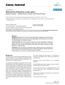
Fig. 1. Trochanteric fracture in right hip.
the same thigh a great hematoma was observed, painful to pressure. The extremity was neither rotated nor shortened. A radiological study of the hip was requested where a trochanteric fracture was observed through the points of entrance of the screws (Fig. 1). An ultrasound of the thigh was also carried
http://dx.doi.org/10.1016/j.ijscr.2017.02.047 2210-2612/© 2017 The Authors. Published by Elsevier Ltd on behalf of IJS Publishing Group Ltd. This is an open access article under the CC BY-NC-ND license (http:// creativecommons.org/licenses/by-nc-nd/4.0/).
CASE REPORT – OPEN ACCESS 98
J. Gómez et al. / International Journal of Surgery Case Reports 33 (2017) 97–98
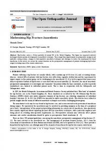
Fig. 2. TC.
In our case, the osteoporotic hip is subject to great vectors of force caused by isometric contractions and is fractured on the weakest part, the entrance point of the previous screws, like in the only case found in the literature, the patient had a stress fracture in that femur nine years before [4]. In these cases the trochanter consumes great part of the traumatic energy and it does not produce damages in more common sites like the neck or the acetabulum. 4. Conclusion We propose that the propofol sedation should be complemented with skeletal muscle relaxants to avoid these injuries in patients with osteoporosis and risk factors like these before the cardioversion therapy. The hip fracture in osteoporotic bones should be considered after muscle violent contraction that causes hip pain and functional impotence. Conflict of interest The authors declare that there is no conflict of interests regarding the publication of this paper. Funding None. Ethical approval The study was approved by the Deontological Committee of Lozano Blesa Clinical Hospital, Zaragoza. Consent
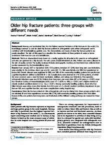
Fig. 3. Nowadays.
out where a partial break of the quadriceps and a hematoma of 16 × 7 mm was appreciated. In order to take a better therapeutic decision, a TC was carried out where great comminution of the major trochanter and perilesional hematoma were appreciated, giving acute entity to the injury (Fig. 2). Due to the minimum movement of the fracture and to the patient status it decided a conservative treatment. After two months the patient begins walking with two crutches leaning her weight partially. She is currently asymptomatic and she walks with a cane without problems (Fig. 3). 3. Discussion This type of hip fracture is produced by an uncontrolled violent, simultaneous contraction of pelvic and trochanteric muscles. It occurs during convulsion from an electric cardioversion [1]. Femoral neck fracture associated with osteoporosis [2] and central dislocation of the hip after electro-convulsive therapy [3] have been reported in the literature, and only in one case after cardioversion therapy with 200 J [4]. A few articles report sternal fractures, but this complication occurred in the absence of conditions that predispose to fractures, e.g., osteoporosis [5].
Patient consent has been obtained to publish this case report. Authors’ contribution All authors contributed significantly and in agreement with the content of the manuscript. All authors participated in data Collection and in writing of the manuscript. Guarantor J. Gómez. References [1] E. Rath, O. Levy, N. Liberman, D. Atar, Bilateral dislocation of the hip during convulsions. A case report, J. Bone Jt. Surg. 79B (1997) 304–306. [2] P.T. Remec, C.M. Evarts, Bilateral central dislocation of the hip: a case report, Clin. Orthop. 181 (1983) 118–120. [3] M.R. Nott, J.S. Watts, A fractured hip during electro-convulsive therapy, Eur. J. Anaesthesiol. 16 (1999) 265–267. [4] S. de Ridder, C. Timmermans, H.J. Wellens, An uncommon cause for hip fracture, Europace 9 (2007) 957–958. [5] D. Vollmann, L. Lüthje, J. Seegers, C. Sohns, M. Dorenkamp, A. Vafa, M. Zabel, Sternal fracture after elective electrical cardioversion of atrial fibrillation, Clin. Res. Cardiol. 100 (2011) 261–262. [6] R.A. Agha, A.J. Fowler, A. Saetta, I. Barai, S. Rajmohan, D.P. Orgill, for the SCARE Group, The SCARE statement: consensus-based surgical case report guidelines, Int. J. Surg. 34 (2016) 180–186.
Open Access This article is published Open Access at sciencedirect.com. It is distributed under the IJSCR Supplemental terms and conditions, which permits unrestricted non commercial use, distribution, and reproduction in any medium, provided the original authors and source are credited.