Clinical Chemistry 58:6 1040–1048 (2012)
Proteomics and Protein Markers
Troponin-Specific Autoantibody Interference in Different Cardiac Troponin I Assay Configurations Tanja Savukoski,1* Emilia Engström,1* Janne Engblom,2 Noora Ristiniemi,1 Saara Wittfooth,1 Bertil Lindahl,3 Kai M. Eggers,3 Per Venge,3 and Kim Pettersson1
BACKGROUND: Autoantibodies to cardiac troponins (cTnAAb) can interfere with the measurement of cardiac troponin I (cTnI) by immunoassays. The aim of this study was to explore the degree of cTnAAb interference in different cTnI assay configurations.
Ternary troponin complex was added into samples (serum or plasma, n ⫽ 132, 68% cTnAAb positive) from individuals without known cardiac conditions. The recovery of cTnI was then measured with 6 investigational cTnI assays (2, 3, or 4 antibodies per assay). Three of these assays were then selected for further comparison by use of samples (plasma, n ⫽ 210, 33% cTnAAb positive) from non–ST-elevation acute coronary syndrome patients in the FRISC-II (FRagmin/Fast Revascularisation during InStability in Coronary artery disease) cohort. Finally, these results were compared to those obtained with 3 commercial cTnI assays.
CONCLUSIONS: A novel cTnI assay deviating from the conventional IFCC-recommended midfragment approach substantially improves cTnI detection in samples containing cTnAAbs.
© 2012 American Association for Clinical Chemistry
METHODS:
RESULTS:
Analytical recoveries varied widely among the 6 investigational assays. Notably the low recoveries (median 9%) of the midfragment-targeting reference assay were normalized (median 103%) with the use of the 4-antibody assay construct (3 capture, 1 tracer antibody) with only 1 antibody against a midfragment epitope. Reduced analytical recoveries correlated closely with measured autoantibody amounts. cTnI concentrations from cTnAAb-positive patient samples determined with 3 investigational assays confirmed the reduced concentrations expected from the low analytical recoveries. The results from the commercial cTnI assays with antibody selections representative for contemporary assay constructs revealed a similar underestimation (up to 20-fold) of cTnI in cTnAAb-positive samples.
1
Department of Biotechnology, University of Turku, Turku, Finland; 2 Turku School of Economics, Turku, Finland; 3 Department of Medical Sciences, Clinical Chemistry, University of Uppsala, Uppsala, Sweden. * Address correspondence to: T.S. at Department of Biotechnology, University of Turku, Tykistökatu 6A 6th floor, FI-20520, Turku, Finland. Fax ⫹358-2-3338050; e-mail
[email protected]. E.E. at Department of Biotechnology, University of Turku, Tykistökatu 6A 6th floor, FI-20520, Turku, Finland. Fax ⫹358-2-333-8050; e-mail
[email protected]. Results from the analytical recovery studies have been orally presented by Kim Pettersson at the XXXI Nordic Congress in Clinical Chemistry, June 2008, Helsinki,
1040
The troponin complex, consisting of subunits I, T and C, is part of the myofibril contractile apparatus in muscle cells. Of these, troponin I and troponin T exist as different isoforms in skeletal and cardiac muscle, which has enabled the use of the cardiac-specific isoforms of troponin (cardiac troponin) as biomarkers for myocardial injury. Because of excellent cardiac specificity, the determination of circulating cardiac troponins along with patient symptoms and electrocardiographic abnormalities currently constitutes a cornerstone in the diagnosis of acute myocardial infarction (1 ). Because of the central role of cardiac troponins in clinical assessment, it is important that the assays for cardiac troponin I (cTnI)4 measurement are highly reliable and consistent. The molecular heterogeneity of the cTnI molecule causes challenges in the selection of antibodies for cTnI assays. For instance, the cTnI molecule undergoes different posttranslational modifications, and various cardiac troponin complexes are found in the circulation (2 ). Because both N- and C-terminal parts of cTnI are sensitive to proteolytic degradation, the IFCC recommends the use of antibodies that recognize the midfragment epitopes (amino acids 30 –110) for the development of new cTnI assays (3 ). It has recently been demonstrated that cardiac troponin–specific autoantibodies (cTnAAb) are found in 5%–20% of individuals with or without cardiac dis-
Finland. Received November 13, 2011; accepted March 12, 2012. Previously published online at DOI: 10.1373/clinchem.2011.179226 4 Nonstandard abbreviations: cTnI, cardiac troponin I; cTnAAb, cardiac troponin– specific autoantibody; ACS, acute coronary syndrome; ITC, cardiac troponin complex; Mab, monoclonal antibody; Fab, antigen binding fragment; SA, streptavidin; TSA, Tris-buffered saline with azide; ILII, insulation layer II; NR, normal recovery; LR, low recovery; MR, medium recovery; FRISC-II, FRagmin/ Fast Revascularisation during InStability in Coronary artery disease.
Autoantibody Interference in Troponin I Assays
eases (4 – 8 ). According to a previous report (9 ), circulating cTnI-specific cTnAAbs are most commonly targeted against the stable midfragment of the cTnI molecule, and especially to the C-terminal region of the midfragment. Although the overall and practical impacts are not well established, cTnAAbs remain a potential confounder in cardiac troponin assays. In following the IFCC recommendation most cTnI-assay manufacturers presently use antibodies binding to the midfragment of the cTnI molecule (10, 11 ). We hypothesize that such cTnI assays are likely to suffer from cTnAAb interference and that this interference can be counteracted by choosing the antibodies differently. Therefore we constructed investigational cTnI assays using different epitopes across the cTnI molecule. Plasma/serum samples with low, medium, or normal cTnI recovery were identified with a midfragment-targeting cTnI assay and then analyzed with other assays to monitor the extent of cTnAAb interference. Finally, we compared the performance of 3 investigational cTnI assays to the performance of 3 commercial assays that employ antibodies highly representative for contemporary cTnI assays, with samples from non–ST-elevation acute coronary syndrome (ACS) patients. Materials and Methods The study protocols were approved by the local ethics committee. Informed consent was obtained from all participants and the study was conducted in accordance with the Declaration of Helsinki of 1975 as revised in 2006. REAGENTS
Human cardiac troponin complex (ITC) and all cTnIspecific monoclonal antibodies (Mab) except 8I7 [International Point of Care (www.ipocdx.com)] were kindly provided by HyTest Ltd. [Turku, Finland (www.hytest.fi)]. Recombinant 4C2 antigen-binding fragment (Fab) of Mab 4C2 was cloned from the hybridoma cell line of HyTest Ltd. and produced at the University of Turku as reported previously (12 ). Similarly, 9707 Fab was cloned from the hybridoma cell line of Medix Biochemica (www.medixbiochemica. com) (University of Turku, unpublished data). Normal capacity streptavidin (SA) plates were purchased from Kaivogen Oy (www.kaivogen.com). Highcapacity SH (sulphydryl)–SA plates were produced as described earlier (13 ). Calibrators were prepared by diluting ITC into Tris-buffered saline with azide (TSA) (50 mmol/L Tris-HCl, pH 7.75; 150 mmol/L NaCl; and 0.5 g/L NaN3) containing 75 g/L BSA [Sigma (www. sigmaaldrich.com)]. The cTnI concentration of the
ITC stock reported by the manufacturer was used to assign the cTnI concentrations of the dilutions. LABELING OF MABS AND FAB FRAGMENTS WITH BIOTIN AND LANTHANIDE CHELATE
Most Mabs were labeled with biotin or intrinsically fluorescent europium (Eu) chelate as described previously (4 ). Sugarbiotinylation of 19C7 Mab was achieved by conjugating biotin-caproylhydrazine (Sigma) covalently to aldehyde moieties produced on its sugar residues. The hydroxyl moieties were first oxidized with 10 mmol/L sodium periodate (Sigma) in 10 mmol/L phosphate buffer, pH 7.4. After a 60-min incubation, Mab was purified with an NAP-5 desalting column [GE Healthcare Life Sciences/Amersham Biosciences AB (www.gehealthcare.com)] with 5 mmol/L phosphate buffer, pH 7.4, as an eluent. Biotinylation was performed with a 200-fold molar excess of biotincaproylhydrazine by incubating the reaction for 2 h and by removing free biotin with an NAP-10 column and TSA. Unreacted aldehydes were removed with 33 mmol/L ethanolamine [Avantor Performance Materials (www.avantormaterials.com)]. After a 30-min incubation, biotinylated 19C7 was purified with a PD-10 column (GE Healthcare Life Sciences) by using TSA for elution. All steps were performed protected from light at room temperature. Fab fragments were site-specifically biotinylated with 95 mol/L maleimide–PEO2– biotin [Thermo Fisher Scientific (www.thermofisher.com)] during protein purification performed essentially as described previously (14 ), with 4 mL of running buffer (50 mmol/L phosphate, pH 7.5; 7.5 mmol/L imidazole; 80 g/L glycerol) containing maleimide–PEO2– biotin circulated through an immobilized metal affinity chromatography column for 1.5 h at room temperature. After the column was washed, biotinylated Fabs were released with elution buffer (50 mmol/L phosphate, pH 7; 300 mmol/L imidazole; 80 g/L glycerol) and transferred to TSA. Biotinylated and Eu(III)-labeled Mabs and Fabs were stabilized with BSA (1g/L) and stored at 4 °C. IMMUNOASSAYS FOR cTnI
Biotinylated capture antibodies (12.5–200 ng) were first immobilized to SA- or SH–SA-coated microtiter wells (cTnI assays 1–5 or 6, respectively) in 25 L of Kaivogen buffer solution (Kaivogen Oy) and incubated for 60 min at room temperature. After washings, 20 L of calibrator or serum and 100 ng of the Eu-labeled tracer antibody in 20-L Insulation layer II (ILII) [Radiometer/Innotrac Diagnostics (www.innotrac.fi)] were added into triplicate wells. The wells were incubated for 30 min at 36 °C, 900 rpm in a plate shaker [iEMS incubator/shaker, Thermo Electron CorporaClinical Chemistry 58:6 (2012) 1041
tion/Labsystems, (www.thermoscientific.com)]. The washed wells were dried and the time-resolved fluorescence was measured directly from the surface with a Victor ⫻4 Multilabel Counter [Perkin-Elmer/Wallac, Turku, Finland (www.perkinelmer.com)]. We determined analytical recovery for cTnI assays 1– 6 by measuring the fluorescence signal from each sample with and without ITC addition (30 g/L) and by comparing the ITC-specific signal to the signal of similarly spiked BSA–TSA (100%). Finally, the results were normalized with the median of the normal recovery (NR) group set to 100%. Full analytical performance was determined only for cTnI assays 1, 3, and 6 because these assays were further studied with clinical samples. ITC calibrators (analyzed in duplicate for 8 days) with a specific signal higher than the analytical sensitivity (3 SD of blank) of that assay were used in a calibration curve [nonlinear allometric 1 fitting, Origin 8, OriginLab Corboration (www.originlab.com)]. The obtained equations were used to calculate the cTnI concentrations of individual non–ST-elevation ACS samples. COMPARISON OF CAPTURE EFFICIENCIES OF cTnI ASSAYS 1 AND 6
The difference in the analytical sensitivities of cTnI assays 1 and 6 was further studied by comparing the capture efficiencies of these assays for standards and cTnIcontaining samples. We added 20 L calibrator or plasma and 20 L ILII on the capture surfaces of assay 1 and assay 6, respectively. After a 30-min incubation (36 °C, 900 rpm), the solution with unbound calibrator/endogenous cTnI was transferred to a second set of assay 6 and assay 1 capture surfaces, respectively, and incubated for another 30 min. Bound cTnI was detected from both surfaces by incubating tracer antibodies in 40 L ILII for 30 min before the signal measurements. IMMUNOASSAY FOR HUMAN cTnAAbS
The cTnAAb status of the samples was determined with the previously published cTnAAb assay (6, 15 ). A sample with a positive net signal (Student’s t-test, P ⬍ 0.05) above the background control was considered cTnAAb positive. SAMPLES
To study cTnAAb interference with analytical recovery tests, we used heparin plasma samples collected from patients (n ⫽ 1100) admitted to S:t Görans Hospital [Stockholm, Sweden (www.capio.com)] for noncardiac conditions and 5 apparently healthy volunteers at our department in 2007–2008. The analytical recoveries were first determined with the midfragment targeting cTnI assay 1. Two study groups with lowered ana1042 Clinical Chemistry 58:6 (2012)
lytical recoveries were selected: low recovery (LR) (⬍10%, n ⫽ 49) and medium recover (MR) (10%– 20%, n ⫽ 17). The NR group (n ⫽ 66) was randomly chosen from the remaining samples. The analytical recoveries of these samples were then analyzed with cTnI assays 2– 6. The performance of the investigational cTnI assays was studied with EDTA plasma samples collected in 1996 –1998 from non–ST-elevation ACS patients for the prospective Scandinavian multicenter trial, FRagmin/Fast Revascularisation during InStability in Coronary artery disease (FRISC-II) (16 ). Endogenous cTnI was analyzed with cTnI assays 1, 3, and 6 from 70 cTnAAb-positive and 140 cTnAAb-negative admission samples randomly chosen from 857 patients who participated in the previously reported FRISC-II substudy (17 ). Investigational assays 1 and 3 were selected because they represented antibody configurations of contemporary commercial assays, whereas cTnI assay 6 was selected because of its apparent lack of dependence on the cTnAAb status of samples. Previously determined cTnI concentrations were available from 3 commercial assays of midfragment type design, the firstgeneration AxSYM assay (Abbott Diagnostics), the second-generation AccuTnI assay (Beckman Coulter), and the Liaison cTnI assay (Byk-Sangtec Diagnostica) (18, 19 ). According to manufacturers, the analytical sensitivities were 0.3, 0.01, and 0.005 g/L for the AxSYM cTnI, AccuTnI, and Liaison cTnI, respectively. For capture efficiency comparisons, we used 4 cTnI-containing heparin plasma samples from patients admitted to Central Ostrobothnia Central Hospital (Kokkola, Finland). STATISTICAL ANALYSIS
In all statistical tests, P values ⬍0.05 were considered statistically significant. We analyzed differences in analytical recoveries between study groups with Stat View 5.0 [SAS Institute (www.sas.com)] using Kruskall– Wallis and Mann–Whitney U-tests. Statistical analyses of endogenous cTnI were performed using statistical software SAS 9.2 (SAS Institute). Patients with a cTnI concentration lower than the analytical sensitivity of the assay in question were excluded, and final analyses were completed with 108 cTnAAb-negative and 53 cTnAAb-positive samples. Normalization equations were constructed from the original cTnI concentrations of the cTnAAb-negative cohort by regression analyses. Because of the heteroscedasticity of residuals, cTnI values from the range with lower variance were weighed. Dependence between cTnI assays 3 and 6 was linear, and consequently a simple linear regression model was used. Dependence between assays 3 and 1 was nonlinear. Therefore a quadratic term and a logarithmic transformation were needed for constructing a
Autoantibody Interference in Troponin I Assays
Fig. 1. Schematic representation of the study design.
linear model used for the normalization of cTnI assay 1 values. We compared the original cTnI assay 3 values and the normalized values with cTnI assays 1 and 6 using repeated measures ANOVA and Tukey–Kramer adjustment. Results A schematic representation of the study design is presented in Fig. 1. To evaluate the effect of cTnAAbs on different cTnI assay configurations, ITC recovery was measured with the 6 investigational cTnI assays (Fig. 2). Fig. 3A shows that the analytical interference of cTnAAbs decreased when alternative epitope combinations were used. Compared to the median of the NR group (100%), the medians of assays 1– 6 were 9%, 26%, 32%, 53%, 58%, and 103% in the LR group and 35%, 76%, 78%, 83%, 65%, and 102% in the MR group, respectively. Differences between study groups remained highly significant (P ⬍ 0.0001) in assays 2–5 but not in assay 6 (P ⫽ 0.1649). As shown in Fig. 3B, all LR and MR and 24 NR samples were cTnAAb positive, with the cTnAAb signals of each study group differing significantly (P ⬍ 0.0001) from each other. Whereas median signals in the LR and MR groups were 13 301 and 2690 counts, respectively, half of the cTnAAbpositive samples in the NR group had cTnAAb signals ⱕ100 counts. The analytical sensitivities of cTnI assays 1, 3, and 6, which were chosen for further studies with cardiac patients, were 0.30, 0.06, and 0.02 g/L, respectively. Fig. 4, A and B show the scatter plots of the cTnI concentrations of cTnAAb-positive and -negative ACS patients measured with cTnI assays 1 and 6 with assay 3 as a reference. Assays 1 and 3 performed similarly,
whereas with assay 6 the majority of cTnAAb-positive samples stand out from the scatter because of higher cTnI results with cTnI assay 6 than with assay 3. The mean concentrations of cTnI assays 1, 3, and 6 were 2.77, 2.41, and 1.13 g/L in the cTnAAb-negative cohort, and 2.21, 2.49, and 4.15 g/L in the cTnAAbpositive cohort, respectively. Similar scatter plots were obtained when the investigational assays were compared to 3 commercial assays. Fig. 4, C and D show that comparison to the second-generation AccuTnI assay presents a scatter profile in relation to cTnI assay 6 that is highly similar to that of investigational assay 3 (Fig. 4B). There were significant differences in measured cTnI concentrations in the cTnAAb-negative cohort. Assay 1 gave on average 36% higher and assay 6 86% lower concentrations than cTnI assay 3 (Bland– Altman). When the measured cTnI concentrations of cTnAAb-negative samples of assays 1 and 6 were normalized against cTnI assay 3 (see the linear regression curves in Supplemental Figs. 1 and 2 in the Data Supplement that accompanies the online version of this article at http://www.clinchem.org/content/vol58/ issue6), the cTnI concentrations (mean) in the cTnAAb-positive cohort were not significantly different (P ⫽ 0.8781) between cTnI assays 1 (1.82 g/L) and 3 (2.49 g/L), but the concentration with assay 6 (9.19 g/L) was 5.0- and 3.7-fold higher (P ⬍ 0.0001) than in the other 2 assays. Although no significant correlation (P ⬎ 0.05) was found between cTnI concentration ratios [c(cTnI assay 6)/c(cTnI assay 1 or 3)] and cTnAAb signals owing to an extensive scatter, it is apparent that the higher ratios are found in samples with higher cTnAAb signals (see online Supplemental Fig. 3). Thus Clinical Chemistry 58:6 (2012) 1043
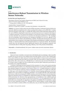
Fig. 2. Epitope map of antibodies used in the 3 commercial and the 6 investigational cTnI assays. The bar next to the assay name represents the linear amino acid sequence of cTnI. The antibody epitopes are marked with shorter lines below the cTnI sequence with respective amino acid locations and Mab codes. Pab, polyclonal antibody; *, Tracer antibody; #, sugar biotinylated.
cTnI assay 6 tended to give higher cTnI results than assays 1 or 3 in samples with higher cTnAAb signals. Because of the clear signal differences seen, especially with cTnI assays 1 and 6, we compared the relative capture efficiencies of assays 1 (19C7) and 6 (4C2, 19C7, 9707). After preincubation on the cTnI assay 6 capture surface, the assay 1 configuration detected only 4%–5% of the ITC calibrator while in the reverse situation, cTnI assay 6 configuration detected almost 40% of the ITC (see online Supplemental Table 1). Similarly, for endogenous cTnI-containing patient samples, assays 1 and 6 detected 16%–31% and 48%– 83% of the cTnI, respectively. Discussion The aim of this study was to explore the extent of cTnAAb interference in cTnI assay configurations by use of different epitopes and to identify antibody combinations that would be minimally affected by this analytical interference. Our results show that several antibody configurations deviating from the conventional midfragment approach improve the cTnI detection in cTnAAb-positive samples. The autoantibody interferences demonstrated by our investiga1044 Clinical Chemistry 58:6 (2012)
tional assays are equally seen with the 3 commercial cTnI kits studied. The analytical recovery tests performed with 6 investigational cTnI assay designs demonstrate that the epitope selection profoundly affects the degree of negative cTnAAb interference, which confirms and extends the conclusions from previous reports (9, 20 ). Thus, selecting a novel 3 ⫹ 1–type antibody configuration (assay 6) with the detector antibody recognizing the N-terminal part of the C-terminal region and 3 capture antibodies for epitopes on the N-terminus, the midfragment, and the C-terminus decisively reduces the cTnAAb interference. Because the standard material used does not truly represent the endogenous cTnI found in circulation, 3 assay configurations (1, 3, and 6) were selected for more detailed characterization with clinical samples obtained from non–ST-elevation ACS patients. Although assay 6 was selected owing to low cTnAAb interference, assays 1 and 3 were selected to exemplify first-generation cTnI assays and more contemporary assays, respectively. Although cTnI assays 1, 3, and 6 were calibrated against the same tissue-derived ITC preparation when the cTnI concentrations of the ACS patients were mea-
Autoantibody Interference in Troponin I Assays
Fig. 3. Analytical recoveries (A) and cTnAAb signals (B) of plasma and serum samples from individuals (n ⴝ 132) without known cardiac conditions. All cTnAAb-negative samples have been given a cTnAAb signal of 1.
sured, assay 6 gave lower cTnI concentrations in the cTnAAb-negative group but higher concentrations in the cTnAAb-positive group compared to assays 1 and 3. This was because the ITC calibrator is poorly recognized by assay 1 relative to assay 6, whereas the differences with patient samples were much lower. To correct the obtained results for this calibrator artifact and enable a comparison of the effect of cTnAAbs, the data were normalized against the cTnI concentrations obtained by cTnI assay 3 for the cTnAAb-negative cohort.
In this manner, the difference between assays 1 and 3, both targeting the midfragment epitopes, was not statistically significant in detecting cTnI in the cTnAAbpositive group. In contrast, cTnI assay 6 provided significantly higher recognition of cTnI in this group. Our results with the investigational assays were also compared to results from 3 commercial cTnI assays, which have been extensively documented both analytically and clinically (18, 19, 21–24 ). The cTnAAb-associated interference is as obvious and sigClinical Chemistry 58:6 (2012) 1045
Fig. 4. Original cTnI concentrations measured with cTnI assays 1 (A) and 6 (B) from cTnAAb-negative (f) and cTnAAb-positive (䡺) non–ST-elevation ACS patient samples with cTnI assay 3 used as a reference, and similarly with cTnI assays 3 (C) and 6 (D) with the commercial AccuTnI assay used as a reference.
nificant in these assays as in the investigational assays 1 and 3 when compared to the new assay 6. In essence this means that significant cTnI concentration underestimations are seen in individual cTnAAb-positive samples in relation to the new assay (Fig. 4D). Tang et al. (25 ) recently reported serious underestimations with 5 commercial cTnI assays in patients having cTnIspecific cTnAAbs, even to the extent that these patients were falsely designated as cTnI negative in relation to the recommended cutoffs. In the light of our results, the possibility for serious underestimation of circulat1046 Clinical Chemistry 58:6 (2012)
ing cTnI amounts remains a valid and reasonable notion. In the present study, all individual samples did not behave similarly. The change of antibody configuration had a stronger effect on the cTnAAb interference of some samples than others. This finding suggests that cTnI-specific cTnAAbs are conceivably even more heterogenous than initially reported (9 ). In addition to the measured autoantibody titers, intraindividual variation in cTnAAb epitope specificities probably affects the degree of interference. In addition, the fine speci-
Autoantibody Interference in Troponin I Assays
ficity of circulating cTnAAbs calls for more precise characterization in the light of a recently reported study (26 ) in which antibodies targeting different cTnI epitopes were associated with different pathological effects in mice. Although a number of manufacturers currently provide immunoassays for cTnI measurement, the results of these assays are not always interchangeable because of the differences in antibody reactivity, calibration materials, and assay formats, and because of the pronounced heterogeneity of the cTnI molecule in the bloodstream. The use of a common calibrator has been found to reduce the bias in measured cTnI values but has not been able to provide acceptable cTnI assay standardization (27, 28 ). Similarly, in our study, despite the use of a common calibrator, the nonnormalized cTnI concentrations deviated significantly in the cTnAAb-negative patient cohort. Although the analytical sensitivities may explain part of this variation, the differences in the epitope specificities, affinities, and formats of chosen antibodies probably have a more significant effect. This is supported by the results of the experiments in which the binding of ITC or endogenous cTnI was studied by transferring them from one capture surface to another before signal measurement. The ITC standard was efficiently bound by the assay 6 capture surface, whereas a remarkable proportion of ITC remained free after preincubation on the assay 1 capture surface. Thus these surfaces recognize the calibrator in a highly variable manner. The results also indicate that the standard material does not truly reflect the cTnI forms in samples of cardiac patients because the difference in signals between the assays for endogenous cTnI was much smaller than for the calibrator. This observation is yet another illustration of the well-known complexity of cTnI standardization. With regard to the different cTnI forms found in the circulation, one limitation of this study may have been the use of plasma samples that had been stored frozen for a substantial length of time and thawed a few times before analysis. Therefore, changes in the cTnI molecular structure affecting the antibody recognition may have occurred. Because our cTnI assay 6 does not recognize the midfragment of cTnI, samples with extensive cTnI fragmentation would not be fully detected. However, as reported recently (20 ), only minor losses of immunoreactivity were seen with clinical samples incubated for 24 h at room temperature using a 2 ⫹ 1–type assay (Radiometer) (11, 29 ) unable to generate signal from the stable midfragment. It is conceivable that the immunoreactivity of archival samples may have altered during storage. However, samples for a critical care marker such as cTnI are not to be stored for extended periods. This study is also limited to a com-
parison of measured cTnI concentrations in the ranges considered analytically reliable by all the included assays. This means that very low cTnI concentrations could not be investigated in this study, although highly sensitive cardiac troponin assays have recently been studied extensively for their clinical use in the triage of ACS and as long-term risk markers (30 ). Because the risk for clinical misclassification due to cTnAAb interference may be even more pronounced at lower cTnI concentrations, a highly sensitive assay suitable for routine clinical use of the novel assay 6 design is presently under development to enable comparison of its clinical utility in relation to contemporary or ultrasensitive commercial cardiac troponin assays. In conclusion, this study establishes the significant interference of circulating cTnAAbs in representative cTnI assays used in clinical practice. The novel assay 6 design of this study circumvents this particular analytical interference and challenges the official recommendation for cTnI assay development. In light of the high frequency of autoantibody-positive individuals also reported elsewhere and the dramatic inhibitory effect they may exert, we strongly feel that future recommendations on cTnI assay designs will have to acknowledge these facts.
Author Contributions: All authors confirmed they have contributed to the intellectual content of this paper and have met the following 3 requirements: (a) significant contributions to the conception and design, acquisition of data, or analysis and interpretation of data; (b) drafting or revising the article for intellectual content; and (c) final approval of the published article. Authors’ Disclosures or Potential Conflicts of Interest: Upon manuscript submission, all authors completed the author disclosure form. Disclosures and/or potential conflicts of interest: Employment or Leadership: None declared. Consultant or Advisory Role: B. Lindahl, Beckman Coulter, Siemens, Radiometer, Philips, bioMeriux, and Roche Diagnostics; K.M. Eggers, Abbott Laboratories. Stock Ownership: None declared. Honoraria: None declared. Research Funding: T. Savukoski, DIA-NET, the Graduate School of Advanced Diagnostic Technologies and Applications. Expert Testimony: None declared. Other: K. Pettersson, patent US2008153109/EP1473567 for cTnI assays. Role of Sponsor: The funding organizations played no role in the design of study, choice of enrolled patients, review and interpretation of data, or preparation or approval of manuscript. Acknowledgments: We gratefully acknowledge Pirjo Laaksonen, Heini Saarima¨ki, and Nina Elonen (Department of Biotechnology, University of Turku) for technical assistance, and Tom Pettersson (Capio AB) and Pa¨ivi Laitinen (Central Ostrobothnia Central Hospital) for providing heparin plasma samples.
Clinical Chemistry 58:6 (2012) 1047
References 1. Thygesen K, Alpert JS, White HD, Joint ESC/ACCF/ AHA/WHF Task Force for the Redefinition of Myocardial Infarction. Universal definition of myocardial infarction. Eur Heart J 2007;28:2525–38. 2. Katrukha A, Bereznikova A, Filatov V, Esakova T. Biochemical factors influencing measurement of cardiac troponin I in serum. Clin Chem Lab Med 1999;37:1091–5. 3. Panteghini M, Gerhardt W, Apple FS, Dati F, Ravkilde J, Wu AH. Quality specifications for cardiac troponin assays. Clin Chem Lab Med 2001; 39:175–9. 4. Eriksson S, Junikka M, Laitinen P, Majamaa-Voltti K, Alfthan H, Pettersson K. Negative interference in cardiac troponin I immunoassays from a frequently occurring serum and plasma component. Clin Chem 2003;49:1095–104. 5. Eriksson S, Hellman J, Pettersson K. Autoantibodies against cardiac troponins. N Engl J Med 2005; 352:98 –100. 6. Eriksson S, Halenius H, Pulkki K, Hellman J, Pettersson K. Negative interference in cardiac troponin I immunoassays by circulating troponin autoantibodies. Clin Chem 2005;51:839 – 47. 7. Adamczyk M, Brashear RJ, Mattingly PG. Circulating cardiac troponin-I autoantibodies in human plasma and serum. Ann NY Acad Sci 2009;1173: 67–74. 8. Adamczyk M, Brashear RJ, Mattingly PG. Prevalence of autoantibodies to cardiac troponin T in healthy blood donors. Clin Chem 2009;55: 1592–3. 9. Eriksson S, Junikka M, Pettersson K. An interfering component in cardiac troponin I immunoassays-its nature and inhibiting effect on the binding of antibodies against different epitopes. Clin Biochem 2004;37:472– 80. 10. Jaffe AS. The 10 commandments of troponin, with special reference to high sensitivity assays. Heart 2011;97:940 – 6. 11. Apple FS, Collinson PO, IFCC Task Force on Clinical Applications of Cardiac Biomarkers. Analytical characteristics of high-sensitivity cardiac troponin assays. Clin Chem 2012;58:54 – 61. 12. Ylikotila J, Hellstrom JL, Eriksson S, Vehniainen
1048 Clinical Chemistry 58:6 (2012)
13.
14.
15.
16.
17.
18.
19.
20.
M, Valimaa L, Takalo H, et al. Utilization of recombinant fab fragments in a cTnI immunoassay conducted in spot wells. Clin Biochem 2006; 39:843–50. Ylikotila J, Valimaa L, Takalo H, Pettersson K. Improved surface stability and biotin binding properties of streptavidin coating on polystyrene. Colloids Surf B Biointerfaces 2009;70:271–7. Korpima¨ki T, Hagren V, Brockmann E, Tuomola M. Generic lanthanide fluoroimmunoassay for the simultaneous screening of 18 sulfonamides using an engineered antibody. Anal Chem 2004;76: 3091– 8. Pettersson K, Eriksson S, Wittfooth S, Engstrom E, Nieminen M, Sinisalo J. Autoantibodies to cardiac troponin associate with higher initial concentrations and longer release of troponin I in acute coronary syndrome patients. Clin Chem 2009;55: 938 – 45. Lagerqvist B, Husted S, Kontny F, Stahle E, Swahn E, Wallentin L, et al. 5-year outcomes in the FRISC-II randomised trial of an invasive versus a non-invasive strategy in non-ST-elevation acute coronary syndrome: a follow-up study. Lancet 2006;368:998 –1004. Lindahl B, Venge P, Eggers KM, Gedeborg R, Ristiniemi N, Wittfooth S, et al. Autoantibodies to cardiac troponin in acute coronary syndromes. Clin Chim Acta 2010;411:1793– 8. Venge P, Lagerqvist B, Diderholm E, Lindahl B, Wallentin L. Clinical performance of three cardiac troponin assays in patients with unstable coronary artery disease (a FRISC II substudy). Am J Cardiol 2002;89:1035– 41. Venge P, Johnston N, Lagerqvist B, Wallentin L, Lindahl B, FRISC-II Study Group. Clinical and analytical performance of the liaison cardiac troponin I assay in unstable coronary artery disease, and the impact of age on the definition of reference limits. A FRISC-II substudy. Clin Chem 2003; 49:880 – 6. Eriksson S, Ilva T, Becker C, Lund J, Porela P, Pulkki K, et al. Comparison of cardiac troponin I immunoassays variably affected by circulating autoantibodies. Clin Chem 2005;51:848 –55.
21. Apple FS, Maturen AJ, Mullins RE, Painter PC, Pessin-Minsley MS, Webster RA, et al. Multicenter clinical and analytical evaluation of the AxSYM troponin-I immunoassay to assist in the diagnosis of myocardial infarction. Clin Chem 1999;45:206 –12. 22. Uettwiller-Geiger D, Wu AH, Apple FS, Jevans AW, Venge P, Olson MD, et al. Multicenter evaluation of an automated assay for troponin I. Clin Chem 2002;48:869 –76. 23. Pagani F, Stefini F, Chapelle JP, Lefe`vre G, Graı¨ne H, Luthe H, et al. Multicenter evaluation of analytical performance of the Liaison® troponin I assay. Clin Biochem 2004;37:750 –7. 24. James S, Flodin M, Johnston N, Lindahl B, Venge P. The antibody configurations of cardiac troponin I assays may determine their clinical performance. Clin Chem 2006;52:832–7. 25. Tang G, Wu Y, Zhao W, Shen Q. Multiple immunoassay systems are negatively interfered by circulating cardiac troponin I autoantibodies. Clin Exp Med. 2012;12:47–53. 26. Kaya Z, Goser S, Buss SJ, Leuschner F, Ottl R, Li J, et al. Identification of cardiac troponin I sequence motifs leading to heart failure by induction of myocardial inflammation and fibrosis. Circulation 2008;118:2063–72. 27. Christenson RH, Duh SH, Apple FS, Bodor GS, Bunk DM, Panteghini M, et al. Toward standardization of cardiac troponin I measurements part II: assessing commutability of candidate reference materials and harmonization of cardiac troponin I assays. Clin Chem 2006;52:1685–92. 28. La’ulu SL, Roberts WL. Performance characteristics of five cardiac troponin I assays. Clin Chim Acta 2010;411:1095–101. 29. Hedberg P, Valkama J, Suvanto E, Pikkujamsa S, Ylitalo K, Alasaarela E, et al. Evaluation of innotrac aio! second-generation cardiac troponin I assay: the main characteristics for routine clinical use. J Autom Methods Manag Chem 2006;2006: 39325. 30. Mohammed AA, Januzzi JL Jr. Clinical applications of highly sensitive troponin assays. Cardiol Rev 2010;18:12–9.