APPLIED AND ENVIRONMENTAL MICROBIOLOGY, May 2007, p. 2777–2786 0099-2240/07/$08.00⫹0 doi:10.1128/AEM.00984-06 Copyright © 2007, American Society for Microbiology. All Rights Reserved.
Vol. 73, No. 9
Two Host-Induced Ralstonia solanacearum Genes, acrA and dinF, Encode Multidrug Efflux Pumps and Contribute to Bacterial Wilt Virulence䌤† Darby G. Brown,‡ Jill K. Swanson, and Caitilyn Allen* University of Wisconsin—Madison, Department of Plant Pathology, Madison, Wisconsin 53706 Received 26 April 2006/Accepted 19 February 2007
Multidrug efflux pumps (MDRs) are hypothesized to protect pathogenic bacteria from toxic host defense compounds. We created mutations in the Ralstonia solanacearum acrA and dinF genes, which encode putative MDRs in the broad-host-range plant pathogen. Both mutations reduced the ability of R. solanacearum to grow in the presence of various toxic compounds, including antibiotics, phytoalexins, and detergents. Both acrAB and dinF mutants were significantly less virulent on the tomato plant than the wild-type strain. Complementation restored near-wild-type levels of virulence to both mutants. Addition of either dinF or acrAB to Escherichia coli MDR mutants KAM3 and KAM32 restored the resistance of these strains to several toxins, demonstrating that the R. solanacearum genes can function heterologously to complement known MDR mutations. Toxic and DNA-damaging compounds induced expression of acrA and dinF, as did growth in both susceptible and resistant tomato plants. Carbon limitation also increased expression of acrA and dinF, while the stress-related sigma factor RpoS was required at a high cell density (>107 CFU/ml) to obtain wild-type levels of acrA expression both in minimal medium and in planta. The type III secretion system regulator HrpB negatively regulated dinF expression in culture at high cell densities. Together, these results show that acrAB and dinF encode MDRs in R. solanacearum and that they contribute to the overall aggressiveness of this phytopathogen, probably by protecting the bacterium from the toxic effects of host antimicrobial compounds. ses have revealed that MDRs are widely distributed in both pathogenic and nonpathogenic bacteria (55). This ubiquity underscores the importance of MDRs in bacterial life cycles. Despite the genomic abundance of apparent MDR-encoding genes, we have only begun to understand the contribution of MDRs to plant-pathogenic and environmental bacteria. Plants respond to microbial attack with sophisticated defenses that include the production of antimicrobial peptides and secondary metabolites, such as flavonoids, isoprenoids, and alkaloids (19, 45, 57). It has been hypothesized that plant pathogens must neutralize the toxicity of these compounds to succeed in the hostile host environment (67). In fact, MDRs contribute significantly to the virulence of several plant-associated fungi, including Botrytis cinerea, Giberella pulcaris, and Mycosphaerella graminicola (18, 59, 71). Recent work has demonstrated that efflux pumps in the plant-associated bacteria Pseudomonas syringae, Agrobacterium tumefaciens, Rhizobium etli, and Bradyrhizobium japonicum are important for establishing a successful plant-bacterium interaction, but the specific role of MDRs in these interactions is still unclear (23, 32, 35, 46, 50). In addition, MDRs also contribute to the pathogenic fitness of enterobacterial phytopathogens. For example, Erwinia amylovora, which causes fire blight of apples and pears, requires AcrAB (a member of the RND superfamily) for successful colonization and pathogenesis in apple rootstock (12). Another recently characterized MDR in E. amylovora, NorM (a MATE superfamily member), is essential for successful competition with other bacteria in the same niche (11). In the soft rot pathogen Erwinia chrysanthemi, inactivation of tolC, which encodes the outer membrane protein of the tripartite AcrAB-TolC pump, impaired the ability of the bacterium to macerate endive leaves and to grow in planta.
Many bacteria can survive and even grow in the presence of toxic compounds (48). One of the means by which bacteria survive in toxic environments is by extruding toxins through membrane-bound efflux pumps (7, 69). These efflux proteins, called multidrug resistance efflux pumps (MDRs), transport a broad range of structurally unrelated compounds out of the cell and can confer resistance to a wide variety of toxins, including antibiotics (7, 36). The following five MDR families have been characterized: (i) the ATP binding cassette (ABC) superfamily (13), (ii) the major facilitator superfamily (54, 55, 66), (iii) the resistance nodulation-cell division (RND) superfamily (66), (iv) the small multidrug resistance (SMR) superfamily (47), and (v) the multidrug and toxic compound extrusion (MATE) superfamily (10). These superfamilies vary in the mechanism of transport, the number of transmembrane domains, and substrate specificity. The SMR superfamily has been found only in prokaryotes, while members of the RND, major facilitator, ABC, and MATE superfamilies are present in all domains of life (48). The role of MDRs in human and animal pathogens in association with the emergence of antibiotic-resistant strains has been well studied (36). However, comparative genomic analy* Corresponding author. Mailing address: Department of Plant Pathology, University of Wisconsin—Madison, 1630 Linden Dr., Madison, WI 53706. Phone: (608) 262-9578. Fax: (608) 263-2626. E-mail:
[email protected]. † Supplemental material for this article may be found at http://aem .asm.org/. ‡ Present address: School of Biological Sciences, University of Auckland, Private Bag 92019, Auckland 1020, New Zealand. 䌤 Published ahead of print on 2 March 2007. 2777
2778
BROWN ET AL.
APPL. ENVIRON. MICROBIOL. TABLE 1. Strains and plasmids used in this study
Strain or plasmid
Characteristicsa
Reference or source
E. coli strains DH5␣ KAM3 KAM32
F⫺ endA1 relA 80 lacZ⌬M15 hsdR17 supE44 thi-1 recA1 gyrA96 Nalr ⌬acrA strain derived from E. coli TG1 ⌬acrA ⌬yghE strain derived from E. coli TG1
24 40 30
R. solanacearum strains K60 KDF K1833 K60-phcA K999 K200
Wild-type race 1 biovar 1 tomato isolate K60 dinF::Gmr K60 acrA::Gmr K60 phcA::⍀, Smr K60 rpoS Kmr K60 hrpB::Gmr
33 This study This study 1 9 51
Apr Tcr, replicates in R. solanacearum acrA-containing cosmid (7-43), Tcr dinF-containing cosmid (6-43), Tcr AT cloning vector, Apr Kmr SOE PCR product with introduced BglII restriction site acrA ORF disrupted by Gmr cassette dinF ORF disrupted by Gmr cassette Gentamicin resistance cassette (aacC1) acrA ORF in pSTBlue-1 dinF ORF in pSTBlue-1 dinF ORF in pBluescript Promoter probe plasmid with promoterless uidA gene, Kmr Promoter probe plasmid with promoterless uidA gene, derived from pVO155, Smr dinF promoter and 5⬘ region (600 bp) in pVO155 dinF promoter and 5⬘ region (600 bp) in pDB155 acrA promoter and 5⬘ region (1,200 bp) in pVO155 acrA promoter and 5⬘ region (1,200 bp) in pDB155 pilA promoter and 5⬘ region in pVO155 in forward orientation
Stratagene 61 This study This study Novagen, Inc. This study This study This study 60 This study This study This study 44 9
Plasmids pBluescript SK(⫺) pLAFR3 PLAFacrA PLAFdinF pSTBlue-1 pSTacrA SOE pSTacrA::Gm pSTdinF::Gm pUCGm pSTacrA pSTdinF pBSdinF pVO155 pDB155 pVOdinF pDBdinF pVOacrA pDBacrA pPilA-FOR a
This This This This 9
study study study study
Ap, ampicillin; Tc, tetracycline; Gm, gentamicin; Km, kanamycin; Sm, streptomycin; Nal, nalidixic acid.
The TolC mutant also exhibited increased sensitivity to toxic compounds from a variety of plant species (5). Ralstonia solanacearum is a gram-negative soilborne phytopathogen that has an unusually wide host range, and it causes bacterial wilt disease on important food crops, such as banana, tomato, and potato (26). This pathogen enters the host through wounded roots and rapidly colonizes the xylem vessels, reaching densities of 1010 CFU/ml of xylem fluid. The leaves of plants infected with R. solanacearum wilt due to impaired water transport; once wilt symptoms appear, the infected plant quickly succumbs to the bacterium. The high incidence of plant mortality, coupled with the scarcity of effective control methods, makes R. solanacearum one of the world’s most destructive bacterial plant pathogens (26, 52). The genome sequences of two R. solanacearum strains (20, 56) have facilitated molecular analysis of virulence in this pathogen. To date, studies have demonstrated that R. solanacearum virulence is complex and depends on diverse, environmentally regulated, quantitative virulence factors, including extracellular polysaccharide, extracellular enzymes, and the cumulative effects of multiple type III secretion system-dependent effectors (21, 58). Previously, we performed an in vivo expression technology screen for R. solanacearum, which identified 153 genes that are up-regulated during growth and pathogenesis in tomato (9). The following two genes encoding putative MDRs were induced in plants: acrA and dinF (encoding DNA damage-induc-
ible protein F, a member of the MATE superfamily). Because two MDRs belonging to distinct protein families are upregulated, we hypothesized that R. solanacearum must actively expel diverse toxic compounds to successfully cause disease in tomato. To test this hypothesis, we cloned and mutated the genes encoding these two MDRs (acrA and dinF). Here we describe characterization of the acrA- and dinF-encoded MDRs, their regulation, and their role in bacterial wilt virulence.
MATERIALS AND METHODS Bacterial strains and growth conditions. All R. solanacearum mutant strains used in this study were derived from wild-type race 1 biovar 1 strain K60 (33). R. solanacearum cells were grown at 28°C in CPG broth (27) or on CPG agar plates supplemented with 0.05% (wt/vol) 2,3,5-triphenyltetrazolium chloride (33). Boucher’s minimal medium (BMM) (8) was supplemented with 0.2% glucose when minimal medium was required. When needed, antibiotics were added to the media at the following final concentrations: 25 to 50 mg/liter kanamycin, 12.5 to 25 mg/liter gentamicin, 30 mg/liter streptomycin, 100 mg/liter ampicillin, and 20 mg/liter nalidixic acid. The growth rates of wild-type and mutant strains in BMM broth amended with 0.2% (wt/vol) glucose, in CPG broth, and in tobacco leaf tissue (cultivar Bottom Special) were compared as previously described (63). Escherichia coli strains were grown in Luria-Bertani or Mueller-Hinton broth supplemented with the appropriate antibiotic (4). The bacterial strains and plasmids used in this study are shown in Table 1. DNA manipulations. Isolation of plasmid and chromosomal DNA, restriction site analysis, cloning, and PCR were performed using standard methods (4). R. solanacearum and E. coli were transformed by electroporation as previously
VOL. 73, 2007
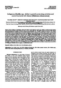
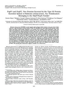
FIG. 1. Generation of R. solanacearum acrA and dinF mutants. Mutations were created by inserting a gentamicin resistance cassette (GmR) into the ORF of either acrA (A) or dinF (B), as indicated. The arrows indicate the directions of transcription. Flanking genes are included for reference and are annotated as follows: RSc0009, encoding a drug efflux lipoprotein (NodT/FusA family); RSp0282, encoding a hypothetical amino acid permease; and RSp0284, encoding a hypothetical transmembrane protein. The asterisk indicates that the BglII site was introduced.
described (1). DNA sequencing was performed at the University of WisconsinMadison Biotechnology Center, Madison. Oligonucleotides were obtained from Sigma Genosys (The Woodlands, TX). The DNA sequence analysis was performed using Biology Workbench (http://workbench.sdsc.edu/), NCBI (http://www.ncbi.nlm.nih .gov/), and NEB cutter (www.neb.com), and the GMI1000 genomic database (http: //bioinfo.genopole-toulouse.prd.fr/annotation/iANT/bacteria/ralsto/index.html) was also used. Cloning, mutagenesis, and complementation of acrA and dinF. To mutate the acrA gene of R. solanacearum, an approximately 1,200-bp fragment containing the acrA open reading frame (ORF) was PCR amplified using primers 5⬘CCG TTATCACCAACTCGCTG3⬘ and 5⬘GTCGACGATCACCTTGTCAC3⬘ designed by using the GMI1000 sequence. The resulting PCR product was AT cloned into pSTBlue-1 (Novagen) to create pSTacrA. Splicing by overlap (SOE) PCR (29) was used to introduce a unique BglII site into pSTacrA. For the SOE PCR experiment, primers were designed to amplify the 5⬘ and 3⬘ halves of the cloned acrA fragment in pSTacrA, such that a BglII site was introduced into the center of the acrA ORF. The final SOE PCR product was AT cloned into pSTBlue-1 (Kmr; Novagen) to obtain pSTacrASOE. The primers used for the SOE PCR included the forward and reverse primers described above, as well as 5⬘GGAGATCTTCCTCTAGAAAATGGGCGAGGTGACGGT3⬘ and 5⬘TCTA GAGGAAGATCTTCCAATCTACCTGACCTTCACGCA3⬘ (the BglII site is underlined). The gentamicin resistance cassette aacC1 from pUCGM (60) was inserted into the introduced BglII site. The acrA::Gmr construct pSTacrA::GM was introduced into R. solanacearum by electroporation, and double recombinants (Gmr Kms) were selected. Correct insertion of the acrA mutagenesis construct was confirmed by Southern blot hybridization analysis (data not shown). Since the R. solanacearum acrABR locus is very similar to the E. coli locus and acrA and acrB are separated by only 29 nucleotides, it is likely that the two genes are cotranscribed. Also, since the proteins must function together, a mutation in acrA and a mutation in acrB should result in the same phenotype. To create a DinF mutant, a 2.1-kb fragment containing the entire dinF ORF was PCR amplified using primers designed by using the GMI1000 database (5⬘AGCATCGACCAGACTTGG3⬘ and 5⬘ ATGTGCAGCATCCAC3⬘). The PCR product was AT cloned into pSTBlue-1 (Kmr; Novagen) to create pSTDinF. The 2.1-kb fragment containing the dinF gene was cloned into pBluescript (Stratagene) to obtain pBSDinF. The aacC1 (Gmr) gene cassette was introduced into the dinF ORF at a unique PstI site in pBSDinF to obtain pDinF::Gm, which was introduced into R. solanacearum by electroporation. Cells were selected on agar plates containing gentamicin and screened for double recombination on agar plates containing double-strength ampicillin. Correct insertion of the dinF mutagenesis construct was confirmed by Southern blot hybridization analysis (data not shown). For in planta complementation, we used cosmids pLAFacrA and pLAFdinF, which contained the full-length acrA and dinF genes, respectively (as well as flanking sequences) (Fig. 1). These cosmids were obtained by screening the R.
R. SOLANACEARUM EFFLUX PUMPS
2779
solanacearum strain K60 genomic library by Southern hybridization using the acrA and dinF PCR products as probes. Cosmids harboring the acrA and dinF loci were introduced into the corresponding mutant strains by electroporation. Plant assays to confirm restoration of the mutant phenotypes to the wild-type phenotype were performed as described below. Construction of GUS reporter strains. For transcription expression analysis of dinF and acrAB using -glucuronidase (GUS) as a reporter, we inserted the 5⬘ regions of both the acrA and dinF ORFs upstream of the promoterless uidA gene (encoding GUS) into plasmid pVO155 (Kmr) or pDB155 (Smr) (9). The dinF::uidA reporter construct was created by cloning a 600-bp EcoRI/PstI fragment from pSTdinF into pBluescript (Ampr; Stratagene) to create pBSdinF. The promoter region and 5⬘ end of dinF were removed from pBSdinF using XbaI and XhoI and directionally cloned into both pVO155 and pDB155 upstream of the promoterless uidA genes in these vectors to create pVOdinF and pDBdinF, respectively. The acrA::uidA reporter construct was created by cloning a 1,200-bp BamHI/XbaI fragment (containing approximately 300 bp of upstream sequence and most of the acrA ORF) from pSTacrA into pVO155 and pDB155, which were digested with BamHI and SpeI to create pVOacrA and pDBacrA. The resulting GUS reporter constructs were introduced by triparental conjugation into the following R. solanacearum strains: K60 (wild type), K200 (hrpB) (51), and K999 (rpoS) (9). Since neither pVO155 nor pDB155 could replicate in R. solanacearum under antibiotic selection conditions, integration into the chromosome must have occurred at the site of homology to the cloned gene fragment. This integration introduced a single copy of the promoterless GUS reporter gene, uidA, and created a cis merodiploid of the gene of interest, leaving an intact functional copy of the gene. Determination of MICs. The MICs of various compounds were determined by a standard twofold serial dilution method (12). For R. solanacearum strains, MIC assays were carried out using BMM supplemented with 0.2% glucose containing different compounds at various concentrations. For E. coli MDR mutant strains KAM3 and KAM32, MIC assays were performed using Mueller-Hinton broth (Becton-Dickinson, Germany). MIC assays were performed in triplicate by using final volumes of 3 ml for R. solanacearum and 200 l for KAM3 and KAM32. The MIC corresponded to the concentration in the tube that showed no growth, as determined by comparison to an uninoculated control both visually and spectrophotometrically (absorbance at 600 nm). When appropriate, controls containing only the drug solvent were included in the MIC determinations to ensure that the solvent did not affect bacterial growth. GUS assays. The in planta GUS assays were performed as described by Brown and Allen (9). Briefly, R. solanacearum cells harboring the reporter gene constructs were grown overnight in CPG medium, washed twice with sterile deionized water, and diluted to obtain an optical density at 600 nm of 0.1 (corresponding to 1 ⫻ 108 CFU/ml). Two-microliter portions of each diluted bacterial suspension were applied to cut petioles of 23-day-old susceptible (cultivar Bonny Best) or resistant (cultivar Hawaii 7996 [H7996]) tomato plants in order to deliver ⬃105 CFU to each petiole. Bacteria were harvested from a pool of six inoculated plants at several times (to obtain bacteria at different cell densities) by removing each infected petiole and 0.5 cm of the stem above and below the petiole, weighing the plant material, and then crushing the infected tissue in a mortar and pestle with 500 l of water. A 50-l sample was removed for enumeration of bacteria by dilution plating, and 450 l of GUS extraction buffer (31) was added to the ground tomato tissue, which was then stored at ⫺80°C until it was used for the GUS assays. To allow comparisons of GUS activity in planta and in culture, the number of CFU per gram of plant tissue was multiplied by 13.75 to obtain the equivalent number of CFU per milliliter of xylem fluid, as previously described (64). Strains carrying the integrated, constitutively expressed pilA promoter region cloned into pIVETDGB (a derivative of pVO155) were used as a positive control for GUS expression (data not shown). For the in planta GUS assays, 25-day-old cultivar Bonny Best and H7996 seedlings (11 days after transplanting) were used. Each assay was repeated three times. For GUS assays in culture, bacteria were grown overnight in CPG medium, washed with sterile deionized water, and then resuspended in BMM (supplemented with 0.2% glucose unless indicated otherwise) to obtain an optical density at 600 nm of 1.0 (corresponding to 1 ⫻ 109 CFU/ml). The bacterial suspension was diluted to obtain various cell densities in BMM containing 0.2% glucose and incubated on an environmental shaker at 28°C at 250 rpm for 6 h (about ⬃1.5 bacterial generations). After this incubation, 100-l samples were removed to determine the cell density. Bacteria were collected by centrifugation (6,000 ⫻ g) for 3 to 30 min (depending on the culture volume), and cell pellets were stored at ⫺80°C until they were used for GUS assays. To assess whether dinF or acrA expression is induced by toxic compounds, K60 cells harboring either the dinF::uidA or acrA::uidA GUS reporter constructs were resuspended to obtain a final cell density of 1 ⫻ 108 CFU/ml in BMM supple-
2780
BROWN ET AL.
APPL. ENVIRON. MICROBIOL.
mented with toxic compounds at sublethal concentrations, as determined by MIC analysis as described above. Each bacterial suspension was incubated at 28°C with shaking (250 rpm) for 6 h; after incubation, the bacteria were harvested by centrifugation and stored at ⫺80°C until they were used for measurement of GUS activity. Measurement of GUS activity. Cells were permeabilized with lysozyme (200 g/ml, 37°C, 20 min) in 1 ml of GUS extraction buffer, and GUS activity was assayed with the substrate methylumbeliferyl-glucuronide as previously described (31, 64). The level of fluorescence of the released methylumbelliferone (MU) was determined using a Hoefer fluorometer calibrated with a known concentration of MU. GUS activity was expressed in nanomoles of MU produced per minute per cell and was graphed against the cell density (expressed in CFU/ml) in either liquid culture or tomato xylem fluid. Plant growth conditions and assays. The susceptible heirloom tomato cultivar Bonny Best and the resistant cultivar H7996 (25) were grown in a growth chamber at 28 to 30°C by using a cycle consisting of 12 h of light and 12 h of darkness. Sixteen-day-old Bonny Best plants were used for soil soak virulence assays as previously described (63). Briefly, 50 ml of a bacterial suspension containing ⬃5 ⫻ 107 CFU/ml was poured onto tomato seedlings in 80 g of soil, resulting in a concentration of ⬃3.1 ⫻ 107 CFU/g of soil. Diseased plants were rated daily using a disease index scale ranging from 0 to 4 as previously described (63). Plant assays were repeated at least three times, using 16 plants per treatment in each assay.
RESULTS In E. coli, acrA encodes the periplasmic subunit of the tripartite AcrAB-TolC drug efflux pump, a member of the RND MDR superfamily (37). The R. solanacearum AcrA protein sequence (NCBI accession number NP 518132.1) was 71% similar to the E. coli AcrA protein sequence. Also, the genomic organization of the acrAB operon and the adjacent acrR locus (encoding the transcriptional repressor of the acrAB operon) was conserved in E. coli and R. solanacearum. The only difference between the R. solanacearum and E. coli acrABR loci was that R. solanacearum has a gene encoding a putative outer membrane channel belonging to the NodT/FusA family (RSc0009) that is present at or near the end of the acrAB operon (Fig. 1A), while E. coli lacks a gene that encodes an outer membrane channel at this locus (70). DinF belongs to a distinct branch of the MATE superfamily of MDRs (10). BLASTP analysis showed that the R. solanacearum DinF protein (NCBI accession number NP 521844.1) resembles DinF homologs in E. coli (58% similarity) and Streptococcus pneumoniae (35% similarity). Yet the genomic synteny of dinF-flanking regions in R. solanacearum and other bacteria was not conserved (data not shown). In E. coli and S. pneumoniae, dinF is located in operons with lexA and recA, respectively (34, 41). However, in R. solanacearum, dinF is located on the organism’s 2.1-Mb megaplasmid and is flanked by genes encoding a hypothetical amino acid permease (RSp0282) and a putative transmembrane protein (RSp0284) (Fig. 1B), while both lexA and recA are on the 3.7-Mb chromosome in R. solanacearum. Two highly conserved motifs, NIILDPLFI and GAAIATVIA, which are characteristic of the DinF/VmrA cluster of the MATE superfamily (10, 30), are present in the R. solanacearum DinF protein sequence, as follows: NMVAVLGLV and GIGAATAVA (conserved and similar residues are underlined and in boldface type, respectively). The presence of these conserved residues suggested that R. solanacearum DinF is a member of the DinF branch of the MATE superfamily rather than the NorM branch, which lacks this conserved sequence. To investigate the overall relationship between the R. so-
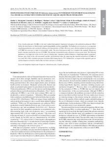
lanacearum AcrA and DinF proteins and homologues in other bacteria, we performed a ClustalW analysis, and a phylogenetic tree was generated using Tree puzzle 5.2 (www.tree-puzzle.de) for 35 MATE superfamily sequences and 28 AcrA sequences derived from a BLASTP search (2) (see Fig. S1 and S2 in the supplemental material). The AcrA protein sequence was highly conserved in all bacterial strains tested. However, the R. solanacearum DinF protein sequence was less similar to the sequences of other DinF homologs and MATE superfamily proteins (including four NorM sequences and two Arabidopsis thaliana MATE superfamily proteins). Importantly, the R. solanacearum DinF sequence clustered with the sequences of other proteins in the DinF branch of the MATE superfamily. Also, the DinF and AcrA proteins from R. solanacearum clustered in the same branch as orthologues from other phylogenetically related proteobacteria, such as Burkholderia cepacia (DinF) and Ralstonia metallidurans (AcrA). We created site-directed mutants with the K60 wild-type background, including K1833 (acrAB) and KDF (dinF) (see Materials and Methods) (Fig. 1). Both K1833 and KDF grew at wild-type levels in minimal medium supplemented with 0.2% glucose, indicating that they had no general in vitro growth defect and were not auxotrophs. To ensure that the loss of DinF or AcrA did not prevent R. solanacearum from multiplying in plant tissue, which is a prerequisite for virulence in this pathogen, we also measured the multiplication of both mutants following infusion into leaves of susceptible tobacco cultivar Bottom Special. This tobacco assay is a rapid and reliable test for the ability of R. solanacearum cells to multiply in planta but is not a means by which pathogenicity can be tested since the bacteria are injected into the leaf apoplast, which is not the usual niche for R. solanacearum in the plant. In contrast, the numbers of pathogen cells in tomato xylem reflect disease progress and are correlated with virulence, as well as the general fitness in planta. The growth of K1833 and KDF in tobacco leaves was indistinguishable from the growth of the wild-type parent strain (data not shown). To measure the contributions of acrAB and dinF to R. solanacearum virulence, we performed a soil inoculation assay in which bacterial suspensions were poured into pots containing unwounded susceptible tomato seedlings. This inoculation method mimics the natural R. solanacearum infection process, where the bacteria must locate and enter the roots from the soil. Both K1833 and KDF were significantly attenuated for the ability to cause wilt symptoms in tomato compared to wild-type strain K60 (Fig. 2). The virulence phenotypes of K1833 and KDF were complemented by introducing cosmids (with the pLAFR3 vector backbone) that harbored the full-length acrAB operon and the complete dinF ORF, respectively, each flanked by at least 1 kb of DNA on each side of the locus. For both K1833 and KDF, addition of the cosmids restored virulence to the mutant strains (Fig. 2). MDR mutants of other plant-pathogenic bacteria are less resistant to various toxins (5, 11, 12, 50). Therefore, we determined the relative MICs of several toxic antibiotics, phytoalexins, and detergents for K1833 and KDF in minimal medium (Table 2). As expected, both mutants exhibited low but measurable sensitivities to various toxic compounds, including caffeic acid and tomatine, which are antimicrobials produced by
R. SOLANACEARUM EFFLUX PUMPS
VOL. 73, 2007
2781
TABLE 2. MICs of various compounds for R. solanacearum acrA and dinF mutant strains MICa Compound
Acriflavine Ampicillin Berberine Biochanin A Caffeic acid Cephalexin Chloramphenicol DAPI Esculetin Ethidium bromide Fusaric acid Gossypol Hydrogen peroxide Mitomycin C Nalidixic acid Plumbagin Pyrithione Resorcinol Rhein Salicylic acid Sodium dodecyl sulfate Tomatineb TPPCl FIG. 2. Virulence of R. solanacearum acrA and dinF strains on susceptible tomato cultivar Bonny Best is significantly reduced. The curves are disease progress curves for R. solanacearum acrA (A) and dinF (B) mutant strains. A bacterial suspension was added to the soil of 14-day-old tomato seedlings to obtain a bacterial density of approximately 3.1 ⫻ 107 CFU/g of soil, and plants were rated on a disease index scale ranging from 0 to 4. (A) F, wild-type strain K60; ■, acrA strain K1833; Œ, complemented strain K1833(pLAFacrA). (B) E, wild-type strain K60; ‚, dinF strain KDF; 䊐, complemented strain KDF(pLAFdinF). The symbols indicate the averages for six plant assays, each performed with 16 plants per treatment; the error bars indicate the standard errors of the means.
tomato plants. Neither mutant exhibited increased sensitivity to biochanin A, gossypol, mitomycin C, plumbagin, pyrithione, rhein, or salicylic acid. acrAB mutant K1833 was sensitive to a greater number of compounds than KDF, while KDF was sensitive to only two compounds (4⬘,6⬘-diamidino-2-phenylindole [DAPI] and tetraphenylphosphonium chloride [TPPCl]) to which K1833 was resistant. These results suggest that the protein products of acrAB and dinF have overlapping substrate specificities but that some compounds are specifically transported by only one MDR. To verify the biological function of the R. solanacearum dinF and acrAB gene products, we examined whether either product could heterologously restore toxin resistance to the E. coli AcrAB deletion mutant KAM3 (40). A plasmid containing either dinF or acrAB was introduced to the E. coli mutant strain in trans, and the MICs of several toxic compounds for the resulting hybrid strain were determined (Table 3). The presence of R. solanacearum acrAB or dinF increased KAM3 resistance to ethidium bromide and acriflavine twofold and eightfold, respectively. The presence of acrAB also resulted in a twofold increase in resistance to berberine, while the presence of dinF resulted in a fourfold increase in resistance. R. solanacearum dinF also increased KAM3 resistance to nalidixic
Wild-type strain K60
acrA strain
dinF strain
62.50 62.50 62.50 ⬎1,000 250.0 500.0 31.30 3.130 31.30 62.50 125.0 ⬎1,000 3.125 25 62.50 62.50 3.910 250.0 31.30 1,000 250.0 ⬎250 1,000
15.60 15.60 15.60 ⬎1,000 125.0 250.0 15.60 3.130 15.60 7.813 31.30 ⬎1,000 3.125 25 15.60 62.50 3.910 62.50 31.30 1,000 125.0 62.50 1,000
15.60 62.50 62.50 ⬎1,000 250.0 500.0 31.30 1.560 15.60 31.30 125.0 ⬎1,000 2.081 25 62.50 62.50 3.910 250.0 31.30 1,000 250.0 125.0 125.0
a The MICs are the minimum concentrations that inhibited bacterial growth. Most MICs are expressed in g/ml; the only exceptions are the MICs of hydrogen peroxide, which are expressed in mM. The assays were performed in triplicate, and the optical densities at 600 nm were normalized to 0.001 at the beginning of the MIC assay. MICs that differ from the MIC for the wild-type strain are indicated by boldface type. b For tomatine, BMM was buffered with 0.1 M morpholineethanesulfonic acid (MES) to pH 7.0 for the MIC assay.
acid and berberine twofold. There was no difference in the resistance of KAM3 to esculetin or TPPCl in the presence of either R. solanacearum protein. In addition, the presence of dinF in the KAM32 mutant (which lacked both AcrAB and the MATE superfamily protein YdhE, which we hypothesize functions like DinF) (14) doubled its resistance to TPPCl and berberine, quadrupled its resistance to acriflavine, increased its resistance to ampicillin eightfold, and increased its resistance to ethidium bromide 16-fold (Table 3). We examined the effect
TABLE 3. MICs for E. coli strains KAM3 and KAM32 harboring acrA or dinF from R. solanacearum MIC (mg/ml)a Compound
KAM3b
Acriflavine 0.49 Ampicillin 3.9 Berberine 31.3 Esculetin 500 Ethidium bromide 3.9 Nalidixic acid 1.9 TPPCl 3.9
KAM3 KAM3 KAM32 KAM32b (pLAFacrA) (pLAFdinF) (pLAFdinF) 3.9 7.8 62.5 500 7.8 1.9 3.9
3.9 3.9 125.0 500 7.8 3.9 3.9
1.9 3.9 15.6 NDc 0.95 ND 15.6
7.8 31.3 31.3 ND 15.6 ND 31.3
a The MICs are the minimum concentrations required to inhibit bacterial growth. The assays were performed in triplicate to ensure consistent results. The optical densities at 600 nm were normalized to 0.001 at the beginning of the MIC assay. MICs that differ from the MICs for the parent mutant strains are indicated by boldface type. b KAM3 and KAM32 with empty plasmid pLAFR3 gave identical results. c ND, no data.
2782
APPL. ENVIRON. MICROBIOL.
BROWN ET AL.
FIG. 3. dinF and acrA are highly expressed in planta. The GUS activities produced by strains carrying a dinF::uidA or acrA::uidA transcriptional fusion were measured for cells grown to different cell densities in planta (susceptible tomato) and in vitro (minimum broth). The cell density (expressed in CFU/ml of broth or xylem fluid) is indicated on the x axis. The data are the averages ⫾ standard errors of the means for three independent experiments performed with six plants per treatment. (A) acrA expression (in R. solanacearum strain K60 with pVOacrA); (B) dinF expression (in R. solanacearum strain K60 with pVOdinF).
of adding dinF to KAM32 because this strain lacks both AcrAB and the MATE superfamily protein YdhE. This construct allowed us to more precisely determine the effect of heterologous expression of dinF, which is itself predicted to be a MATE superfamily protein. We included the dinF gene in the MIC tests for KAM3 in order to examine the functional redundancy between dinF and acrAB, but not in the MIC tests for KAM32 since it is unlikely that addition of acrAB to KAM32 would alter the MICs from those observed with KAM3. To measure the expression of the acrAB and dinF genes in different environments, we created cis merodiploid reporter strains using a promoterless GUS gene (uidA) fused to the promoter and the 5⬘ region of each gene (see Materials and Methods). The strains harboring the acrA::uidA and dinF::uidA transcriptional fusions were collected at various cell densities from cultures growing in minimal medium and in the xylem tissue of whole tomato plants. The expression of both dinF and acrAB increased more than 100-fold in the tomato xylem compared to the expression in minimal medium at low cell densities (105 to 106 CFU/ml) (Fig. 3). However, at higher cell densities, the differences in gene expression in BMM and in planta were smaller, suggesting that MDRs play a critical role during the initial stages of plant colonization by R. solanacearum. Expression of dinF varied as a function of cell density
both in planta and in culture, and the highest levels of expression were observed at lower cell densities. In contrast, acrAB expression was cell density dependent in both the plant and in minimal medium. Intriguingly, the expression in minimal medium increased about fivefold as the cell density increased, while the -glucosidase activity decreased with increasing cell density in planta until the cell density reached ⬃108 CFU/ml; then the expression started to increase. The resistant tomato cultivar H7996 has been used to study R. solanacearum population dynamics in root and stem tissues (38, 43, 68). In H7996, R. solanacearum is largely confined to the primary xylem and poorly colonizes the root parenchyma cells. It has been hypothesized that the inability of R. solanacearum to effectively colonize H7996 is due to inhibitory compounds present in the resistant plants (43, 68). We reasoned that if H7996 produced toxins, then the expression of acrAB or dinF in R. solanacearum would increase during growth in this tomato cultivar. To test this possibility, we measured the GUS activities of both acrA::uidA and dinF::uidA fusion strains in H7996. We found that the expression of the dinF::uidA fusion doubled at low to moderate cell densities (105 to 108 CFU/ml) in H7996 compared to the expression in the susceptible tomato cultivar, while the expression of the acrA::uidA fusion was about 1.5-fold higher at similar cell densities compared to the expression in susceptible cultivar Bonny Best. However, at high cell densities (109 CFU/ml), the relative levels of gene expression in H7996 decreased significantly (Table 4). These results showed that during the initial host colonization MDR gene expression was moderately increased in H7996 but at high cell densities the MDR gene expression in the resistant tomato cultivar was significantly lower than the MDR gene expression in the susceptible tomato cultivar. We measured the relative levels of expression of acrAB or dinF in the presence and absence of sublethal concentrations of several toxins (Table 5). All of the compounds tested increased the expression of acrAB and dinF between 1.3- and 5.3-fold. The DNA-damaging toxin mitomycin C was the most potent inducer for both acrAB and dinF. The plant defense signaling molecule salicylic acid induced acrAB expression and dinF expression 1.3- and 2.1-fold, respectively. This is interest-
TABLE 4. acrA and dinF expression in resistant tomato cultivar H7996 compared to expression in susceptible tomato cultivar Bonny Best Fold induction of GUS expressiona
SE
acrA::uidA strain 105 106 108 109
1.511 1.385 1.807 0.079
0.247 0.411 0.329 0.009
dinF::uidA strain 106 108 109
2.449 2.104 0.698
0.507 0.001 0.071
Concn (CFU/ml)
a The fold induction was determined by dividing the GUS activity observed in resistant tomato cultivar H7996 by the expression in the susceptible tomato cultivar Bonny Best.
R. SOLANACEARUM EFFLUX PUMPS
VOL. 73, 2007 TABLE 5. Relative induction of acrA and dinF in the presence of various compoundsa Induction of:
Compoundb
Acriflavine Berberine Esculetin H2O2 Mitomycin C Salicylic acid BMM alone
acrA
dinF
2.9 (0.03)c 1.3 (0.09) 1.5 (0.02) 3.2 (0.02) 5.3 (0.07) 1.3 (0.01) 1.0
1.6 (0.06) 1.9 (0.02) 2.0 (0.03) 1.6 (0.17) 5.1(0.10) 2.1 (0.03) 1.0
a Relative induction was measured by determining GUS activity from acrA::uidA and dinF::uidA reporter gene fusions. The levels of expression are the levels relative to basal gene expression in minimal medium plus glucose (BMM). b Compounds were added at the following sublethal concentrations: acriflavine, 31.25 g/ml; berberine, 31.25 g/ml; esculetin, 16.25 g/ml; H2O2, 5 mM; mitomycin C, 1.0 g/ml; and salicylic acid, 500 g/ml. c The data are means of three determinations; the values in parentheses are the standard errors.
ing since high concentrations of mitomycin C and salicylic acid did not inhibit the growth of either mutant (Table 2). RpoS is an alternative sigma factor involved in bacterial stationary-phase and stress response pathways (28). Flavier and coworkers (17) showed that the virulence of an R. solanacearum RpoS mutant was attenuated and that this mutant was unable to survive under carbon limitation conditions and at a low pH. Also, the RpoS mutant produced less of the quorum-sensing autoinducer acyl-homoserine lactone (17). To determine if RpoS is required for expression of acrAB in R. solanacearum, we introduced the acrA::uidA reporter fusion into rpoS mutant strain K999 (9) and measured acrAB expression during growth in minimal medium and in planta. We found that an intact copy of rpoS was necessary for wild-type levels of acrAB expression at cell densities greater than 107 CFU/ml (Fig. 4). The same pattern of rpoS-dependent acrAB expression at high cell densities was observed in minimal me-
FIG. 4. RpoS is required for full acrA expression in planta at a high cell density. GUS activity in cells grown in the susceptible tomato cultivar Bonny Best was determined. The acrA::uidA fusion was introduced into wild-type K60 cells and the rpoS mutant strain (K999). The adjusted cell density (CFU/ml of xylem fluid) is indicated on the x axis. The data are the means ⫾ standard errors of the means for three independent measurements obtained using six plants per treatment.
2783
TABLE 6. acrA::uidA expression in wild-type strain K60 and the phcA and rpoS mutant strains increased in the absence of a carbon source Bacterial strain (plasmid)
Wild-type K60 (pVOacrA) phcA (pVOacrA) rpoS (pDBacrA)
GUS activity (fmol MU released per cell per min) in BMM witha: Carbon source
No carbon source
0.0474 (0.003390) 0.0854 (0.010700) 0.0120 (0.000015)
0.210 (0.05370) 1.60 (0.20300) 0.023 (0.00035)
a The carbon source added was 0.2% glucose. Assays were performed in triplicate, and the values in parentheses are standard errors. Significantly different values, as determined by Student’s t test at a P value of ⬍0.001 with a 95% confidence interval, are indicated by boldface type.
dium (data not shown). We did not measure the expression of the dinF::uidA fusion in the rpoS mutant because, as shown in Fig. 3, dinF was expressed at a low cell density and not during the stationary phase. In E. coli, slow growth and carbon starvation increase acrAB expression (53). To determine whether acrAB expression was affected by the absence of carbon in R. solanacearum, we measured acrA::uidA expression at a cell density of approximately 108 CFU/ml after 4 h of growth (which corresponded to one generation) in minimal medium containing either no glucose (carbon starvation) or 0.2% glucose. Expression of acrAB increased ⬃4-fold in the absence of glucose (Table 6). The expression of the dinF::uidA fusion also increased slightly under these conditions (data not shown). In a preliminary experiment in which we assessed the dependence of acrAB expression on seven regulators (9), we observed that in cells growing on minimal medium agar, expression of acrAB was affected by the stationary-phase sigma factor RpoS and by the global virulence gene regulator PhcA. Therefore, we also measured the carbondependent expression of acrAB in mutants lacking PhcA or RpoS. In medium lacking a carbon source, acrAB expression was ⬃19-fold higher in the phcA mutant strain and ⬃8-fold higher in the wild-type genetic background. The absence of RpoS resulted in a twofold increase in acrAB expression in medium lacking carbon (Table 6). Overall, these data suggest that PhcA inhibits acrAB expression, while RpoS stimulates acrAB expression. Cunnac et al. reported that the promoter region of dinF in R. solanacearum strain GMI1000 contained an HrpII box (TTCG N16TTCG), which is recognized by HrpB (15). HrpB is an AraC-type transcriptional activator required for expression of the type III secretion system, which delivers effector proteins to the host cell (3). An hrpB mutant is avirulent on susceptible tomatoes and does not produce a hypersensitive response in resistant hosts (22). To experimentally determine if dinF expression is indeed dependent on HrpB, we introduced the dinF::uidA reporter construct pVOdinF into an hrpB mutant of strain K60, K200 (51), and measured the relative GUS activity during growth in minimal medium. In the absence of HrpB, dinF expression increased 3.4-fold at a cell density of 107 CFU/ ml, 8.7-fold at a cell density of 108 CFU/ml, and 6.1-fold at a cell density of 109 CFU/ml. However, there was no difference in dinF expression in the hrpB mutant at lower cell densities. In planta, dinF expression was similar in both wild-type and hrpB strains at cell densities less than 5 ⫻ 106 CFU/ml (data not
2784
BROWN ET AL.
FIG. 5. dinF expression is increased in an hrpB strain at a high cell density. The GUS activity of the dinF::uidA fusion introduced into wild-type strain K60 and the hrpB mutant strain (K200) was measured using cells grown to different cell densities in BMM supplemented with 0.2% glucose. The cell density (CFU/ml) is indicated on the x axis. The data are the averages ⫾ standard errors of the means for three independent determinations.
shown); we were not able to measure the expression of dinF in the hrpB mutant at high cell densities in planta since this mutant cannot grow well in the tomato stem. Our results suggest that HrpB negatively regulates dinF expression (Fig. 5). DISCUSSION Putative toxin efflux pump genes are a prominent feature of the R. solanacearum genome, and we show here that two of these genes, acrAB and dinF, are upregulated during growth in host plants and that they are required for full virulence in susceptible tomato plants. Furthermore, both acrAB and dinF functioned heterologously as efflux pumps in toxin-sensitive E. coli strains. These results suggest that these genes do encode efflux pumps and have critical roles in the biology of this soil-dwelling plant pathogen. This is the first report describing mechanisms underlying the intrinsic resistance of R. solanacearum to various toxic compounds, and to our knowledge, this is the first characterization of a member of the DinF subfamily of MATE MDRs in a phytopathogenic bacterium. Plants produce a battery of antimicrobial compounds that function synergistically to limit pathogen colonization and spread. Indeed, inactivation of either acrAB or dinF resulted in reduced virulence and increased sensitivity to several antimicrobial compounds produced by plants, including caffeic acid, resorcinol, esculetin, tomatine, and berberine. However, the concentrations of the compounds tolerated were relatively high compared to the concentrations that inhibit animal or human pathogens (65). The difference may be explained by the discovery of a class of plant-derived compounds known as MDR inhibitors, which can significantly increase the antimicrobial properties of plant secondary metabolites (6, 62, 65). The sensitivity of R. solanacearum to these compounds in the presence of MDR inhibitors and to the full repertoire of toxins present in host plants, such as tomato, remains to be determined. The fact that both mutants were less tolerant to toxins that are produced by tomato (caffeic acid and tomatine), as
APPL. ENVIRON. MICROBIOL.
well as toxins that are produced by nonhost plants (resorcinol, esculetin, and berberine), implies that resistance to such toxins reflects a general bacterial defense response that is not limited to a particular host. During the initial bacterial colonization, nutrients in the plant are scarce, especially in the xylem (49). In E. coli, acrAB expression increases in carbon-starved cells, probably because starvation triggers a general bacterial stress response, resulting in increased MDR expression (53). It is therefore not surprising that R. solanacearum acrAB expression also increases under carbon starvation conditions; dinF expression also increased slightly in the absence of carbon (data not shown). An R. solanacearum global virulence gene regulator, PhcA, inhibits acrAB expression; as PhcA is expressed at higher cell densities (58), this correlates with our observation that AcrA expression generally decreases as cell density increases, at least in planta. Additionally, we found that the stress-responsive sigma factor RpoS was required for acrAB expression in minimal medium and when cell densities approached 108 CFU/ml in planta, which is equivalent to the stationary phase. This finding is in contrast to acrAB expression in E. coli, which increases during the stationary phase and is not dependent on RpoS (53). We found that the type III secretion regulator HrpB was required for maximal dinF expression at high cell densities in minimal medium, but at low cell densities in minimal medium and in planta loss of HrpB had no effect on dinF expression. This finding should be examined further because HrpB regulation of non-type III secretion genes is not well documented, even though the HrpB regulon is apparently large (16, 42). Although dinF is not physically near genes known to be involved in either SOS repair or competence in R. solanacearum, we found that expression of dinF was induced by the DNA-damaging compounds mitomycin C and hydrogen peroxide at sublethal concentrations in culture (Table 5), even though there were not great differences in the MICs of these compounds (Tables 2 and 5). However, these compounds also induced expression of acrAB, which is not associated with the SOS response, suggesting that a general function of MDRs may be to expel DNA-damaging toxins from the bacterial cell, independent of a specific SOS response. Furthermore, such induction may reflect a general protective response to compounds that can potentially compromise the integrity of the bacterial DNA. This hypothesis is supported by the recent finding that in E. coli -lactam antibiotics trigger the SOS response (39). It has been hypothesized that polyphenols produced by the moderately resistant tomato cultivar H7996 might restrict pathogen spread through the root (68). Thus, we measured the expression of acrAB and dinF in H7996 and found that the expression of both acrAB and dinF was slightly induced at low cell densities but significantly reduced at high cell densities in this resistant cultivar compared to the expression in susceptible tomato plants. This finding was unexpected, but it is possible that high bacterial cell densities caused the resistant host to produce an as-yet-unknown factor that suppresses MDR expression. The fact that acrAB and dinF are required for bacterial wilt virulence does not preclude the possibility that these pumps have an additional function in facilitating rhizosphere survival
R. SOLANACEARUM EFFLUX PUMPS
VOL. 73, 2007
and competition. The E. amylovora NorM efflux pump contributes to the ability of this bacterium to compete with other bacteria that occupy the same niche (11). MDRs may be required for fitness in rhizosphere environments containing plant root exudates and antimicrobial compounds produced by competitors. R. solanacearum is a successful inhabitant of tropical and subtropical soils, and the biology of its soil survival is poorly understood. Could the acrAB- and dinF-encoded MDRs in R. solanacearum also play a role in niche competition? We found that loss of either of two R. solanacearum MDRs measurably increased sensitivity to various toxins. The moderate change in sensitivity likely reflected the presence of additional MDRs in the bacterium. Indeed, the R. solanacearum GMI1000 genome annotation suggests that there are at least 56 MDR genes, 32 of which appear to be organized in 12 separate operons. Five of these operons are located on the 3.7-Mb chromosome, and seven are on the 2.1-Mb megaplasmid (acrAB is on the chromosome, and dinF is on the megaplasmid). These genes include genes encoding MDRs belonging to the MATE superfamily (4), the RND superfamily (24), a group of acrR-type transcription regulators (9), the major facilitator superfamily (7), the ABC superfamily (4), and a group of unclassified drug efflux transporters (7). Only one gene encodes a protein that belongs to the SMR family; this gene was also identified in our recent in vivo expression technology screen, but it has not been studied yet. The presence of so many likely MDR-encoding genes throughout the genome implies that R. solanacearum must confront and export a wide range of toxic compounds, which may contribute to this pathogen’s ability to infect such a broad range of plant hosts and survive saprophytically in the soil. The contribution to virulence of MDRs in a nonhuman pathogen highlights the importance of MDRs in a natural (versus nosocomial) environment and indicates that these pumps increase bacterial fitness and are maintained by natural selection pressures. Furthermore, the study of MDRs in R. solanacearum can help us understand this pathogen’s unusually wide plant host range and may suggest biocontrol measures. Additional research with this and other plant-bacterium systems will likely add to the accumulating body of evidence that plant- and microbe-derived toxins affect bacterial survival and virulence. ACKNOWLEDGMENTS We thank T. Tsuchiya for providing E. coli strains KAM3 and KAM32 and Mark Schell and Michael San Francisco for useful discussions. We thank E. M. Hong for providing technical assistance and J. L. Tans-Kersten and J. Handelsman for critical reading of the manuscript. This work was supported by the UW—Madison College of Agricultural and Life Sciences, by NSF grant IBN-0090692, and by USDANRI grant 03-35319-13851. D. G. Brown gratefully acknowledges financial support from the Howard Hughes Medical Institute, the Ruth Dickie Scholarship Fund, and the Storkan-Hanes-McCaslin Foundation. REFERENCES 1. Allen, C., Y. Huang, and L. Sequeira. 1991. Cloning of genes affecting polygalacturonase production in Pseudomonas solanacearum. Mol. PlantMicrobe Interact. 4:147–154. 2. Altschul, S. F., T. L. Madden, A. A. Schaffer, J. Zhang, Z. Zhang, W. Miller, and D. J. Lipman. 1997. Gapped BLAST and PSI-BLAST: a new generation of protein database search programs. Nucleic Acids Res. 25:3389–3402.
2785
3. Arlat, M., C. L. Gough, C. Zischeck, P. A. Barberis, A. Trigalet, and C. Boucher. 1992. Transcriptional organization and expression of the large hrp gene cluster of Pseudomonas solanacearum. Mol. Plant-Microbe Interact. 5:187–193. 4. Ausubel, F., R. Brent, R. Kingston, D. Moore, J. Seidman, J. Smith, and K. Struhl. 1995. Short protocols in molecular biology, 3rd ed. John Wiley and Sons, New York, NY. 5. Barabote, R. D., O. L. Johnson, E. Zetina, S. K. San Francisco, J. A. Fralick, and M. J. D. San Francisco. 2003. Erwinia chrysanthemi tolC is involved in resistance to antimicrobial plant chemicals and is essential for phytopathogenesis. J. Bacteriol. 185:5772–5778. 6. Belofsky, G., D. Percivill, K. Lewis, G. P. Tegos, and J. Ekart. 2004. Phenolic metabolites of Dalea versicolor that enhance antibiotic activity against model pathogenic bacteria. J. Nat. Prod. 67:481–484. 7. Blackmore, C. G., P. A. McNaughton, and H. W. van Veen. 2001. Multi-drug transporters in prokaryotic and eukaryotic cells: physiological functions and transport mechanisms. Mol. Membr. Biol. 18:97–103. 8. Boucher, C., P. Barberis, A. Trigalet, and D. Demery. 1985. Transposon mutagenesis of Pseudomonas solanacearum: isolation of Tn5-induced avirulent mutants. J. Gen. Microbiol. 131:2449–2457. 9. Brown, D. G., and C. Allen. 2004. Ralstonia solanacearum genes induced during growth in tomato: an inside view of bacterial wilt. Mol. Microbiol. 53:1641–1660. 10. Brown, M. H., I. T. Paulsen, and R. A. Skurray. 1999. The multidrug efflux protein NorM is a prototype of a new family of transporters. Mol. Microbiol. 31:394–395. 11. Burse, A., H. Weingart, and M. S. Ullrich. 2004. NorM, an Erwinia amylovora multidrug efflux pump involved in in vitro competition with other epiphytic bacteria. Appl. Environ. Microbiol. 70:693–703. 12. Burse, A., H. Weingart, and M. S. Ullrich. 2004. The phytoalexin-inducible multidrug efflux pump AcrAB contributes to virulence in the fire blight pathogen, Erwinia amylovora. Mol. Plant-Microbe Interact. 17:43–54. 13. Chang, G. 2003. Multidrug resistance of ABC transporters. FEBS Lett. 555:102–105. 14. Chen, J., Y. Morita, M. N. Huda, T. Kuroda, T. Mizushima, and T. Tsuchiya. 2002. VmrA, a member of a novel class of Na⫹-coupled multidrug efflux pumps from Vibrio parahaemolyticus. J. Bacteriol. 184:572–576. 15. Cunnac, S., C. Boucher, and S. Genin. 2004. Characterization of the cisacting regulatory element controlling HrpB-mediated activation of the type III secretion system and effector genes in Ralstonia solanacearum. J. Bacteriol. 186:2309–2318. 16. Cunnac, S., A. Occhialili, P. Barberis, C. Boucher, and S. Genin. 2004. Inventory and functional analysis of the large Hrp regulon in Ralstonia solanacearum: identification of novel effector proteins translocated to plant host cells through the type III secretion system. Mol. Microbiol. 53:115–128. 17. Flavier, A. B., M. A. Schell, and T. P. Denny. 1998. An RpoS (sigma S) homologue regulates acylhomoserine lactone-dependent autoinduction in Ralstonia solanacearum. Mol. Microbiol. 28:475–486. 18. Fleißner, A., C. Sopalla, and K.-M. Weltring. 2002. An ATP-binding cassette multi-drug-resistance transporter is necessary for tolerance of Gibberella pulicaris to phtyoalexins and virulence on potato tubers. Mol. Plant-Microbe Interact. 15:102–108. 19. Fritig, B., T. Heitz, and M. Legrand. 1998. Antimicrobial proteins produced in plant defense. Curr. Opin. Immunol. 10:16–22. 20. Gabriel, D. W., C. Allen, M. Schell, T. P. Denny, J. T. Greenberg, Y. P. Duan, Z. Flores-Cruz, Q. Huang, J. M. Clifford, G. Presting, E. T. Gonza ´lez, J. Reddy, J. Elphinstone, J. Swanson, J. Yao, V. Mulholland, L. Liu, W. Farmerie, M. Patnaikuni, B. Balogh, D. Norman, A. Alvarez, J. A. Castillo, J. Jones, G. Saddler, T. Walunas, A. Zhukov, and N. Mikhailova. 2006. Identification of open reading frames unique to a select agent: Ralstonia solanacearum race 3 biovar 2. Mol. Plant-Microbe Interact. 19:69–79. 21. Genin, S., and C. Boucher. 2004. Lessons learned from the genome analysis of Ralstonia solanacearum. Annu. Rev. Phytopathol. 42:107–134. 22. Genin, S., C. L. Gough, C. Zischek, and C. A. Boucher. 1992. Evidence that the hrpB gene encodes a positive regulator of pathogenicity genes from Pseudomonas solanacearum. Mol. Microbiol. 6:3065–3076. 23. Gonzales-Pasayo, R., and E. Martinez-Romera. 2000. Multiresistance genes of Rhizobium etlii CFN42. Mol. Plant-Microbe Interact. 13:572–577. 24. Hanahan, D. 1983. Studies on transformation of Escherichia coli with plasmids. J. Mol. Biol. 166:557–580. 25. Hanson, P. M., J.-F. Wang, O. Licardo, Hanudin, S. Y. Mah, G. L. Hartman, Y.-C. Lin, and J. Chen. 1996. Variable reactions of tomato lines to bacterial wilt evaluated at several locations in Southeast Asia. HortScience 31:143– 146. 26. Hayward, A. C. 1991. Biology and epidemiology of bacterial wilt caused by Pseudomonas solanacearum. Annu. Rev. Phytopathol. 29:65–87. 27. Hendrick, C., and L. Sequeira. 1984. Lipopolysaccharide-defective mutants of the wilt pathogen Pseudomonas solanacearum. Appl. Environ. Microbiol. 48:94–101. 28. Hengge-Aronis, R. 2002. Signal transduction and regulatory mechanisms involved in control of the sigma (s) (RpoS) subunit of RNA polymerase. Microbiol. Mol. Biol. Rev. 66:373–395.
2786
BROWN ET AL.
29. Horton, R. M., H. D. Hund, S. N. Ho, J. K. Ullen, and L. R. Pease. 1989. Engineering hybrid genes without the use of restriction enzymes; gene splicing by overlap extension. Gene 77:61–68. 30. Huda, M. N., J. Chen, Y. Morita, T. Kuroda, T. Mizushima, and T. Tsuchiya. 2003. Gene cloning and characterization of VcrM, a Na⫹-coupled multidrug efflux pump, from Vibrio cholerae non-O1. Microbiol. Immunol. 47:419–427. 31. Jefferson, R. A. 1987. Assaying chimeric genes in plants: the GUS gene fusion system. Plant Mol. Biol. Rep. 5:387–405. 32. Kang, H. J., and D. C. Gross. 2005. Characterization of a resistance-nodulation-cell division transporter system associated with the syr-syp genomic island of Pseudomonas syringae pv. syringae. Appl. Environ. Microbiol. 71: 5056–5065. 33. Kelman, A. 1954. The relationship of pathogenicity of Pseudomonas solanacearum to colony appearance in a tetrazolium medium. Phytopathology 44:693–695. 34. Krueger, J. H., S. J. Elledge, and G. C. Walker. 1983. Isolation and characterization of Tn5 insertion mutations in the lexA gene of Escherichia coli. J. Bacteriol. 153:1368–1378. 35. Krummenacher, P., and F. Narberhaus. 2000. Two genes encoding a putative multi-drug efflux pump of the RND/MFP family are cotranscribed with an rpoH gene in Bradyrhizobium japonicum. Gene 241:247–254. 36. Li, X. Z., and H. Nikaido. 2004. Efflux-mediated drug resistance in bacteria. Drugs 64:159–204. 37. Ma, D., D. N. Cook, M. Alberti, N. Pon, H. Nikaido, and J. E. Hearst. 1993. Molecular cloning and characterization of acrA and acrE genes of Escherichia coli. J. Bacteriol. 175:6299–6313. 38. McGarvey, J. A., T. P. Denny, and M. A. Schell. 1999. Spatial-temporal and quantitative analysis of growth and EPSI production by Ralstonia solanacearum in resistant and susceptible tomato cultivars. Phytopathology 89:1233– 1239. 39. Miller, C., L. E. Thomsen, C. Gaggero, R. Mosseri, H. Ingmer, and S. N. Cohen. 2004. SOS response induction by -lactams and bacterial defense against antibiotic lethality. Science 305:1629–1631. 40. Morita, Y., K. Kodama, S. Shiota, T. Mine, A. Kataoka, T. Mizushima, and T. Tsuchiya. 1998. NorM, a putative multidrug efflux protein, of Vibrio parahaemolyticus and its homolog in Escherichia coli. Antimicrobial Agents Chemother. 42:1778–1782. 41. Mortier-Barriere, I., A. De Saizieu, J. P. Claverys, and B. Martin. 1998. Competence-specific induction of recA is required for full recombination proficiency during transformation in Streptococcus pneumoniae. Mol. Microbiol. 27:159–170. 42. Mukaihara, T., T. Naoyuki, Y. Murata, and M. Iwabuchi. 2004. Genetic screening of Hrp type III-related pathogenicity genes controlled by the HrpB transcriptional activator in Ralstonia solanacearum. Mol. Microbiolol. 54: 863–875. 43. Nakaho, K., I. Hiroyochi, T. Takayama, and H. Miyagawa. 2004. Distribution and multiplication of Ralstonia solanacearum in tomato plants with resistance derived from different origins. J. Gen. Plant Pathol. 70:115–119. 44. Oke, V., and S. R. Long. 1999. Bacterial genes induced within the nodule during the Rhizobium-legume symbiosis. Mol. Microbiol. 33:837–849. 45. Osbourn, A. E. 1996. Preformed antimicrobial compounds and plant defense against fungal attack. Plant Cell 8:1821–1831. 46. Palumbo, J. D., C. I. Kado, and D. A. Phillips. 1998. An isoflavonoidinducible efflux pump in Agrobacterium tumefaciens is involved in competitive colonization of roots. J. Bacteriol. 180:3107–3113. 47. Paulsen, I. T., M. H. Brown, and R. A. Scurray. 1996. Proton-dependent multidrug efflux systems. Microbiol. Rev. 60:575–608. 48. Paulsen, I. T., J. H. Park, P. S. Choi, and M. H. J. Saier. 1997. A family of Gram-negative bacteria outer membrane factors that function in the export of proteins, carbohydrates, drugs and heavy metals from Gram-negative bacteria. FEMS Microbiol. Lett. 156:1–8. 49. Pegg, G. F. 1985. Life in a black hole: the micro-environment of the vascular pathogen. Trans. Br. Mycol. Soc. 85:1–20. 50. Peng, W.-T., and E. W. Nester. 2001. Characterization of a putative RNDtype efflux system in Agrobacterium tumefaciens. Gene 270:245–252. 51. Pfund, C., J. K. Tans-Kersten, M. Dunning, C. Allen, and A. F. Bent. 2004. Flagellin is not a major defense elicitor in Ralstonia solanacearum cells or
APPL. ENVIRON. MICROBIOL.
52. 53.
54.
55.
56.
57. 58.
59.
60.
61.
62.
63. 64.
65.
66.
67.
68.
69. 70. 71.
extracts applied to Arabidopsis thaliana. Mol. Plant-Microbe Interact. 17: 696–706. Prior, P., C. Allen, and J. Elphinstone (ed.). 1998. Bacterial wilt disease: molecular and ecological aspects. Springer Verlag, Berlin, Germany. Rand, J. D., S. G. Danby, D. L. A. Greenway, and R. R. England. 2002. Increased expression of the multidrug efflux genes acrAB occurs during slow growth of Escherichia coli. FEMS Microbiol. Lett. 207:91–95. Saier, M. H., Jr., I. T. Paulsen, M. K. Sliwinski, S. S. Pao, R. A. Skurray, and H. Nikaido. 1998. Evolutionary origins of multidrug and drug-specific efflux pumps in bacteria. FASEB J. 12:265–274. Saier, M. H., Jr., J. T. Beatty, A. Goffeau, K. T. Harley, W. H. Heijne, S. C. Huang, D. L. Jack, P. S. Jahn, K. Lew, J. Liu, S. S. Pao, I. T. Paulsen, T. T. Tseng, and P. S. Virk. 1999. The major facilitator superfamily. J. Mol. Microbiol. Biotechnol. 1:257–279. Salanoubat, M., S. Genin, F. Artiguenave, J. Gouzy, S. Mangenot, M. Arlat, A. Billault, P. Brottier, J. Camus, L. Cattolico, M. Chandler, N. Choisne, C. Claudel-Renard, S. Cunnac, N. Demange, C. Gaspin, M. Lavie, A. Moisan, C. Robert, W. Saurin, T. Schiex, P. Siguier, P. Thebault, M. Whalen, P. Wincker, M. Levy, J. Weissenbach, and C. A. Boucher. 2002. Genome sequence of the plant pathogen Ralstonia solanacearum. Nature 415:497–502. Scheel, D. 1998. Resistance response physiology and signal transduction. Curr. Opin. Plant Biol. 1:305–310. Schell, M. A. 2000. Control of virulence and pathogenicity genes of Ralstonia solanacearum by an elaborate sensory array. Annu. Rev. Phytopathol. 38: 263–292. Schoonbeek, H., G. Del Sorbo, and M. A. De Waard. 2001. The ABC transporter BcatrB affects the sensitivity of Botrytis cinerea to the phytoalexin resveratrol and the fungicide fenpiclonil. Mol. Plant-Microbe Interact. 14: 562–571. Schweizer, H. P. 1993. Small broad-host-range gentamycin resistance gene cassettes for site-specific insertion and deletion mutagenesis. BioTechniques 15:831–833. Staskawicz, B., D. Dahlbeck, N. Keen, and C. Napoli. 1987. Molecular characterization of cloned avirulence genes from race 0 and race 1 of Pseudomonas syringae pv. glycinea. J. Bacteriol. 169:5789–5794. Stermitz, F. R., P. Lorenz, J. N. Tawara, L. A. Zenewicz, and K. Lewis. 2000. Synergy in a medicinal plant: antimicrobial action of berberine potentiated by 5⬘-methoxyhydnocarpin, a multidrug pump inhibitor. Proc. Natl. Acad. Sci. USA 97:1433–1437. Tans-Kersten, J., H. Huang, and C. Allen. 2001. Ralstonia solanacearum needs motility for invasive virulence on tomato. J. Bacteriol. 183:3597–3605. Tans-Kersten, J. K., D. Brown, and C. Allen. 2004. Swimming motility, a virulence factor of Ralstonia solanacearum, is regulated by FlhDC and by the plant host environment. Mol. Plant-Microbe Interact. 17:686–695. Tegos, G., F. R. Stermitz, O. Lomovskaya, and K. Lewis. 2002. Multidrug pump inhibitors uncover remarkable activity of plant antimicrobials. Antimicrobial Agents Chemother. 46:3133–3141. Tseng, T. T., K. S. Gratwick, J. Kollman, D. Park, D. H. Nies, A. Goffeau, and M. H. J. Saier. 1999. The RND permease superfamily: an ancient, ubiquitous and diverse family that includes human disease and development proteins. J. Mol. Microbiol. Biotechnol. 1:107–125. VanEtten, H., E. Temporine, and C. Wasmann. 2001. Phytoalexin (and phytoanticipin) tolerance as a virulence trait: why is it not required by all pathogens? Physiol. Mol. Plant Pathol. 59:83–93. Vasse, J., S. Danoun, and A. Trigalet. 2005. Microscopic studies of root infection in resistant tomato cv. Hawaii7996, p. 275–291. In C. Allen, P. Prior, and A. C. Hayward (ed.), Bacterial wilt: the disease and the Ralstonia solanacearum species complex. APS Press, St. Paul, MN. Walsh, C. 2000. Molecular mechanisms that confer antibacterial drug resistance. Nature 406:775–781. Zgurskaya, H. I., and H. Nikaido. 1999. AcrA is a highly asymmetric protein capable of spanning the periplasm. J. Mol. Biol. 285:409–420. Zwiers, L. H., I. Stergiopoulos, M. M. Gielkens, S. D. Goodall, and M. A. DeWaard. 2003. ABC transporters of the wheat pathogen Mycosphaerella graminicola function as protectants against biotic and xenobiotic toxic compounds. Mol. Genet. Genomics 269:499–507.