May 14, 2007 - were assembled using Photoshop Illustrator (Adobe Systems. Incorporated). FrzSâGFP fluorescence intensity was quanti- fied using Image J ...
Molecular Microbiology (2007) 65(2), 363–372
doi:10.1111/j.1365-2958.2007.05789.x First published online 21 June 2007
Two localization motifs mediate polar residence of FrzS during cell movement and reversals of Myxococcus xanthus Tâm Mignot,†‡ John P. Merlie Jr‡ and David R. Zusman* Department of Molecular and Cell Biology, University of California, Berkeley, CA 94720-3204, USA. Summary Myxococcus xanthus utilizes two motility systems for surface locomotion: A-motility and S-motility. S-motility is mediated by extension and retraction of type IV pili. Cells exhibiting S-motility periodically reverse by switching the assembly of type IV pili from the old leading pole to the new leading pole. These cellular reversals involve regulated pole-to-pole oscillations of the FrzS protein. We constructed and characterized in-frame deletion mutations in several FrzS domains to determine their roles in protein localization. We found that FrzS has distinct domains required for residence at the leading cell pole, poleto-pole transport and lagging cell pole. Our results are consistent with a model whereby S-motility reversals are mediated by a protein translocation system that delivers motility proteins such as FrzS from the leading cell pole to the lagging cell pole.
Introduction Bacterial proteins are often localized and functional at specialized subcellular sites (Shapiro et al., 2002). Thus, processes such as cell division, morphological differentiation and cell motility may require pole-to-pole oscillation of spatial regulators. For example, in Escherichia coli, Min proteins regulate placement of division sites by dynamic oscillations during the cell cycle (Kruse et al., 2007). In Caulobacter crescentus, the signalling protein DivK monitors cytokinesis by shuttling between poles where kinases and phosphatases are localized (Matroule et al., 2004). We have been studying motility in Myxococcus xanthus and have discovered that directed motility requires that Accepted 14 May, 2007. *For correspondence. E-mail zusman@ berkeley.edu; Tel. (+1) 510 642 2293; Fax (+1) 510 643 6334. †Present address: Laboratoire de Chimie Bactérienne, Centre CNRS, Marseille 13009, France. ‡These authors made equal contributions to this work.
© 2007 The Authors Journal compilation © 2007 Blackwell Publishing Ltd
several motility proteins exhibit regulated pole-to-pole oscillations (Mignot et al., 2005; 2007). Here, we extend our findings by analysing FrzS, a component of the social motility (S-motility) system. We show that its localization dynamics result from a novel mechanism involving distinct polar targeting motifs. Myxococcus xanthus utilizes two motility systems to move over solid surfaces: A-motility and S-motility (Hodgkin and Kaiser, 1979). S-motility is similar to twitching motility from Pseudomonas aeruginosa and depends on the retraction of polar type IV pili (TFP) to pull cells forward (Wall and Kaiser, 1999; Sun et al., 2000). When cells reverse their direction of movement, S-motility proteins must reverse their polarity within the cell so that the new leading cell pole contains active TFP (Mignot et al., 2005). Cell reversals in M. xanthus are controlled by the Frz chemosensory pathway. Mutants lacking the Frz system very rarely reverse their direction of movement or switch the polarity of their active TFP (Blackhart and Zusman, 1985; Sun et al., 2000). Previously, Ward et al. (2000) identified FrzS as an essential component of the S-motility system. The specific role of FrzS in S-motility is not known but we observed that FrzS is found in cytoplasmic clusters that localize to the cell poles in a dynamic manner over the course of the cell reversal cycle (Mignot et al., 2005). In addition, this localization pattern appears to parallel that of the active TFP in the cell, suggesting that FrzS is part of an S-motility complex that localizes, along with TFP, at the new leading pole upon cell reversal. The period of the cycle is a few minutes and is regulated by the signalling activity of the Frz pathway (Mignot et al., 2005). Bleaching experiments suggested that FrzS oscillations are not driven by rapid diffusion and transient localization at the poles, as proposed for MinCD or DivK, but rather result from regulated release at the leading cell pole and active transport to the lagging pole (Mignot et al., 2005). In this article, we investigated the mechanism leading to spatial oscillations of FrzS. We analysed five putative FrzS domains for their roles in S-motility and FrzS localization as cells moved forward and reversed. We found that three of these domains are responsible for the observed localization patterns during M. xanthus motility.
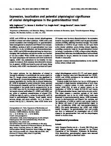
364 T. Mignot, J. P. Merlie Jr and D. R. Zusman Fig. 1. Three regions of FrzS are required for M. xanthus social motility. A. Soft agar colony expansion phenotypes of the FrzS domain deletion mutants. FrzS–GFP (TM3), DFrzS (DZ4526), DRec (DZ4533), DDBD (DZ4534), DN (DZ4535), DCoil (DZ4541), DCt (DZ4542) and DRecDCt (DZ4543) were spotted at 4 ¥ 109 cells ml-1 on nutrient-rich CYE media containing 0.5% agar and incubated for 48 h at 32°C. Bar = 1 cm. B. SDS-PAGE analysis of the FrzS domain deletion mutants. Whole-cell lysates were prepared from the strains in (A) after growth in nutrient-rich CYE media. An anti-GFP mAb was used to detect the GFP epitope on each fusion protein. C. Summary of FrzS domain deletion mutants. Cartoons show the domain organization of the wild-type FrzS–GFP fusion protein and the domain deletion mutants. ‘REC’ indicates the receiver domain and ‘DBD’ indicates the HTH-like domain. ‘Social Motility’ refers to the soft agar colony expansion phenotype for each protein listed: ‘+’ indicates a wild-type rate of colony expansion, ‘–’ indicates no colony expansion, and ‘–/+’ indicates severely defective colony expansion. ‘Subcellular Localization’ refers to the localization pattern observed for each protein listed: ‘Polar-Leading’ and ‘Polar-Lagging’ each indicate the pole of greatest FrzS enrichment, ‘ND’ indicates not done.
Results The N-terminal receiver domain of FrzS is required for function and localization The sequence of FrzS reveals five domains of interest (Fig. 1C). (i) At the N-terminus (from amino acids 1 to 116) there is a receiver domain that is 19% identical to CheY. The sequence of this domain is notable for the lack of conservation among residues that make up the canonical phosphorylation pocket (see Fraser et al., 2007). (ii) The second domain, from residues 130 to 237, shows homology, in part, to the winged helix–turn–helix (HTH) DNAbinding motif common to many response regulators. (iii) Residues 238–277 are very rich in alanine and proline, suggesting that they form a flexible linker (Perham, 2000). (iv) Residues 278–536 are predicted to form a large coiledcoil region (Ward et al., 2000). (v) Finally, residues 537– 562 constitute a C-terminal tail region (Mignot et al., 2005). The N-terminus of FrzS is essential for S-motility (Fig. 1A). However, a deletion of residues 130–237,
encompassing the HTH-like domain, produced no effect on M. xanthus motility (Fig. 1A). Based on these results, we constructed an in-frame deletion of the receiver domain in an M. xanthus strain that expressed FrzS–GFP (Mignot et al., 2005). This mutant, frzS DRec–gfp, showed as severe a defect in S-motility as a mutant with a deletion of the entire gene. Importantly, this motility defect was not due to protein instability as FrzSDRec–GFP was expressed in this strain at levels similar to FrzS–GFP in the control strain (Fig. 1B). These results show that the receiver domain of FrzS is essential for S-motility swarming. To study the effect of this mutation on FrzSDRec–GFP localization, we used a live cell imaging assay to follow the GFP fusion protein within M. xanthus cells during cell movements. Mignot et al. (2005) showed that in this assay, FrzS–GFP exhibits a complex and dynamic localization pattern (see Fig. 2A and B): (i) FrzS is primarily located at the leading cell pole as cells move forward, (ii) as movement continues, FrzS gradually localizes to the lagging pole so that FrzS is now found at both cell
© 2007 The Authors Journal compilation © 2007 Blackwell Publishing Ltd, Molecular Microbiology, 65, 363–372
Distinct FrzS domains drive polar localization 365
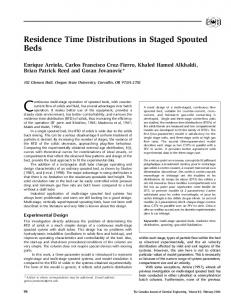
Fig. 2. The N-terminal receiver domain is required for retention of FrzS at the leading cell pole. A. Time-lapse image sequence showing the localization of FrzS–GFP during the movement of a FrzWT M. xanthus cell (TM3). The top panel shows cell location and a schematic of the fluorescent signal from the bottom panel. The bottom panel shows the GFP fluoresence signal. The red arrowhead represents the initial lagging cell pole while the green arrowhead represents the initial leading pole. Arrows indicate the direction of cell movement. ‘R’ indicates the frames in which a reversal took place. Scale bar equals 5 mm. B. Quantification of polar GFP fluoresence in the cell shown in (A). The red line (crosses) indicates the level of GFP fluoresence at the initial lagging cell pole at each time point in the image series. The green line (open boxes) indicates GFP fluoresence levels at the initial leading cell pole. ‘R’ indicates time points at which a reversal took place. Fluoresence levels at all points were normalized to the highest level observed during the time-course (initial lagging pole at 4.5 min). C. Time-lapse image sequence showing the localization of FrzSDRec–GFP during the movement of an M. xanthus cell (DZ4533). Scale bar equals 5 mm. D. Quantification of polar GFP fluoresence in the cell shown in (C). Fluoresence levels at all points were normalized to the highest level observed during the time-course (initial lagging pole at 3 min).
poles, and (iii) when cells reverse, the remaining FrzS from the old leading pole rapidly localizes to the new leading pole. In contrast to the FrzS–GFP control strain, deletion of the FrzS receiver domain caused the
truncated GFP fusion protein to mislocalize in the majority of the cells (Table 1). As these cells moved forward, FrzSDRec–GFP localized preferentially to the lagging cell pole rather than the leading cell pole (Fig. 2C and D).
Table 1. Polar localization phenotypes of FrzS–GFP variants. Straina WT
Frz FrzS–GFP FrzWT FrzSDRec–GFP DfrzE FrzS–GFP DfrzE FrzSDRec–GFP frzCD C FrzS–GFP frzCD C FrzSDRec–GFP
% leadingb 96.6 35.5 100 3.3 100 95.8
% laggingc
% mixedd
56.1
8.4
93.3
3.3 4.2
% bipolare
Cells counted
3.4
59 107 26 30 17 24
a. FrzWT FrzS–GFP (TM3), FrzWT FrzSDRec–GFP (DZ4533), DfrzE FrzS–GFP (TM4), DfrzE FrzSDRec–GFP (DZ4536), frzCD C (TM5) and frzCD C FrzSDRec–GFP (DZ4537) characterized in a live cell imaging assay for FrzS–GFP subcellular localization. Cells were filmed for 10 min on 0.5 CTT containing 1.5% agar. Only moving cells were counted. b. The percentage of filmed cells that had the strongest focus of FrzS–GFP fluorescence at the leading pole of the cell during the entire filming period. c. The percentage of filmed cells that had the strongest focus of FrzS–GFP fluorescence at the lagging pole during the entire filming period. d. The percentage of cells in which the strongest focus of FrzS–GFP fluorescence switched from the lagging pole to the leading pole during filming. e. The percentage of cells that had equal foci of fluorescence at the leading and lagging poles during the entire filming period.
© 2007 The Authors Journal compilation © 2007 Blackwell Publishing Ltd, Molecular Microbiology, 65, 363–372
366 T. Mignot, J. P. Merlie Jr and D. R. Zusman Fig. 3. The subcellular localization of FrzSDRec–GFP is regulated by the Frz system. A. Time-lapse image sequence showing the localization of FrzSDRec–GFP in a frzCD c genetic background (DZ4537). The top panel shows cell location and a schematic of the fluorescent signal from the bottom panel. The bottom panel shows the GFP fluoresence signal. The red arrowhead represents the initial lagging cell pole while the green arrowhead represents the initial leading pole. Arrows indicate the direction of cell movement. ‘R’ indicates the frames in which a reversal took place. Scale bar equals 5 mm. B. Quantification of polar GFP fluoresence in the cell shown in (A). The green line (open boxes) indicates the level of GFP fluoresence at the initial leading cell pole at each time point in the image series. The red line (crosses) indicates GFP fluoresence levels at the initial lagging cell pole. Fluoresence levels at all points were normalized to the highest level observed during the time-course (initial leading pole at 0 min). ‘R’ indicates time points at which a reversal took place.
Upon reversal, FrzSDRec–GFP initially localized to the new leading pole, but within 1–2 min, redistributed to the lagging pole (Fig. 2C and D). These results suggest that in the absence of the receiver domain, FrzSDRec–GFP is still targeted to the new leading pole; however, without the receiver domain, the truncated FrzS protein cannot stably localize at this site and the protein rapidly redistributes to the lagging pole. FrzS is not required for the control of TFP-based cell reversals by the Frz chemosensory system (J. Merlie, unpubl. data); however, the Frz system does regulate the polar localization dynamics of FrzS (Mignot et al., 2005). We therefore examined the effect of mutations in the Frz system on the subcellular localization of FrzSDRec–GFP. Previously, it was shown that DfrzE mutants very rarely reverse, while frzCD C mutants signal constitutively and reverse once every 1–2 min (Blackhart and Zusman, 1985; Bustamante et al., 2004). We tracked the GFP fluorescent clusters in the DfrzE frzS DRec–gfp double mutant, and the frzCD CfrzS DRec–gfp double mutant as cells moved forwards and reversed. In cells with a wild-type Frz system (FrzWT), FrzS–GFP preferentially accumulated at the leading cell pole throughout the reversal cycle (Table 1). In contrast, in DfrzE cells, FrzSDRec–GFP localized preferentially at the lagging pole (Table 1 and data not shown). Indeed, very few cells showed FrzSDRec–GFP localized to the leading pole, probably due to infrequent cell reversals. On the other hand, FrzSDRec–GFP localized
primarily to the leading pole of cells in the constitutive frzCD c mutant (Table 1 and Fig. 3A). These results suggest that the transient leading pole localization of FrzSDRec–GFP seen in reversing FrzWT cells results from signalling from the Frz pathway and that the receiver domain acts not to direct FrzS to the new leading pole but to maintain its residence there over the course of the reversal cycle. The C-terminal domain of FrzS is involved in lagging cell pole localization The localization pattern observed with the frzS DRec–gfp mutant suggested that another FrzS motif is responsible for targeting FrzS to the lagging pole. We therefore used a genetic screen designed to isolate additional mutations in frzS that are essential for its function. We first constructed an expression system in which frzS was cloned downstream of Ptet, a tetracycline-inducible promoter in a plasmid vector (Fig. 4A). The inducible frzS gene was integrated into the chromosome ectopically at the car locus (Lopez-Rubio et al., 2002) in a DfrzS mutant background. This strain showed anhydrotetracyclinedependent expression of FrzS and complementation of the swarming defect associated with DfrzS mutants (Fig. 4A and data not shown). Although integration of this expression system disrupted synthesis of carotenoid pigments, it did not effect swarming or development (data not
© 2007 The Authors Journal compilation © 2007 Blackwell Publishing Ltd, Molecular Microbiology, 65, 363–372
Distinct FrzS domains drive polar localization 367
Fig. 4. A C-terminal motif targets FrzS to the lagging cell pole. A. Tetracycline-inducible complementation of a frzS mutant. Design and integration of the construct in the car locus (top). Anydrotetracycline (AT)-dependent complementation of the soft agar colony swarming defect of the frzS mutant after transformation and integration of the inducible construct (frzS frzSind; bottom). B. The 537–548 motif is located downstream from the coiled-coil domain. The probability of each FrzS residue to be involved in coiled coil formation as calculated by the multicoil algorithm was plotted as a function of their location within the FrzS sequence. FrzS residues within the 537–548 motif show probabilities to be folded as a coiled coil below 20%. C. The 537–548 motif is conserved in a FrzS homologue from A. dehalogenans. M. xanthus FrzS [FrzS (Mx)] and A. dehalogenans FrzS [FrzS (Ad)] were aligned using the Sim algorithm. The most conserved regions between the two proteins are the pseudo-receiver domains and the C-terminal tail. The 10-residue motif is boxed in pink. D. FrzSDCt–GFP is localized at the leading cell pole and switches to the new leading pole upon cellular reversals. Fluorescence micrographs of two reversing cells captured every 30 s are shown. The white arrows show the direction of respective cells and the white arrowheads highlight switching of FrzSDCt–GFP to the new leading pole. Scale bar = 2 mm. E. Accumulation of FrzSDCt–GFP at the leading cell pole. Mean relative fluorescence at the leading cell pole for FrzS–GFP (1) and FrzSDCt–GFP (2) measured for 100 cells in each case.
shown). The FrzS expression plasmid was then subjected to random mutagenesis and transformed into the frzS mutant strain by electroporation. Clones that were drug resistant but failed to complement FrzS expression were analysed further. Surprisingly, almost all of the selected clones were found to produce unstable FrzS protein or to contain mutations in the promoter region. Nevertheless, we did identify one nonsense mutation that resulted in the production of a 483-residue polypeptide that was very stable but showed complete loss of function (data not shown).
To further define the extent of the C-terminus that is important for FrzS function, we constructed strains containing FrzS truncated after residues 517 (FrzS517), 537 (FrzS537) and 548 (FrzS548). FrzS517 and FrzS537 were nonfunctional, but FrzS548 was fully functional (data not shown), indicating that the region encompassing residues 537–548 were critical for function. To confirm that the results were not due to C-terminal processing of the truncated versions of FrzS, we constructed a strain expressing a FrzS–GFP variant deleted only in residues 537–548 (FrzSDCt) (described as FrzSD537–548 in Mignot et al., 2005).
© 2007 The Authors Journal compilation © 2007 Blackwell Publishing Ltd, Molecular Microbiology, 65, 363–372
368 T. Mignot, J. P. Merlie Jr and D. R. Zusman Fig. 5. Distinct roles of the FrzS domains. A. Localization phenotypes of FrzSDCt–GFP, FrzSDRecDCt–GFP and FrzSDCoil–GFP. Inset: Localization of wild-type (WT) FrzS–GFP. Numbers in black reflect the number of cells from each subpopulation for each mutant. The numbers in red are the values obtained for WT FrzS–GFP. Scale bar = 2 mm. B. Transient localization of FrzSDRecDCt–GFP at the leading cell pole. Fluorescence micrographs of two cells captured every 30 s are shown. The white arrows show the direction of respective cells and the white arrowheads highlight FrzSDRecDCt–GFP localizing transiently to the leading cell pole in one of the cells. Scale bar = 2 mm. C. FrzSDRecDCt–GFP localizes within a coiled filament. Deconvolved images of three different z planes of a cell expressing FrzSDRecDCt–GFP stained with a highly specific a-FrzS antibody (Processed). The Raw image is the overlay of the unprocessed FrzS-specific signal (green) and the cell membrane stain FM464 (red). Scale bar = 1 mm. The insets show IFM images obtained in similar conditions for WT (processed) or frzS mutant cells (unprocessed). Note the change in scale: scale bar = 2 mm.
As expected, FrzSDCt–GFP was fully stable and severely defective in S-motility (Fig. 1A and B). It is likely that residues 538–547 contain a specific functional motif because they are unlikely to be structural components of the coiled-coil domain (Fig. 4B). Moreover, the motif is also present in the FrzS homologue from Anaeromyxobacter dehalogenans where 8 out of 10 residues are conserved (Fig. 4C). This is especially striking because although both proteins show a conserved domain structure, their primary sequences outside of this region show a significant level of divergence (Fig. 4C). The fusion of FrzSDC-term to GFP allowed us to investigate
its subcellular localization and dynamics in moving cells. FrzSDCt–GFP either was localized at one cell pole only (80% of the cells; Fig. 5A) or was completely mislocalized (20% of the cells; Fig. 5A). In moving cells, FrzSDCt–GFP localized to the leading pole and upon cellular reversal, its localization switched to the new leading pole (Fig. 4D). These results suggest that the FrzS motif between residues 537 and 548 is essential for targeting to the lagging pole. Interestingly, the 537–548 motif was also required for optimal binding at the leading pole as the level of FrzSDCt– GFP fluoresence was reduced about twofold at that pole compared with FrzS–GFP (Fig. 4E).
© 2007 The Authors Journal compilation © 2007 Blackwell Publishing Ltd, Molecular Microbiology, 65, 363–372
Distinct FrzS domains drive polar localization 369 Distinct roles for the domains of FrzS in protein localization To test whether the receiver domain and the C-terminal tail are the only regions necessary for polar localization, we constructed a strain that expresses a FrzS protein lacking both the receiver domain and the C-terminal tail fused to GFP at the C-terminus (FrzSDRecDCt–GFP, Fig. 1C). The swarming defect of the resulting strain was similar to that of the FrzSDRec–GFP-expressing strain (Fig. 1A). This phenotype was not the result of protein instability or lack of expression, as FrzSDRecDCt–GFP was fully stable (Fig. 1B). As expected, FrzSDRecDCt–GFP was more severely mislocalized than FrzSDCt–GFP with complete absence of polar localization in ~95% of the cells (Fig. 5A). These results show that stable polar residence of FrzS is mostly dependent on the combined actions of the receiver domain and the C-terminal tail. The fact that FrzSDRec–GFP and FrzSDCt–GFP still switch to the new leading pole upon reversal suggests that transport of the FrzS protein between the cell poles is mainly mediated by the coiled-coil domain. Therefore, active transport of FrzSDRecDCt–GFP would still be expected to occur. Indeed, polar localization of FrzSDRecDCt–GFP could be observed in 5% of the cells (Fig. 5A). In these cases, FrzSDRecDCt–GFP was localized very transiently at the leading pole suggesting that it could be transported but failed to reside at its destination (Fig. 5B). To further confirm this, we constructed a strain expressing a FrzS variant lacking the coiled-coil domain fused to GFP at the C-terminus (FrzSDCoil–GFP; Fig. 1C). Again, the resulting strain was completely deficient at swarming on soft agar (Fig. 1A). The level of expressed FrzSDCoil–GFP is lower than the level of FrzS–GFP (Fig. 1B). However, it is unlikely that this is responsible for the swarming defect, as lower levels of induced Ptet-dependent FrzS expression are sufficient to fully restore swarming to the frzS mutant strain (data not shown). FrzSDCoil–GFP was completely mislocalized with 100% of the cells showing absence of any detectable polar localization, even when protein dynamics were monitored in moving cells (Fig. 5A and data not shown). These results suggest that FrzS is transported between poles via the coiled-coil domain. Also, as FrzSDCoil–GFP is essentially a fusion between the receiver domain and the C-terminal tail, we conclude that these motifs are necessary but not sufficient to target FrzS to the cell poles. Finally, we have shown that non-polarly localized FrzSDCt–GFP appears to decorate a coiled filament that can be resolved by immunofluorescence (IFM) and deconvolution microscopy (Mignot et al., 2005). This filament was proposed to be involved in transporting FrzS between the cell poles (Mignot et al., 2005). FrzSDRecDCt– GFP also localized along a coiled filament (Fig. 5C). That
structure was not observed when FrzSDCoil–GFP was analysed by IFM (data not shown). However, this result is not definitive as the lower levels of FrzSDCoil–GFP could prevent detection of the filament. These results, in toto, suggest that pole-to-pole transport of FrzS is mediated through the coiled coil. Discussion Cell polarity is involved in many bacterial processes including cell division, chromosome segregation, pilus and flagella assembly, chemoreceptor clusters, cell differentiation and actin-driven bacterial motility in the cytoplasm of eukaryotic host cells (Shapiro et al., 2002). As the cell poles are sites of multiple specialized cellular functions, they exhibit some of the characteristics of subcellular compartments (Shapiro et al., 2002). Thus, the targeting of proteins to the cell poles is important, although our understanding of polar protein localization is limited. M. xanthus S-motility is an especially attractive model to study the dynamics of protein localization because cells periodically reverse their direction of movement and the biochemical identity of the leading and lagging cell poles. Here, we analyse the FrzS protein as a probe to investigate the molecular mechanisms that control these dynamics. Previously, we showed pole-to-pole trafficking of FrzS as cells moved forward and reversed and that this trafficking is most likely achieved by active transport along a cytoskeletal filament (Mignot et al., 2005). In this article, we analysed the various domains of FrzS to identify regions critical for function and localization. We identified three domains of interest. (i) The N-terminal receiver domain of FrzS appears to be required for maintenance of FrzS at the leading cell pole; mutants lacking this domain were still targeted to the new leading pole, but the protein rapidly redistributed to the lagging pole. (ii) The C-terminal domain of FrzS was required for localization at the lagging cell pole; mutants lacking this motif, from residues 537 to 548, localized primarily to the leading pole. (iii) The coiledcoil domain, from residues 278 to 536, appears to be important for protein stability and for all polar localization events; mutants lacking this motif displayed cytoplasmic FrzS and the complete absence of any polar localization. Our results are compatible with a model whereby each FrzS domain has a specific role in trafficking (Fig. 6). We propose that the receiver domain of FrzS docks with an anchor specifically located at the leading pole. The C-terminal domain may also contribute to residence at the leading pole because localization at that pole is reduced by 50% when this motif is deleted. Regulation at the level of the anchor or the FrzS receiver domain would result in the detachment of FrzS from the leading pole (see below and Fraser et al., 2007). Released FrzS would then bind
© 2007 The Authors Journal compilation © 2007 Blackwell Publishing Ltd, Molecular Microbiology, 65, 363–372
370 T. Mignot, J. P. Merlie Jr and D. R. Zusman
Fig. 6. Role of the FrzS domains in pole-to-pole oscillations across the reversal cycle. 1. FrzS binds to a leading pole-specific anchor (purple) by the pseudo-receiver domain (yellow diamond). The C-terminal tail (orange star) is probably also involved in leading pole residence but to a lesser extent. Following regulated release FrzS is transported to the lagging pole along a putative cytoskeletal filament via the coiled-coil domain (blue coil) and its C-terminal tail binds a lagging pole-specific anchor. 2. At the time of reversal the respective anchors are switched from one pole to the other and all available FrzS molecules are rapidly transported to the new leading pole. 3. The cycle resumes. The different sizes of FrzS in the cartoons illustrate the relative subpopulations of FrzS at each stage.
to the lagging pole by docking of the C-terminal tail to a lagging pole-specific anchor (Fig. 6). Upon cellular reversal, the polar localization of each specific anchor would be switched and the cycle would resume. We hypothesize that FrzS is transported from one cell pole to the other via the coiled-coil domain. Indeed, a mutant lacking both the pseudo-receiver domain and the C-terminal tail was still transiently localized at the cell poles. We suggest that this double mutant reaches the cell poles but cannot reside there in the absence of the receiver and C-terminal motifs. Indeed, a FrzS mutant lacking the coiled-coil domain never localized to the cell poles, suggesting that it was not being transported. These results are consistent with the model that FrzS is transported along a filament by way of the coiled-coil domain and delivered to the cell poles where it resides through the combined actions of the receiver domain and the C-terminal tail (Fig. 6). The coiled coil is also essential for polar localization of FrzS, either because transport between poles is required for the docking of FrzS at the poles or because the coiled coil itself is also involved in binding at the cell poles. We could not discriminate between these possibilities because disruption of the coiled coil led to complete loss of function and localization. Pole-to-pole oscillation has been shown for the DivK response regulator in C. crescentus (Matroule et al.,
2004). In that case, the oscillations result from covalent modification of DivK by a kinase and a phosphatase, each localized at an opposite cell pole (Lam et al., 2003). However, the catalytic site for receiver domain phosphorylation is not conserved in FrzS, suggesting that regulation of the pseudo-receiver domain of FrzS does not involve phosphorylation (Fraser et al., 2007). Instead, detachment from the leading pole may be triggered by modification of the polar anchor as is the Min system, where ATP hydrolysis by polar MinD releases MinE from the cell pole (Fig. 5; Rothfield et al., 2005). Unlike DivK that continuously shuttles from one pole to the other, FrzS resides at both poles and is only ‘shed’ slowly from the leading pole before it is transported in bulk to the lagging pole upon cell reversal (Mignot et al., 2005). These dynamics are possible because FrzS has evolved two distinct motifs to ensure selective affinity for the leading pole and the lagging pole. The exact function of FrzS remains enigmatic. Proper spatial regulation of FrzS is essential for S-motility yet our analysis (Fig. 6) predicts that the oscillations of FrzS rely on prior switching of distinct polar anchors, and therefore, FrzS may not act to trigger cellular reversals. Indeed, we recently found that FrzS is dispensable for S-motilitydependent reversals in a hyper-reversing frzCD c strain (unpublished data). The FrzS phenotype is reminiscent of Myosin II mutations in Dictyostelium discoidum resulting in slight defects at the single cell level but leading to a complete block at the mound stage of development (Chisholm and Firtel, 2004). Within the social swarm, proteins such as FrzS may be required for pili to produce strong enough forces to move in three dimensions between other cells, often breaking tight cell–cell interactions. It is likely that the Frz system does not regulate FrzS oscillations directly but rather acts upstream to regulate pole-to-pole oscillations of proteins that regulate motility, for example, by signalling the general release of polar motility proteins. Several lines of evidence suggest that FrzS is part of a downstream module that is transported between cell poles. (i) The localization defect of FrzSDRec–GFP was mostly rescued in a frzCD c strain, suggesting that enhanced Frz signalling results in hyperoscillation of binding proteins that drag FrzSDRec–GFP to the leading pole. (ii) Similarly, FrzS mutants lacking the receiver domain, the C-terminal tail or both were still delivered to the cell poles suggesting that these motifs are important for polar residence but not polar targeting which may happen because FrzS is transported along with other motility components. In conclusion, our experiments identified several domains of FrzS that are required for proper dynamic localization of FrzS as cells move forward and reverse. Elucidating the molecular identity of the polar anchors and the mechanism of transport between poles will further
© 2007 The Authors Journal compilation © 2007 Blackwell Publishing Ltd, Molecular Microbiology, 65, 363–372
Distinct FrzS domains drive polar localization 371 advance our understanding of subcellular localization and trafficking in the bacterial cell. Experimental procedures Strains and growth conditions All M. xanthus strains were cultured on nutrient-rich CYE media (10 mM MOPS pH 7.6, 1% Casitone, 0.5% yeast extract and 4 mM MgSO4) containing 1.5% agar at 32°C. Liquid cultures were grown in CYE at 32°C with shaking (225 r.p.m.). Standard colony-level S-motility assays were performed on CYE containing 0.5% agar. Live cell imaging assays were performed with cells spotted on 0.5 CTT (10 mM Tris pH 7.6, 0.5% Casitone, 1 mM KH2PO4, 8 mM MgSO4) containing 1.5% agar. MMC buffer (10 mM MOPS pH 7.6, 4 mM MgSO4, 2 mM CaCl2) was used for cell re-suspensions. E. coli strains were grown in LB media (Sambrook et al., 1989). Kanamycin and ampicillin were used at 100 mg ml-1 and oxytetracycline was used at 10 mg ml-1.
Construction of strains In-frame deletions in frzS were generated by the selection/ counter-selection method as previously described (Ueki et al., 1996). Briefly, all plasmids used for in-frame deletions were generated by two-step overlap PCR extension from M. xanthus DZ2 purified chromosomal DNA. Regions (1 kb) upstream and downstream of the region to be deleted or mutagenized were amplified by an initial round of PCR and fused in a subsequent round by overlap extension. Inserts were cloned into the XbaI and HindIII sites of the selection/ counter-selection plasmid pBJ113. PBJ113 contains a kanamycin resistance gene (KmR) and a galactose sensitivity (galK) counter-selection gene. Deletion plasmids were electroporated into the relevant strains, and integrants were selected with kanamycin. Plasmids were then looped out via counter-selection with galactose to generate the desired mutation. All strains were verified with a combination of at least two of the following methods: PCR, Southern blotting or Western blotting.
Inducible expression of frzS and random mutagenesis The pCT2 integration vector was constructed as follows: the tet repressor and promoter region from E. coli plasmid RP4 (Sakanyan et al., 1978) was flanked on each side by 1 kb M. xanthus DNA regions from the car locus encoding carotenoid biosynthetic enzymes (Lopez-Rubio et al., 2002) and cloned in pBJ113. frzS was then cloned in pCT2 immediately downstream from the tet promoter region. A detailed map of pCT2 is available upon request. The resulting construct allows integration of the frzS-inducible gene within the car locus by a single integration event. Removal of the pBJ113 plasmid backbone after integration can be obtained by a standard galactose resistance selection procedure (Ueki et al., 1996). All possible integrations of inducible frzS perturbed carotenoid biosynthesis but did not affect motility and development as measured under standard laboratory conditions. Induction of frzS was obtained in CYE supplemented by Anhydrotetracycline
(Sigma) in concentrations ranging between 10 and 100 ng ml-1 without noticeable toxic effect. Random mutagenesis of inducible frzS was obtained by transforming the inducible frzS plasmid into the mutator strain E. coli XL1-Red (Stratagene). A library of mutated plasmids was then transformed into the M. xanthus frzS mutant strain. All tranformants that failed to display motility on soft 0.3% agar CYE were then selected and, in each case, frzS was amplified by PCR and sequenced.
Analysis of motility phenotypes Colony-level social motility analysis was performed by spotting 4 ¥ 107 cells on nutrient-rich CYE media containing 0.5% agar and incubating for 24–48 h at 32°C. Phenotypes were recorded an Alpha Innotech gel documentation system (Alpha Innotech Corporation).
Live cell imaging of FrzS–GFP Imaging was performed as described by Mignot et al. (2005). Briefly, cells were harvested and spotted on thin 0.5 CTT 1.5% agar pads on glass slides and covered directly with a glass coverslip. Cells were incubated for 15 min at room temperature in the dark before imaging on a Deltavision fluorescence microscope with a FITC filter set and a 100¥ oil-immersion objective (Applied Precision). Cells were filmed for 10 min with 30 s time-lapse. Time-lapse montage images were assembled using Photoshop Illustrator (Adobe Systems Incorporated). FrzS–GFP fluorescence intensity was quantified using Image J (NIH) and graphed using Kalidagraph.
Immunoblot analyses Whole-cell lysates were prepared from liquid cultures harvested at 4 ¥ 108 cells ml-1 and proteins were resolved by SDS-PAGE on 8% polyacrylamide (Sambrook et al., 1989). Proteins were transferred to nitrocellulose membranes (BioRad) using a tank transfer apparatus (Bio-Rad) in TrisGlycine buffer containing 20% methanol; membranes were blocked for 30 min in PBS with 0.1% Tween 20 and 5% powdered milk and probed with rabbit anti-FrzS antibody (Mignot et al., 2005) at 1:10 000, or mouse anti-GFP monoclonal antibody (mAb) (Jackson Immunoresearch) at 1:2000. Anti-mouse or anti-rabbit horseradish peroxidase-conjugated secondary antibodies (Pierce) were used at 1:5000. Immunoreactive complexes were detected by Western Lightning chemiluminescent reagents (NEN) and Kodak Biomax Light film.
Acknowledgements We would like to thank Kyungyung Cho for the initial experiments with the inducible tet promoter in M. xanthus. We also thank Emilia Mauriello and Ansley Scott for critical comments on this manuscript and the entire Zusman lab for stimulating discussion. This research was supported by a grant from the National Institute of Health to DRZ (GM20509). J.P.M. was the recipient of a NSF Graduate Fellowship.
© 2007 The Authors Journal compilation © 2007 Blackwell Publishing Ltd, Molecular Microbiology, 65, 363–372
372 T. Mignot, J. P. Merlie Jr and D. R. Zusman References Blackhart, B.D., and Zusman, D.R. (1985) ‘Frizzy’ genes of Myxococcus xanthus are involved in control of frequency of reversal of gliding motility. Proc Natl Acad Sci USA 82: 8767–8770. Bustamante, V.H., Martinez-Flores, I., Vlamakis, H.C., and Zusman, D.R. (2004) Analysis of the Frz signal transduction system of Myxococcus xanthus shows the importance of the conserved C-terminal region of the cytoplasmic chemoreceptor FrzCD in sensing signals. Mol Microbiol 53: 1501–1513. Chisholm, R.L., and Firtel, R.A. (2004) Insights into morphogenesis from a simple developmental system. Nat Rev Mol Cell Biol 5: 531–541. Fraser, J.S., Merlie, J.P., Echols, N., Weisfield, S.R., Mignot, T., Wemmer, D.E., et al. (2007) An atypical receiver domain controls the dynamic polar localization of the Myxococcus xanthus social motility protein FrzS. Mol Microbiol, 65, in press. doi: 10.1111/j.1365-2958.2007.05785.x. Hodgkin, J., and Kaiser, D. (1979) Genetics of Gliding motility in Myxococcus xanthus (Myxobacteriales): two gene systems control movement. Mol Gen Genet 171: 177–191. Kruse, K., Howard, M., and Margolin, W. (2007) An experimentalist’s guide to computational modelling of the Min system. Mol Microbiol 63: 1279–1284. Lam, H., Matroule, J.Y., and Jacobs-Wagner, C. (2003) The asymmetric spatial distribution of bacterial signal transduction proteins coordinates cell cycle events. Dev Cell 5: 149–159. Lopez-Rubio, J.J., Elias-Arnanz, M., Padmanabhan, S., and Murillo, F.J. (2002) A repressor-antirepressor pair links two loci controlling light-induced carotenogenesis in Myxococcus xanthus. J Biol Chem 277: 7262–7270. Matroule, J.Y., Lam, H., Burnette, D.T., and Jacobs-Wagner, C. (2004) Cytokinesis monitoring during development; rapid pole-to-pole shuttling of a signaling protein by
localized kinase and phosphatase in Caulobacter. Cell 118: 579–590. Mignot, T., Merlie, J.P., and Zusman, D.R. (2005) Regulated pole-to-pole oscillations of a bacterial gliding motility protein. Science 310: 855–857. Mignot, T., Shaevitz, J.W., Hartzell, P.L., and Zusman, D.R. (2007) Evidence that focal adhesion complexes power bacterial gliding motility. Science 315: 853–856. Perham, R.N. (2000) Swinging arms and swinging domains in multifunctional enzymes: catalytic machines for multistep reactions. Annu Rev Biochem 69: 961–1004. Rothfield, L., Taghbalout, A., and Shih, Y.L. (2005) Spatial control of bacterial division-site placement. Nat Rev Microbiol 3: 959–968. Sakanyan, V.A., Yakubov, L.Z., Alikhanian, S.I., and Stepanov, A.I. (1978) Mapping of RP4 plasmid using deletion mutants of pAS8 hybrid (RP4–ColE1). Mol Gen Genet 165: 331–341. Sambrook, J., Fritsch, E.F., and Maniatis, T. (1989) Molecular Cloning: A Laboratory Manual. Cold Spring Harbor, NY: Cold Spring Harbor Laboratory Press. Shapiro, L., McAdams, H.H., and Losick, R. (2002) Generating and exploiting polarity in bacteria. Science 298: 1942– 1946. Sun, H., Zusman, D.R., and Shi, W.Y. (2000) Type IV pilus of Myxococcus xanthus is a motility apparatus controlled by the frz chemosensory system. Current Biology 10: 1143– 1146. Ueki, T., Inouye, S., and Inouye, M. (1996) Positive-negative KG cassettes for construction of multi-gene deletions using a single drug marker. Gene 183: 153–157. Wall, D., and Kaiser, D. (1999) Type IV pili and cell motility. Mol Microbiol 32: 1–10. Ward, M.J., Lew, H., and Zusman, D.R. (2000) Social motility in Myxococcus xanthus requires FrzS, a protein with an extensive coiled-coil domain. Mol Microbiol 37: 1357– 1371.
© 2007 The Authors Journal compilation © 2007 Blackwell Publishing Ltd, Molecular Microbiology, 65, 363–372