Research Article
Ultrasonographic Measurement of the Femoral Cartilage Thickness in Patients with Juvenile Idiopathic Arthritis Levent Ozcakar1, Aslı Celebi2, Murat Kara1, Turker Turker3, Adem Polat4, Victoria Akse-Onal2, Erkan Demirkaya4, Seza Ozen2; FMF Arthritis Vasculitis and Orphan disease Research in Paediatric Rheumatology (FAVOR). 1 Department of Physical Medicine and Rehabilitation; 2 Division of Pediatric Nephrology and Rheumatology, Hacettepe University Medical School; 3Division of Epidemiology; 4Division of Pediatric Nephrology and Rheumatology, Gülhane Military Medical Academy, Ankara, Turkey
Corresponding author: Murat Kara, Hacettepe Üniversitesi Hastaneleri Zemin Kat FTR AD, Sıhhıye, Ankara, e-mail:
[email protected] Received: November 03, 2011 Accepted: January 02, 2012 Ann Paediatr Rheum 2012; 1: 54-57 DOI: 10.5455/apr.010220120434
Abstract Objective: To evaluate the femoral cartilage thicknesses of oligoarticular JIA patients (affected vs unaffected) by using ultrasonography (US), and to find out whether those US measurements were related with the clinical parameters of the patients. Methods: Twenty-six children (12 boys, 14 girls) with mono-articular (knee involvement) juvenile idiopathic arthritis (JIA), with a mean age of 8.84±4.06 years were enrolled. Demographic and clinical features of the subjects including age, sex, body mass index, disease duration, age at onset, medical treatment, laboratory evaluations and presence of uveitis were all noted. The femoral cartilage thicknesses of both knees were measured with a 7-12 MHz linear probe while subjects’ knees were held in maximum flexion. Three mid-point measurements were taken from both knees (lateral condyle, intercondylar area and medial condyle) and the values relating to the unaffected knees were taken as controls. Results: Femoral cartilage thickness values were found to be similar (affected vs. non-affected sides) for all the three measurement sites (p values >0.05) and cartilage thickness values did not correlate with the clinical features of the patients. Conclusion: According to our preliminary findings, the femoral cartilage seems to be unaffected in JIA; however taking into account the limited number of patients with inhomogenous disease course, we suggest that further studies would be necessary to confirm our results. Key words: Juvenile idiopathic arthritis, knee, cartilage, thickness, ultrasonography
Introduction Juvenile idiopathic arthritis (JIA) is the most common rheumatic disorder in childhood and it is characterized by chronic inflammatory lesions of the joints and the nearby soft tissues.
The disease process mainly starts with the involvement of the synovium whereby proliferative synovitis may lead to articular cartilage damage as well. Overall, the knee is the most commonly affected joint.
55
Ultrasonographic Measurement in JIA
Having many advantages in the diagnosis and follow up of several diseases in childhood (cheap, time efficient, does not require sedation or radiation exposure), ultrasound (US) helps to understand several structural changes in JIA. Further, in recent years, measurement of the femoral cartilage thickness by using US has been shown as a valid and reliable method [1-3]. Accordingly, the purpose of our study was two-fold; first we aimed to compare the femoral cartilage thicknesses of oligoarticular JIA patients (affected vs. unaffected knees) by using US and second, we aimed to find out whether those US measurements were related with the clinical parameters of the patients.
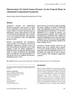
Methods Participants Twenty-six children with mono-articular (knee involvement) JIA (12 boys, 14 girls) with a mean age of 8.84±4.06 years (range 2.20-16.22) were enrolled in this study. Inclusion criteria were a diagnosis of JIA based on the 2001 revised ILAR criteria [4] and a disease category of persistent oligoarthritis and extended oligoarthritis. Patients were excluded if they met the ILAR criteria for systemic arthritis, enthesitisrelated arthritis, polyarthritis psoriatic arthritis, or undifferentiated arthritis. All subjects and parents were informed about the study procedure and the parents of all the participants gave informed consent. Local Ethics Committee approved the study protocol. Protocol Demographic and clinical features of the subjects including age, sex, body mass index (BMI), disease duration, age at onset, medical treatment, laboratory evaluations (complete blood count, erythrocyte sedimentation rate, C-reactive protein, anti-nuclear antibody, liver/renal functions test) and presence of uveitis were all noted. The US measurements of the femoral cartilage were performed bilaterally with a linear probe (7-12 MHz Logiq P5, GE Medical Systems, USA). While subjects comfortably sat on the examination table with their knees in maximum flexion, the probe was placed in an axial position on the suprapatellar area; the distal femoral cartilage was visualized as a strongly anechoic structure between the sharp bony (femur) cortex and the suprapatellar fat (Figure 1). Three mid-point DOI:10.5455/apr.010220120434
Figure 1. Ultrasonographic image (suprapatellar axial view) demonstrating the femoral cartilage measurements (LC: Lateral cond-
yle, IC: Intercondylar area, MC: Medial condyle, F: Femur)
measurements (at lateral condyle, intercondylar area and medial condyle) were taken from each knee. Statistical analysis was performed by using SPSS version 15.0. Data were expressed as mean ± standard deviation or percentages. Paired-samples t test was used to compare the mean cartilage thickness values between the affected and non-affected knees. Spearman correlation coefficients were calculated to evaluate relations between the disease duration and cartilage measurements. Statistical significance was set at p0.05). Cartilage thickness values did not correlate with the clinical features of the patients except a weak negative correlation between the medial femoral condyle cartilage thickness (affected knee) and disease duration (r=-0.389, p=0.06). Table 2. Ultrasonographic measurements of the femoral cartilage thickness (mm) Affected side
Contralateral side
p
Lateral condyle
3.47±0.71
3.57±0.71
>0.05
Intercondylar area
3.44±0.78
3.32±0.63
>0.05
Medial condyle
3.57±0.78
3.55±0.75
>0.05
Discussion Juvenile idiopathic arthritis (JIA) is a heterogeneous group of childhood disorders. The destructive pannus may cause significant deformity and morbidity whereby early and proper treatment is required to limit the disability. Although the disease course can be variable, with periods of activity followed by remission, nearly up to 70% of children continue to report disability and limitation of their activities into adulthood [5-10]. Herein, as the synovial proliferation may affect the cartilage as well -resulting in joint space narrowing and leading poor quality of life and disability- we considered that US would to be a convenient and prompt way of monitoring such changes in these patients. Normal cartilage is seen as a hypoechoic structure with a smooth outline over the bony surfaces [1]. In the initial stages of JIA, ultrasound yields thickening of the cartilage due to edema. On the contrary, as the inflammation goes on, synovium begins to grow progressively over the articular cartilage -causing blurring of the otherwise sharp marginsand produces irregular defects on the cartilage and the bones [11-13]. Therefore, the cartilage is usually eroded and thinned in chronic diseases course. In this regard, instead of a single measurement, we imply that serial US imaging might be useful in the follow up of any disease impact on the cartilage. Our finding of indifference between the cartilage thicknesses (affected vs. unaffected) could be attributed to the fact that oligoarticular type is relatively the most benign Annals of Paediatric Rheumatology
form of JIA. Yet, in contrast to female gender, polyarticular and symmetrical joint involvement, elevated inflammatory markers and rheumatoid factor positivity as being the consistent predictors of a poor outcome [10]; oligoarticular type has always been associated with a more favorable outcome when compared with other subtypes [14-16]. Thus, a possible impact of JIA on knee cartilage could have been masked with the favorable disease course of this type. Increase of cartilage thickness in the acute phase, followed by marked reduction in the chronic stage, has been described on US in patients with JIA [13]. This could have also confounded our results since the patients did not have a homogenous disease activity at the time of enrollment. On the other hand, during growth, epiphyseal cartilage transforms to bone, gradually narrowing the radiographic joint space. For that reason, we have taken the healthy contralateral limbs of JIA patients as controls in our study. Further, it has been also shown that there was no difference between femoral cartilage thickness values of the left and right extremities in healthy children [17]. Other than the limited number of subjects in our study, measuring only the cartilage thickness could be considered as a limitation; whereas it has been also shown that differences in cartilage volume result primarily from a difference in joint size rather than cartilage thickness [18]. Overall, in the light of our preliminary findings, we may conclude that knee cartilage seems to be unaffected in monoarticular JIA patients. However, taking into account the limited number of patients with inhomogenous disease course in our study population, we suggest that further studies with larger groups and longer disease durations would be necessary to establish a better conclusion. Competing interests: We have no competing interest. Funding: None. Provenance and peer review: Not commissioned; externally peer reviewed.
References 1. Spannow AH, Pfeiffer-Jensen M, Andersen NT, Stenbøg E, Herlin T. Inter and intraobserver variation of ultrasonographic cartilage thickness assessments in small and large joints in healthy children. Pediatr Rheumatol Online J 2009;7:12. Year 2012 Volume:1 Issue:1 54-57
Ultrasonographic Measurement in JIA
2. Spannow AH, Stenboeg E, Pfeiffer-Jensen M, Herlin T. Ultrasound measurement of joint cartilage thickness in large and small joints in healthy children: a clinical pilot study assessing observer variability. Pediatr Rheumatol Online J 2007;5:3. 3. Castriota-Scanderbeg A, De MV, Scarale MG, Bonetti MG, Cammisa M. Precision of sonographic measurement of articular cartilage: inter- and intraobserver analysis. Skelet Radiol 1996;25:545-549. 4. Petty RE, Southwood TR, Manners P, Baum J, Glass DN, Goldenberg J, et al. International League of Associations for Rheumatology classification of juvenile idiopathic arthritis:second revision, Edmonton, 2001. J Rheumatol 2004;31:390-392. 5. Oen K, Malleson PN, Cabral DA, Rosenberg AM, Petty RE, Cheang M. Disease course and outcome of juvenile rheumatoid arthritis in a multicenter cohort. J Rheumatol 2002;29:1989-1999. 6. Packham JC, Hall MA. Long-term follow-up of 246 adults with juvenile idiopathic arthritis: functional outcome. Rheumatology (Oxford) 2002;41:1428-1435. 7. Packham JC, Hall MA. Long-term follow-up of 246 adults with juvenile idiopathic arthritis: social function, relationships and sexual activity. Rheumatology (Oxford) 2002;41:1440-1443. 8. Packham JC, Hall MA. Long-term follow-up of 246 adults with juvenile idiopathic arthritis: education and employment. Rheumatology (Oxford) 2002;41:14361439. 9. Foster HE, Marshall N, Myers A, Dunkley P, Griffiths ID. Outcome in adults with juvenile idiopathic arthritis: a quality of life study. Arthritis Rheum 2003;48:767-775.
DOI:10.5455/apr.010220120434
57
10. Adib N, Silman A, Thomson W. Outcome following onset of juvenile idiopathic inflammatory arthritis: II. predictors of outcome in juvenile arthritis. Rheumatology (Oxford) 2005;44:1002-1007. 11. Sureda D, Quiroga S, Arnal C, Boronat M, Andreu J, Casas L. Juvenile rheumatoid arthritis of the knee: evaluation with US. Radiology 1994;190:403-406. 12. Johnson K. Imaging of juvenile idiopathic arthritis. Pediatr Radiol 2006;36:743-758. 13. El-Miedany YM, Housny IH, Mansour HM, Mourad HG, Mehanna AM, Megeed MA. Ultrasound versus MRI in the evaluation of juvenile idiopathic arthritis of the knee. Joint Bone Spine 2001;68:222-230. 14. Flato B, Aasland A, Vinje O, Forre O. Outcome and predictive factors in juvenile rheumatoid arthritis and juvenile spondyloarthropathy. J Rheumatol 1998;25:366375. 15. Minden K, Kiessling U, Listing J et al. Prognosis of patients with juvenile chronic arthritis and juvenile spondyloarthropathy. J Rheumatol 2000;27:2256-2263. 16. Peterson LS, Mason T, Nelson AM, O’Fallon WM, Gabriel SE. Psychosocial outcomes and health status of adults who have had juvenile rheumatoid arthritis: a controlled, population-based study. Arthritis Rheum 1997; 40: 2235-2240. 17. Spannow AH, Pfeiffer-Jensen M, Andersen NT, Herlin T, Stenbøg E. Ultrasonographic measurements of joint cartilage thickness in healthy children: age- and sexrelated standard reference values. J Rheumatol 2010; 37: 2595-2601. 18. Jones G, Glisson M, Hynes K, Cicuttini F. Sex and site differences in cartilage development: a possible explanation for variations in knee osteoarthritis in later life. Arthritis Rheum 2000; 43: 2543-2449.
www.aprjournal.org